The cat leg anatomy comprises the bones, muscles, vessels, and nerves. These structures are somewhat different from that of the ruminant. Here, I will show all the bone, muscle, and nerve anatomical facts from both the front and back legs with a diagram.
I will also show you the major joints from the cat’s front and back legs. So, if you want to get the basic idea of the cat leg anatomy, you may continue this article till the end.
Cat leg anatomy
If you are a veterinary student, I hope you have a good idea of the term leg. Generally, the term leg means the tibia and fibula region of the hind limb of any animal. But, here, you will find the detailed guide on the front and back leg anatomy of a cat.
Anatomically, you will find some bones, muscles, vessels, and nerves in both the front and back legs of a cat-like dog. The front leg possesses a clavicle, scapula, humerus, radius, ulna, carpal, and manus. You know, the manus includes the metacarpals, phalanges, and sesamoid bones.
Again, the cat’s back leg consists of os coxa, femur, tibia, fibula, tarsal, pes, patella, and other sesamoid bones. The pes comprises the metatarsals, phalanges, and associated sesamoid bones.
You will find certain muscles like acrominodeltoid and spinodeltoid in a cat’s shoulder region compared to the ruminant. The most important muscles from the cats’ brachium region are coracobrachialis, biceps brachii, and brachialis.
Again, the forearm of a cat consists of an extensor and flexor group of muscles. You will find a special muscle (lumbrical) in the manus of a cat.
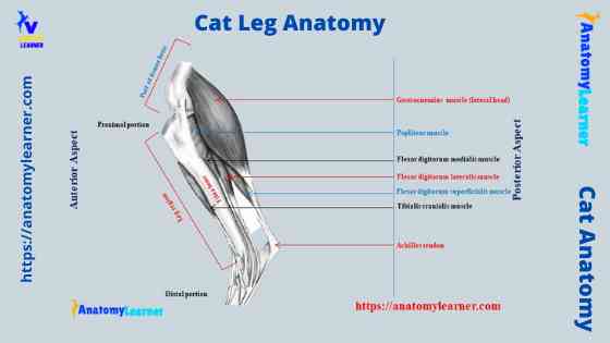
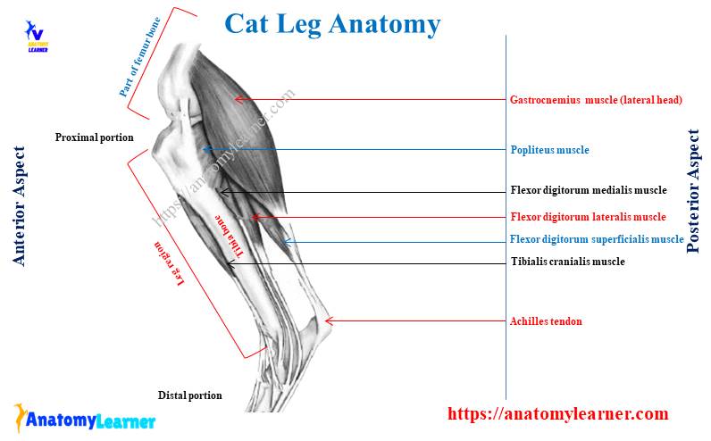
The most important muscles from the cat thigh are the biceps femoris, semitendinosus, semimembranosus, and abductor. Again, the leg region of a cat consists of both extensor and flexor muscles. Most of these extensor and flexor muscles supply to the pes of the cat.
The branches of the axillary artery supply to the front leg of a cat. In contrast, the external iliac artery branches supply the cat’s hind leg.
You will find almost 15 nerves in a cat’s brachial plexus, which supplies to the front leg. The branches of the lumbosacral plexus innervate the hind leg of a cat.
Special features of cats leg bones
I will not describe all the features of the cat leg bones. Rather, I prefer to provide the basic and special features from the cat’s leg bones. You will learn more about these cat bones in the next section of this article.
- The clavicle of a cat is a slender, rod-shaped curved bone with two extremities.
- You will find a triangular flat scapula laterally divided into two halves by spine.
- The coracoid process and supraglenoid tubercle are well-developed in the cats’ scapula.
- There is a very conspicuous ovoid slit occurring proximal to the medial epicondyle of the cat femur bone (supracondyloid foramen).
- The radius and ulna are separated bones in the cat’s front leg. You will find a deep radial notch in the proximal extremity of the ulna bone.
- There are seven small and irregular carpal bones present in a cat’s front leg.
- You will find five metacarpal bones in the manus, consisting of four developed and one ill-developed digit.
Now, let’s know some of the important osteological features from the hind limb bone. Here, I will enlist the most particular anatomical facts of the hind limb bones from cat skeleton anatomy.
- The os coxae of the cat are irregular, elongated, and complex structures that consist of ilium, ischium, and pubis bones.
- Ilium of both sides run parallel to each other as in dog. You will find a concave lateral surface in cats’ ilium bone.
- The posterior end of the cat ischium bone is rough, thick, but not trifid like the cattle.
- The tibia and fibula are two separated bones in the cat’s hind leg.
- There are seven tarsal and five metatarsal bones in the cat’s hind leg.
Muscles of cats front legs
Here, I will enlist some of the important muscles from both a cat’s front and hind legs. But, you will learn the detailed anatomical facts of these muscles from the cat leg anatomy. From the foreleg of a cat, I will show you the muscles from the shoulder, upper arm, forearm, and manus.
I will also show you the important muscles from the cat’s back legs’ hip, thigh, leg, and pes region. Okay, first, let’s find the below-mentioned muscles from the shoulder region.
- Supraspinatus muscle of the cat’s shoulder
- Infraspinatus muscle of the cat
- Subscapularis muscle of the cat
- Levator scapulae ventralis muscle
- Acromiodeltoid muscle
- Spinodeltoid muscle
- Teres major and teres minor muscles of a cat
Again, you will find different muscles in the upper arm or brachium of a cat’s front leg. Here, I would like to mention some of the important muscles from the brachium of a cat.
- Coracobrachialis muscle of the cat
- Biceps brachii muscle of a cat
- Triceps brachii muscle of a cat
- Brachialis and anconeus muscles of the front cat leg
- Epithrochlearis muscle of the cat
There are lots of muscles in the forearm of a cat. Mostly they are grouped into extensor and flexor groups. Let’s try to find out the below-mentioned muscles from the forearm of a cat. You will learn the details about these muscles from the next section of this article.
- Extensor carpi radialis longus and brevis muscles
- Extensor digitorum lateralis and communis muscles
- Abductor pollicis longus muscle
- Supinator and pronator muscles
- Flexor carpi radialis and ulnaris muscles
- Flexor digitorum profundus and superficialis muscles
- Brachioradialis muscle
- Pronator teres and quadratus muscles
These muscles will also run over the manus and supply to the digits.
Hind leg muscles of the cat
If you want to identify the back leg muscles, it will be better if you start with the hip region. In the hip region of a cat, there are different clinically important muscles. You know the hind limb divides into thigh, leg, and pes region. So, here I will also show you the most important muscles from these regions of a cat’s hind leg.
From the hip region of a cat, I will show you the following important muscles –
- Gluteus maximus, minimus, and medius muscles
- Pyriformis muscle
- Articularis coaxe muscle
- Gemellus cranialis and caudalis muscles
- Coccygeus muscles
- Obturator internus and externus muscles
- Quadratus femoris muscle of a cat
Again, from the thigh region of the cat’s hind leg, you might identify and learn the details anatomy of the below-mentioned muscles.
- Biceps femoris muscle of a cat
- Sartorius and gracilis muscles
- Tenuissimus muscle
- Semitendinosus and semimembranosus muscles of a cat
- Adductor femoris and longus muscles
- Pectineus muscle of a cat
- Quadriceps femoris muscle of the cat
In the quadriceps femoris muscle of a cat, you will find vastus medialis, lateralis, femoris, and intermediate muscles.
In addition, let’s identify the following muscles from the leg and pes region of a cat’s hind limb.
- Tibialis cranialis and caudalis muscles
- Extensor digitorum longus muscle
- Extensor hallucis longus muscle
- Peroneus longus, tertisu, and brevis of a cat
- Flexor digitorum longus, and brevis muscles
- Flexor hallucis longus muscle
- Popletius and soleus muscles
- Gastrocnemius muscle of a cat
- Plantaris and triceps surae muscles of a cat
Now, you might learn the details anatomical facts of these muscles of a cat’s legs. The cat leg muscle section will discuss the origin, insertion, and action of these muscles.
Blood vessels of cats limb
The arterial supply of the cats’ forelimb and hindlimb are so complicated. You should read them from a details guide dedicated to the anatomy of the arterial supply only. But, don’t worry, here I will show you the major arterial supply from the cat’s fore and hind legs.
First, see the arteries you should identify from the front leg of a cat. Here, I will enlist some of the major arteries from the cat’s front leg –
- Subclavian and its branches (vertebral, costocervical, internal thoracic, and superficial cervical)
- Axillary and branches (external thoracic, lateral thoracic, subscapular, thoracodorsal, cranial and caudal circumflex humeral)
- Brachial and its branches (deep brachial, bicipital, collateral ulnar, superficial brachial, transverse cubital)
- Common interosseous and its branches (ulnar, cranial and caudal interosseous, deep antebrachial)
- Median and its branches (radial and superficial palmar arch)
The branches of the superficial brachial, radial, median, and ulnar artery supply to the digits of the cat’s front leg. You will find the main arterial supply in the hind limb from the external iliac. I will show you all the branches of the external iliac artery of a cat with a labelled diagram.
Okay, first, let’s try to identify all of the following branches of the external iliac artery –
- Deep femoral and branches (pudendoepigastric, medial circumflex)
- Lateral circumflex femoral artery
- Femoral, popliteal, cranial and caudal tibia arteries, and dorsal pedal (same artery, continuation of femoral artery)
- Saphenous and its branches (cranial and caudal)
The branches of the femoral and saphenous arteries run over the pes and supply to the dorsal and palmar aspects of the digits. The digital arterial supply will be shown from the cat legs with labelled diagrams.
Nerves from cat legs
The basic brachial plexus formation is almost similar to the ruminant. But, the branches and distribution of the brachial plexus nerves are somewhat different in a cat’s front leg anatomy.
Here, I will enlist the major nerves from the brachial plexus. Later on, you will learn more about these nerves with a labelled diagram.
- The suprascapular nerve of a cat
- First subscapular nerve
- The axillary nerve of a cat
- The musculocutaneous nerve of a cat
- Second subscapular nerve
- Radial, median, and ulnar nerve (supply up to the digits)
- Posterior ventral thoracic nerve
- Medial cutaneous nerve
- Third subscapular nerve
- Long thoracic nerve
- The lateral thoracic nerve of a cat
- The thoracodorsal nerve of a cat
- Cranial and caudal pectoral nerves of the cat
Again, a cat’s basic lumbosacral plexus formation is similar to the ruminant. But, the branches and distribution of lumbosacral plexus nerves are somewhat different in a cat. Here, I will enlist the major nerves from the lumbosacral plexus.
- Lateral cutaneous femoral and femoral nerve
- The saphenous nerve of a cat
- Obturator nerve of a cat
- Ischiatic nerve and its branches (muscular branches, sural nerve, common peroneal, tibial nerve)
- Cranial and caudal gluteal nerves
- Posterior femoral cutaneous nerve
- The pudendal nerve of a cat
- The caudal hemorrhoidal nerve of a cat
Now, you may identify these nerves from the brachial and lumbosacral plexus and learn more about their anatomical facts.
Cat front leg anatomy
From the cat front leg anatomy, you might have learned the anatomical facts of the bones, muscles, vessels, and nerves. I hope you already got the basic idea of bones, muscles, vessels, and nerves from the cat’s front leg. I will describe all of these cat structures in detail with a labelled diagram.
“If you want to learn the anatomical facts of the bones, muscles, and nerves from the cat with video, you may get help from the video page.”
You will find great similarity in the anatomical facts of cats’ bones, muscles, and nerves with the dogs. Here, in anatomylearner, I have also published an article on anatomical facts of the dogleg. So, you may also read that article to know the similar anatomical features between dog and cat.
Cat leg bone anatomy
I know you have a good piece of knowledge on the name and number of bones from the cat’s front leg. Now, it is better to know their special identifying osteological features.
The cat’s clavicle is a slender, rod-like, curved bone in the skeleton. You will find two extremities in the clavicle of a cat where the sternal end is slightly enlarged.
Cat scapula
The scapula of a cat is a flat triangular bone that articulates with the humerus distally. You will find almost the same features in the cat scapula as you found in the ruminant scapula. There are two surfaces, three borders, and three angles in the cat scapula.
The spine divides the lateral surface of the cat scapula into two halves (anterior supraspinous process and posterior infraspinous process). On the medial aspect, you will find subscapular fossa. The tuberosity of the spine is curved and thicker in a cat. There is a sharp-edged metaacromion process just dorsal to the acromion process.
The dorsal border is curved and continuous with cranial border anteriorly and caudal border posteriorly. You will find a comparatively deep glenoid cavity in the cat scapula at the glenoid angle. There is a beb-like projection at the medial aspect of the anterior border of the glenoid fossa.
Again, the supraglenoid tubercle (laterally) is more prominent in a cat scapula than in a goat.
Cat humerus bone
The most upper long bone of the cat’s forelimb is the humerus. It consists of a slightly curved diaphysis and two epiphyses. You will find a smooth rounded medial head at the proximal epiphysis of the humerus bone.
Medial to the head, there is a prominent but small lesser tuberosity. Again, at the lateral aspect of the head, you will find a large greater tuberosity. There is a bicipital groove in between the lesser and greater tuberosities.
The distal extremity of the cats’ humerus consists of a large medial trochlea and a small lateral capitulum. Proximal to the trochlea, a coronoid fossa is present in the humerus. Again, you will find a radial fossa proximal to the capitulum.
Medial and lateral epicondyles are also present at the distal extremity of the cat’s humerus bone. The conspicuous ovoid slit occurs proximal to the medial epicondyle, known as the supracondyloid foramen.
You will find a deep olecranon fossa at the posterior aspect of the distal end. The supracondyloid ridge is very sharp and extends from the lateral epicondyle to the midpoint of the diaphysis.
Cat radius and ulna bones
The radius and ulna are also the long bones of the cat’s forelimb. They also contain a diaphysis and two extremities. The proximal extremity of the cats’ radius bone contains the head, neck, and articular circumference.
The head of the radius is slightly concave and articulates with the humerus bone’s capitulum. You will find a more prominent bicipital tuberosity at the poster-lateral aspect of the cat radius bone.
There is some interosseous crests just distal to the bicipital tuberosity of the radius bone. You will find a medial styloid process at the distal extremity of the cat’s radius bone.
The ulna is a fully separated bone in a cat’s forelimb consisting of two identifying osteological features. You will find an olecranon process at the proximal end and a styloid process at the distal end of the ulna bone.
Again, some other osteological features like the semilunar notch, radial notch, and coronoid process are present at the proximal end. You will also find the interosseous crest at the anterolateral aspect of the ulna bone of a cat.
Cat carpal and manus bones
The cat has seven small, irregular carpal bones arranged into two rows. Again, the manus consists of five metacarpal bones that connect to the proximal phalanges. You will also find three phalanges and associated sesamoid bones in the structure of a manus.
You will find a detailed guide on the cat carpal and manus bones in another article of anatomy learner. I want to suggest you read that article to get a good concept on the cat carpal and manus bones.
Cat front leg muscles anatomy
As you got the list of muscles from the cat front leg anatomy, I will skip enlisting muscles from the shoulder, brachium, antebrachium, and manus region. Fine, let’s know the anatomical facts of the cat muscles from the different regions of a forelimb.
Shoulder muscles
The supraspinatus muscle is thick in the supraspinous fossa of the cat’s scapula. It originates from the entire surface of the supraspinous fossa and inserts into the greater tuberosity of the humerus.
The infraspinatus muscle is also thick but somewhat smaller, filling the scapula’s infraspinatus fossa. This muscle originates from the surface of the infraspinatus fossa and inserts to the lateral surface of the greater tuberosity of the humerus.
The subscapularis is a large, medial, and triangular muscle that locates at the subscapular fossa of the cat’s scapula. It inserts to the dorsal border of the lesser tuberosity of the humerus.
The levator scapulae ventralis is a band-like muscle that emerges beneath the clavotrapezius. You will find two heads in the levator scapulae ventralis muscle that unite to form a strong band. This band insert into the ventral border of the metaacromion process of the cat’s scapula.
The acromiodeltoid is a flat muscle ventral to the levator scapulae ventralis muscle. This muscle originates from the acromion process and is inserted into the surface of spinodeltoid muscle.
The spinodeltoid lies ventral to acromiotrapezius and levator scapulae ventralis and caudal to acromiodeltoid muscles. It insert to the deltoid ridge of the cats humerus bone.
Teres major is thick, triangular muscle occupying the caudal border of the scapula. This muscle helps to flex and rotate the humerus medially.
On the other hand, teres minor is a small, somewhat triangular muscle between the infraspinatus and long head of triceps brachii muscles. It originates from the caudal border of the scapula near the glenoid fossa and inserts to the greater tuberosity of the humerus.
Brachium muscles
The coracobrachialis is a short, band-like muscle that lies on the medial aspect of the shoulder joint. You will find the origins of this muscle at the coracoid process of the scapula. Again, it inserts at the proximal end of the humerus bone.
Biceps brachii is a thick muscle that lies on the cranial surface of the humerus. This muscle helps to flex the forearm with the brachialis muscle.
You will find a large and lateral muscle (triceps brachii) in the arm region of a cat. This muscle consists of three head that originates from three different sites but inserts at the surface of the olecranon process of the ulna bone.
The three heads of these triceps muscles are – lateral, long, and medial heads. This muscle helps to extend the cat’s forearm.
The anconeus is a small and triangular muscle of the cat’s arm that covers the lateral surface of the elbow. It originates from the lateral epicondyle and inserts the lateral surface of the ulna bone.
There is a lateral muscle (brachialis) located along the humerus’s cranial surface. It lies partly obscured by the lateral head of the triceps brachii muscle. This muscle helps to flex the forearm and work with the biceps brachii.
You will find an epitrochlearis muscle on the medial surface of the cat’s arm. It is a delicate and flat muscle that partly overlies the triceps muscle. It originates from the lateral border of the latissimus dorsi muscle. This muscle works together with the triceps brachii that help extend the cat’s brachium.
Muscles of forearm
There is a narrow, bandlike brachioradialis muscle in a cat that extends along the radial border of the forearm. It inserts into the styloid process of the radius bone.
The extensor carpi radialis longus is a slender muscle that lies on the radial surface of the forearm. You will find the origin of this muscle at the lateral supracondyloid ridge of the humerus. This extensor muscle helps to extend the manus of the cats.
You will also find another slender but short muscle (extensor carpi radialis brevis) just medial to longus muscle. It inserts at the base of the metacarpal bones and also helps to extend the manus.
The extensor digitorum communis is a long, slender dorsal muscle that partially overlies the extensor radialis longus and brevis muscles. This muscle helps to extend the second, third, fourth, and fifth digits. On the other hand, the extensor digitorum lateralis lies lateral to the extensor digitorum communis.
The abductor pollicis longus is a flat muscle that originates from the ventrolateral aspect of the ulnar shaft. It extends and abducts the digits I of the cats’ manus.
There are five heads present in a cat’s flexor digitorum profundus muscle. Again, you will find two parts in the flexor digitorum profundus muscles of the cat. These two flexor muscles of the manus help to flex the digits.
There is also flexor carpi ulnaris that lies along the ulnar side of the ventral aspect of the lower forearm of a cat. Pronator teres is the ventral muscle that runs obliquely over the upper surface of the forearm of a cat. Again, you will find a pronator quadratus muscle that originates from the distal part of the ventral border of the ulna bone.
Special muscle of cats manus
There are lumbrical muscles in the structure of a cat’s manus. These are the small intrinsic muscles of the manus of a cat. They originate from the common tendon of flexor digitorum profundus muscles.
These lumbrical muscles are inserted at the base of the first phalanx of digits II to V. Do you know the function of these lumbrical muscles of the cat’s leg? Well, these lumbrical muscles help to blend the digits.
Cat front leg blood vessels (artery)
All the arterial supplies from the front leg are enlisted before. Before going to a detailed description of these arteries from the cat’s front leg, you may memorize them again. You see, all the arteries that supply to the front leg are the continuation of the axillary artery.
In the cat leg diagram, I tried to show you most of the axillary artery branches. But, here, I will describe only the main branches of the axillary and brachial arteries from the cat’s front leg.
Axillary artery of cat
The axillary artery is the continuation of the subclavian artery that extends from the first rib to the tendon of teres major muscle. You will find four major branches in the axillary artery of a cat –
- The external thoracic artery of the cat
- A lateral thoracic artery of a cat
- The subscapular artery of a cat and
- A cranial circumflex humeral artery of the cat
The external thoracic artery leaves the axillary artery near its origin. It curves around the cranio-medial border of the deep pectoral with the superficial pectoral nerve. This external thoracic artery supplies almost the entire to the superficial pectoral region of the cat.
The lateral thoracic artery of the cat leg runs across the lateral surface of the axillary lymph node. It also runs along the dorsal border of the deep pectoral ventral to the latissimus dorsi.
This lateral thoracic artery supplies to the latissimus dorsi, deep pectoral, and cutaneous trunci muscles of the cat.
The subscapular artery of the cat’s leg is larger and continuation of the axillary artery. It passes caudodorsally between the cat’s front leg’s subscapular and teres major muscles. Again, it becomes subcutaneous near the caudal angle of the scapula.
You will find two major branches in the subscapular artery of a cat –
- The thoracodorsal artery and
- A caudal circumflex humeral artery
The thoracodorsal artery leaves the dorsal surface of the subscapular artery near its origin. This artery supplies the teres major, latissimus dorsi muscles, and skin.
Again, the caudal circumflex humeral artery leaves the subscapular opposite the thoracodorsal artery. This artery courses laterally between the head of the humerus and the teres major muscle.
The cranial circumflex arises from the axillary artery distal to the subscapular artery.
Brachial artery of a cat
The brachial artery of a cat is the continuation of the axillary artery. It courses distally across the body of the humerus and ends at the craniomedial surface of the elbow. At this elbow joint, the brachial artery gives several branches.
You will find the deep brachial and bicipital arteries, which are the muscular branches of the brachial artery. Again, the other branches of the cat’s brachial artery include – collateral ulnar, superficial brachial, and transverse cubital arteries.
The collateral ulnar artery of the cat is a caudal branch of the brachial in the distal third of the arm. This collateral ulnar artery supplies the triceps muscle and elbow joint area.
The superficial brachial artery forms a loop around the cranial surface of the biceps brachii muscle. It continues as the cranial superficial antebrachial artery in the forearm of a cat.
The transverse cubital artery also arises at the level of the elbow joint and supplies to the adjacent muscles of this joint.
The brachial artery of a cat continues as the common interosseous artery at the proximal forearm of a cat. You will also find a small deep antebrachial artery at the forearm. Again, the brachial artery continues as the median artery at the mid of antebrachial of a cat.
You may also learn the detailed distribution of radial, median, and ulnar arteries in cats manus from another article.
Brachial plexus nerves of a cat
The brachial plexus is formed by the ventral branches of the sixth, seventh, and eighth cervical and first and second thoracic spinal nerves. You may get the basic idea of forming the brachial plexus of ox first. Then, it will be better to compare the formation of brachial plexus nerves and their distribution with cats.
The formation and distribution of the brachial plexus nerves in cats are almost similar to the dog. Here, I will describe a little about the different nerves from the brachial plexus.
Nerves of cats front leg
You will find ten to eleven nerves in the brachial plexus of a cat leg anatomy. In the diagram, I will try to show you all these nerves from the brachial plexus.
“All the nerves from cats brachial plexus are enlisted before. So, you may again see these nerves from that list.”
The suprascapular nerve originates and passes with the transverse blood vessels between the supraspinatus and subscapularis muscles. You will also find some of the small branches of the subscapular nerve that supply to the supraspinatus and subscapularis muscles.
The first subscapular nerve originates from the sixth and seventh cervical nerve and innervates the subscapularis muscle. Again, the axillary nerve runs along the posterior edge of the biceps brachii muscle. This axillary nerve supplies to the shoulder muscles (like teres major, deltoids, coracobrachialis).
The musculocutaneous nerve originates from the sixth and seventh cervical nerve in a cat. It supplies to biceps brachii, brachialis, coracobrachialis, and the skin of the forelimb.
You will get the second subscapular nerve that supplies to the teres major muscle of the cat. Again, the third subscapular nerve runs with the thoracodorsal artery and supplies to the latissimus dorsi muscle.
The anterior ventral thoracic nerve arises from the seventh cervical and supplies to a cat’s pectoral muscles. Again, the posterior ventral thoracic nerve runs long thoracic artery and also supply to the pectoral muscles.
You will also find a medial cutaneous nerve that arises from the first thoracic nerve and supplies the skin of the forelimb of a cat. Again, the long thoracic nerve of cat supplies to serratus ventralis muscle.
Nerves that supply to digits
The branches of the radial, median and ulnar nerves of the cat’s front leg supply the digits. So, let’s know about these nerves from the brachial plexus of a cat.
The largest nerve in the brachial plexus of a cat is radial that originates from the ventral branches of the sixth, seventh, eighth cervical, and first thoracic nerve. It runs along the deep brachial artery and innervates to the triceps brachii and extensor muscles of the cat’s forelimb.
The median nerve of the cat arises from the ventral branches of the cervical seven, eight and first thoracic nerve. This median nerve supplies to the flexor muscles of the forearm of a cat.
Again, the ulnar nerve of a cat arises from the eighth cervical and first thoracic nerve. It runs parallel to the median nerve and crosses the medial epicondyle of the humerus. The ulnar nerve of the cat is also supplied to the forelimb’s flexor muscles and in the digits.
Cat hind leg anatomy
From the cat hind leg anatomy, I will describe the anatomical facts of the bones, muscles, blood vessels, and nerves. I hope you got the basic idea of these structures from the hind leg earlier.
You will find great similarity in the osteological features of the cats’ hind leg bones with the dogs. Again, most of the muscles from the cat’s back legs are almost similar to that of a dog.
I already enlisted the bones, muscles, nerves, and vessels from the back leg. So, I will not enlist these bones, muscles, and vessels again from the cat legs.
I suggest you read the specific bone, muscle, nerve, and vessel from the ruminant anatomy section. You will find a specific video for each of these bones from the back leg. If you learn them well, it will be better for you to compare the anatomical features of these structures with cats or dogs.
Fine, let’s start to learn the details of the bones, muscles, vessels, and nerves from the cat’s back legs.
Bones of cats hind leg
The hind limb of a cat also divides into pelvic, thigh, leg, and pes regions like other animals. You will find the same bones (ilium, ischium, and pubis) in the formation of the pelvic girdle in a cat. The thigh of a cat consists of the femur bone.
Again, the leg region of a cat consists of the tibia and fibula bones. There are seven tarsal bones in the cat’s hind limb. The pes region of a cat comprises the metatarsal, phalanges, and associated sesamoid bones.
Bones of cats os coxae
The pelvic girdle is elongated, irregular shaped, and complex that forms with the paired os coxae. These paired os coxae articulate ventrally along the medial surface of the pubis and ischium.
The ilium is a cranially projecting bone of the os coxae of the cat. It is wing-shaped and contains a lateral concave surface. The dorsal portion of the wing is thick and forms a crest.
The ischium of a cat consists of a heavy body and slightly constricted ramus. A thicken, and roughened tuberosity is present at the posterior end of the ischium body. The spine of the ischium is fairly prominent at the posterior border of the ischium bone.
The pubis is the small bone of the cat os coxae that helps form the obturator foramen and the ischium. You might identify the most common and important osteological features from the os coxae bone altogether.
- Wing of the cat ilium bone
- Pubic and ischial symphysis
- Acetabulum of cats hip
- Ileopectineal line and eminence
- The spine of the cat’s ilium bone
- Tuberosity of the ischium bone
- The spine of the cats’ ischium bone
- Ramus of the cat pubis
- Pubic tubercle
- Obturator foramen and acetabular notch
The obturator foramen is the most prominent opening of the hip bone of the cats. This obturator foramen forms by the union of ichium and pubis bones.
All the os coxae bones structures are shown on the cat pelvic girdle labelled diagram.
Cat femur bone
The femur of a cat is a typical long bone that contains a middle shaft and two extremities. At the proximal end of the cat femur, you will find some important structures like the head, greater trochanter, trochanteric fossa, and ridge.
The head of the cat femur is the ball like projection that contains a fovea capitis. At the lateral aspect of the proximal extremity, you will find a greater trochanter. Medial to the greater trochanter, there is a trochanteric fossa. The greater trochanter terminates in a small triangular projection (lesser trochanter).
At the distal end of the cats’ femur, you will find two distinguished projections – lateral and medial condyles. Again, both the condyles possess small projections on their lateral aspect (epicondyles).
An intercondyloid fossa at the posterior aspect separates two condyles of the cats’ femur bone. You will find a smooth surface (trochlea) at the cranial aspect of the distal end of the femur. This smooth surface is for the patella bone.
Cat tibia and fibula bones
The tibia of a cat is the longer and more robust bone of the two lower legs. You will find a triangular diaphysis and two irregular extremities in the cat tibia bone.
There is lateral and medial condyle present in the proximal extremity of the tibia bone. You will also find a double-pointed projection (spine) at the proximal end. The tibial tuberosity is the triangle and continues as the tibial crest.
Again, at the distal extremity of the tibia, you will find two prominent projections – medial malleolus and dorsal projection. There is an articular facet for the fibula on the postero-lateral aspect of the tibia.
The cat fibula is a long slender bone containing the middle diaphysis and two extremities. At the proximal end of the cat fibula, you will find the head whose medial surface bears a smooth facet for articulation with the tibia.
Again, there is a distinguished lateral malleolus on the distal end of the cat fibula bone.
Cat hind leg anatomy muscles
From the cat hind leg muscles anatomy, I will show you the most important muscles from the different regions like hip, thigh, leg, and pes. In the hip region of a cat, there is a thin gluteus maximus muscle that originates from the transverse process of the last sacral and first caudal vertebra.
Again, the gluteus medius is a very thick muscle between the cranial tensor fasciae latae and the caudal gluteus maximus. Both these muscles help to abduct the thigh of a cat.
Pyriformis is a fan-shaped muscle that lies under the superficial gluteus muscle. The articularis coxae is a small flat muscle lying beneath the gluteus minimus muscle.
Gemellus cranailis is a fan-shaped muscle beneath the pyriformis muscle, whereas the gemellus caudalis is a flat muscle that lies caudal to obturator muscle. The obturator interus muscle of a cat originates along the medial surface of the ramus of ischium bone.
The quadratus femoris of a cat is the deepest muscle which is short and thick. It originates from the lateral surface of the ischial tuberosity and inserts along the ventral border of the greater and lesser trochanters.
Cat thigh muscles
The Sartorius is a thick band and narrow muscle in the thigh region of a cat. It originates from the crest and ventral border of the ilium bone and is inserted into the tibia, knee, and patella.
Gracilis is the thin muscle that lies in the caudal half of the medial surface of the thigh. This muscle helps to abduct and retract the legs.
The Biceps femoris is the large, thick muscle that covers the lateral surface of the thigh. It inserts to the proximal one-third of the tibia bone and lateral patella.
Tenuissimus is an extremely slender muscle that attaché to the biceps femoris. Just caudal to biceps femoris, you will find caudofemoralis muscle.
The semitendinosus is the long muscle that forms the caudal border of the cat’s thigh. It originates from the ischial tuberosity and inserts the medial surface of the tibia bone.
The semimembranosus is a thick muscle that lies cranial to the semitendinosus muscle. It inserts to the medial epicondyle of the femur and adjacent medial surface of the tibia bone.
The Adductor femoris is a broad muscle that lies cranial to the semimembranosus muscle that inserts the femur’s shaft. Again, the adductor longus muscle inserts into the middle portion of the femur bone of a cat. These two muscles help to adduct the cat’s thigh.
The tensor fascia latae is the thick and triangular muscle in a cat that originates from the ventral border of the ilium bone. Again, in the quadriceps muscles of a cat, you will find vastus medialis, vastus lateralis, vastus intermediate, and rectus femoris parts. All these parts of the quadriceps of a cat help to extend the leg.
Cat leg muscles
The tibialis cranialis is the large and flat muscle that lies on the craniolateral aspect of the tibia bone. This muscle originates from the proximal end of the tibia and fibula bones and helps to flex the cat pes. The tibialis caudalis is a slender and flat muscle under the flexor digitorum longus muscle.
The extensor digitorum longus is a long muscle that lies beneath the tibialis cranialis. It helps to extend the digits and flexes the pes of the cats.
The extensor hallucis longus lies deep to the tibialis cranialis and originates from the anterior surface of the fibula bone. Peroneus longus is the most superficial muscle on the lateral surface of the leg region of a cat. You will find the slender peroneus tertius muscle just beneath the peroneus longus.
The flexor digitorum longus of the cat leg is a long and slender muscle on the medial aspect. It originates from the head of the fibula and shaft of the tibia bone.
The flexor hallucis longus of a cat is a large muscle that lies lateral to the flexor digitorum longus on the posterior aspect of the leg. You will find two heads (lateral and medial) in the cat’s gastrocnemius muscle. This muscle helps form the powerful Achilles tendon and insert it to the proximal end of the calcaneus.
The planteris of a cat lies beneath the gastrocnemius muscle. Again, the soleus is a flat muscle just beneath the planteris muscles. This muscle helps to extend the cat’s pes with gastrocnemius and planteris muscles.
Special muscle of the pes
Most of the extensor and flexor groups of muscles run and supply to the pes region. The extensor digitorum brevis and flexor digitorum brevis are the most important muscle in the cats’ pes. Again, you will also find the lumbrical muscles in the pes structure of a cat.
The extensor digitorum brevis is a thin muscle that covers the dorsolateral surface of the tarsus and metatarsus of a cat. It originates from the proximal end of metatarsus III to V and helps to extend the cat toes.
Again, the flexor digitorum brevis muscle lies on the plantar surface of the toe and originates from the plantar tendon. This muscle helps to flex the cat’s toes.
Artery of the cat back leg
In a cat, the arterial and venous supply in the hindlimb is very similar. The right and left external iliac arteries arise from the abdominal aorta at the sixth and seventh lumbar vertebrae levels. It runs caudoventrally and is related laterally near its origin to the common iliac vein and psoas minor muscle.
The external iliac artery of a cat passes the abdomen and become continue as the femoral artery. Again, you will find the deep femoral artery, the only branch of the external iliac artery that arises inside the abdomen.
The deep femoral artery gives the small pudendoepigastric trunk and passes cranially on the dorsal surface of the rectus abdominis. It supplies the caudal half of the rectus abdominis and ventral part of the oblique and transverse muscles.
After giving off the pudendoepigastric artery, the deep femoral artery continues as the medial circumflex femoral artery. You will find some muscular branches of the medial circumflex artery.
Again, some of the small branches like lateral circumflex femoral proximal caudal femoral arteries are found in the external iliac artery of a cat.
Cat femoral artery
The cat femoral artery is the continuation of the external iliac artery at the distal two-thirds of the femur bone. You will find several branches in the cat femoral artery like superficial circumflex, lateral circumflex, proximal caudal femoral, saphenous, descending genicular, middle and distal caudal femoral artery.
The superficial circumflex iliac artery is the small branch that arises from the lateral side of the femoral artery. It courses cranially and supplies both parts of the Sartorius, tensor fascia latae, and rectus femoris muscles.
The lateral circumflex femoral artery is a large branch that runs between the rectus femoris and the vastus medialis muscles. This artery of the cat also supplies to the tensor fascia lata, superficial and medial gluteus and the hip joint capsule.
The proximal caudal femoral artery of the cat arise from the caudal surface of the femoral artery distal to the origin of the lateral circumflex artery. This caudal femoral artery of the cat supplies to the pectineus adductor muscles and enter deep into the gracious muscle.
The descending genicular artery arises from the femoral artery distal to the origin of the saphenous artery. This descending genicular of the cat supplies to the medial surface of the stifle joint.
The middle caudal femoral artery of the cat arises distal to saphenous and descending genicular artery and supplies to adductor and semimembranosus muscle. Again, the distal caudal femoral artery arises from the caudal part of the femoral artery.
Cat saphenous artery
The saphenous artery is another important structure in the cat leg anatomy. You will find this saphenous artery and the saphenous vein and nerve in between the Sartorius and gracias muscles.
The saphenous artery of a cat supplies to the skin on the medial aspect of the stifle joint and divides into cranial and caudal branches. The cranial branch of the saphenous artery arises opposite to the proximal end of the tibia bone. It crosses the medial surface of the cat tibia bone and passes distally on the cranial tibial muscle.
At the proximal part of the metatarsus bone of a cat, this cranial tibial artery ends as the dorsal common digital arteries.
Again, the caudal branches of the saphenous artery arise from the proximal end of the tibia bone. This artery lies between the medial head of the gastrocnemius muscle and the tibia bone. This vessel supplies the tarsus and metatarsus of a cat and gives rise to planter common digital arteries.
Cat popliteal artery
The popliteal artery of a cat is the continuation of the femoral artery that passes between two heads of the gastrocnemius muscle. It crosses the medial surface of the superficial digital flexor and runs over the flexor surface of the stifle joint.
The cat popliteal artery supplies the stifle, gastrocnemius muscle, and popliteal muscles of the leg. It ends as the cranial and caudal tibial artery in the cat’s legs.
The cranial tibial artery of a cat passes between the tibia and fibula bones. This cranial tibia artery supplies the fibularis longus, long digital extensor, and cranial tibial muscles.
The cranial tibia artery of a cat continues as the dorsal pedal artery that supplies to the pes region. Again, the cranial branch of the saphenous artery joins with the branch of the cranial tibia artery that also supplies to the pes region (digits).
Cat leg anatomy diagram
Now, I will show you all the structures from the cat leg anatomy with the labelled diagram. Here, I try to show you all the bones from the forelimb and hindlimb of a cat.
Again, all the muscles from both the forelimb and hindlimb of a cat are identified in the labelled diagram. You will find the more labelled diagram on the vessels and nerves from the cat legs in social media of anatomy learners.
I will try to update the cat leg labelled diagrams in future.
Frequently asked questions on cat leg anatomy
Here, I enlisted most of the common questions on cat leg anatomy that the learner or owner asks. I hope you will find the answers to your inquiries.
What are the parts of a cat leg?
In the front leg of a cat, you will find the shoulder, arm, forearm, and manus. The shoulder of the cat includes the scapula bone, whereas the arm consists of the humerus bone. Again, the forearm of a cat consists of radius and ulna bones. The manus of a cat consists of the metacarpal, phalanges, and sesamoid bones.
What are cat-back legs called?
The back legs of the cat are also known as the hindlimb. You will find the different regions in the hind limb of a cat. There are pelvic, thigh, leg, and pes regions found in the cat’s back legs. The os coxae of two sides form the pelvic girdle.
Again, the thigh region of a cat consists of the femur bone. The leg of a cat consists of tibia and fibula bones.
Do cats have bones in their paws?
Do cats have a pelvis?
You may also read some other articles related to cat anatomy from anatomy learner.
Conclusion
I hope all the information will help you acquire basic knowledge of the cat leg anatomy. All the diagrams also might help you get a good concept of the anatomical facts of the cat legs. The most important facts of the cat leg anatomy are the bones, muscles, joints, vessels, and nerves.
You will find some special muscles in the cat legs anatomy compared to a ruminant’s. Again, the nerves’ distribution pattern in cat legs is somewhat different compared to the ruminant.