

The dog cervical vertebrae form the basis of its neck. These cervical vertebrae are somewhat different from these of the ruminant or equine.
Quick summary of a dog’s cervical vertebrae: there are 7 cervical vertebrae in a dog where the first 2 (atlas and axis) are highly modified in their conformity. The third, fourth, and fifth show the typical features of vertebrae, whereas the sixth and seventh possess some peculiar osteological features.
In this article, I will show you all the osteological features of 7 cervical vertebrae from the canine neck. Again, you will find the difference among the cervical vertebrae of canines, horses, and ruminants with diagrams.
So that you will identify and differentiate dog or canine neck vertebrae from the other animals easily.
Dog cervical vertebrae
First, let’s know how many cervical vertebrae the dogs have. In the neck or cervical region of the dogs, you will find 7 vertebrae.
- First cervical vertebra of the dog – is highly modified and termed the atlas,
- Second cervical vertebra – also modified in its conformity and termed as axis,
- The third, fourth, and fifth cervical vertebrae – show the ideal features of the typical vertebra and differ little from each other,
- Sixth cervical vertebra – has expanded sagittal platelike transverse process, and
- The seventh cervical vertebra – possesses an articular facet on the caudal aspect of its body.
So, you may easily identify these dog cervical vertebrae with their special osteological features. As the first and second cervical vertebrae (atlas and axis) are highly modified in their conformity and greatly different from each other, you may easily identify them.
But, it is very hard to differentiate the third, fourth, and fifth cervical vertebrae from each other. This is because they (cervical – 3, 4, and 5) show almost similar osteological features.
On the other hand, the sixth cervical vertebra of a canine shows a unique transverse process. And the seventh shows only one pair of caudal facets for the head of the first pair of ribs.
So, you may also identify and differentiate the sixth and seventh cervical vertebrae from the dog’s neck quickly.

Suggested reading from anatomy learner – if you want to know the details of osteological features from the typical vertebrae, you may read the following article –
- Typical vertebrae of animal – body, arch, and processes with the labelled diagram,
Identification of dog cervical vertebrae
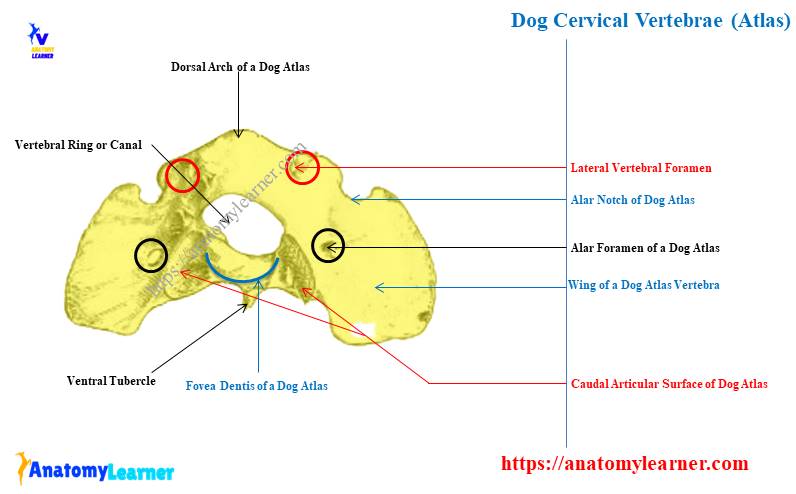
Here, you will identify the different osteological features of the canine cervical vertebrae. First, let’s see the osteological features from the first cervical vertebrae or atlas –
- Cranial oval articular surface,
- Dorsal and ventral arch of the dog atlas vertebra,
- Wing of the dog atlas,
- Dorsal and ventral tubercles of the dog atlas,
- Lateral vertebral foramen,
- Alar foramen of the dog atlas,
- Alar notch,
- Caudal articular fovea of the dog atlas, and
- Fovea dentis of the atlas,
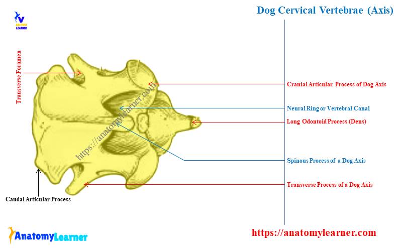
Now, let’s see the main osteological features of the dog’s second cervical vertebra or axis. In the dog axis, you will find the below-mentioned osteological features –
- Long and pointed dens or odontoid process of dog axis,
- Cranial articular process of the dog’s axis,
- Body, arch, and spinous process of the axis,
- Caudal articular process of axis,
- Transverse foramen of dog axis,
- Transverse process of the dog axis bone, and
- Caudal epiphysis of the axis,
Now let’s see the osteological features from the third, fourth, and fifth cervical vertebrae of the dogs. Here, in these three cervical vertebrae, you will find the following osteological features in common –
- Body, arch (lamina and pedicle) of the cervical vertebrae,
- Cranial and caudal articular process,
- Spinous and transverse processes, and
- Vertebral and transverse foramina,
From the sixth and seventh cervical vertebrae of the dogs, you might identify the below-mentioned osteological features –
- Body and different parts of the arch,
- Different processes – spinous, articular, and transverse,
- The sixth cervical present – platelike transverse process, and transverse foramen,
Again, the seventh cervical vertebrae don’t possess any transverse foramen. But, you might identify the caudal costal fovea from the caudal part of the vertebral body.
Unique features of canine cervical vertebrae
If you follow the previous part of this article, you may easily understand the unique features of the dog’s cervical vertebrae. For better understanding, you may also read the below-mentioned article as it provides the basic osteological facts of different (including cervical) vertebrae from animals –
Animal vertebrae identification – anatomy of cervical, thoracic, and lumbar vertebrae,
Okay, let’s enlist the unique features of the canine cervical vertebrae –
- There are 7 cervical vertebrae where the first two greatly differ from each other,
- The first cervical or atlas shows 2 strong curved wings and is considered the most atypical vertebrae,
- Cranial oval-shaped articular process and caudal fovea dentis other most important osteological features of a dog’s atlas,
- The presence of longer and pointed odontoid processes (dens) is the peculiar osteological feature of the dog’s axis vertebra,
- The third to fifth cervical vertebrae of the dogs possess the typical features of a vertebra, and hard to differentiate them from each other,
- Again, the canine sixth cervical has expanded platelike transverse process,
- The unique feature of the canine seventh cervical vertebrae is the presence of a caudal costal facet,
Now, you may learn the details of osteological facts from every single cervical vertebra from the dogs.
Canine cervical vertebrae anatomy
From dog cervical vertebrae, you might describe the feature of the followings structures –
- Osteological facts of the cervical vertebra body,
- Features of the pedicle and lamina of the cervical vertebrae, and
- Osteological features of the different processes of the canine cervical vertebrae,
Okay, first start with the atypical cervical vertebra (atlas and axis) from the canine neck.
Dog atlas anatomy – atypical cervical vertebra
Atlas is the first and atypical cervical vertebra of the dog’s neck. What might you describe here on the dog atlas anatomy? Well, you might describe the following features from the dog atlas anatomy –
- Curved plate or transverse process or wing of the dog atlas vertebra,
- Dorsal and ventral arches of the dog atlas, and
- Different articular surfaces from the dog atlas vertebra,
Let’s see the unique osteological features from the dog atlas vertebra anatomy –
- You will not find any defined body and spinous process in the dog atlas vertebra,
- The dog atlas has a strong ring (vertebral foramen) that is formed by two lateral wings or a modified transverse process,
- The dorsal and ventral arches connect the two lateral masses (wings),
- On the cranial aspect of the dog atlas, you will find two oval articular surfaces that receive the occipital condyle of the skull,
- The caudal articular facet is somewhat saddle-shaped and dorsally separated by a wide area,
- The dorsal tubercle of the dog atlas is tuberous and located on the cranial end of the dorsal arch,
- Again, the ventral tubercle of the dog atlas is projected from the caudal end of the ventral arch,
Here, the dorsal tubercle of the dog atlas is bifid, whereas the ventral tubercle is in the conical form. The dorsal surface of the ventral arch bears the fovea dentis. It is concave from side to side and articulate with the dens (odontoid process) of the dog axis
Now, this caudal part of the dog atlas blends with the articular area of the caudal surface of the lateral masses (wings). This bending part of this vertebra is known as the caudal articular fovea or surface.
Wings and foramina from dog atlas
The thick lateral part of the wing is the lateral masses. From these lateral masses, the modified transverse process arises. Sometimes this modified transverse process of the dog atlas is known as the wings.
On the wings of the dog atlas, you will find different osteological features. You will see 2 important foramina on the wings of the dog atlas bone –
- An alar foramen – is a short canal that passes obliquely through the transverse process or wings. Within this alar foramen, the vertebral artery and vein pass.
- The lateral vertebral foramen – passes through the cranio-dorsal part of the vertebral arch. The first cervical spinal nerve and vertebral artery pass from this lateral vertebral foramen of the dog atlas.
You will also find the alar notch on the cranial border of the base of the transverse process of the canine atlas. The vertebral artery pass through this alar notch of the transverse process.
You will also see the fossae atlantal on the ventral aspect of the canine atlas (wings). This is a deep depression where the vertebral artery and vein traverse.
Now, let’s see the osteological difference between the atlas vertebra from various species like ox and horses. Table 1 shows the main osteological difference among the ox, horse, and dog’s atlas vertebrae –
| Features | Dog Atlas | Ox Atlas | Horse atlas |
| Wings | Flattened wide | Less curved | More curved |
| Ventral arch | Not developed | Thick | More Thick |
| Dorsal arch | Bifid | Larger | Larger |
| Alar formaen | Present | Present | Present |
| Alar notch | Absent | Absent | Present |
| Transverse foramen | Absent | Absent | Present |
Dog axis – second cervical vertebra anatomy
Typically, the dog axis vertebra has the 3 parts of the typical vertebra. But, some of the osteological features from this axis vertebra are modified.
What structures might you describe here on the dog axis vertebra? Well, let’s see the structures that you might describe for the dog axis vertebra –
- Body of the dog axis vertebra,
- Arch of the dog axis vertebra, and
- Three different processes of the dog axis vertebra,
Let’s see the unique osteological features from the dog axis vertebrae with the labelled diagram –
The dog axis vertebrae represent the elongated dorsal spinous process which is bladelike cranially and expanded caudally,
This spinous process of the dog axis vertebra overhang the cranial and caudal articular surfaces,
You will find the most unique cranioventral pointed or peglike eminence (known as the dens or odontoid process) in the dog axis vertebra,
The odontoid process of the dog axis and the cranial articular surface attaches to the preceding vertebra of the dog’s neck. Here, the long odontoid process of the dog axis lies within the vertebral foramen of the atlas.
The odontoid process is held within the vertebral foramen of the dog’s atlas with the help of the transverse ligament. If there is any abnormality in the structure of the odontoid process (dens) of the dog’s axis, subluxation may occur on the neck.
Arches and other processes of the dog axis vertebra
The arches of the dog axis show the typical features of the typical vertebra. Here, the cranial notch occurs on both sides of the pedicle.
These cranial notches on both sides form the larger cranial intervertebral foramina. The second pair of the cervical spinal nerve and spinal vessels pass through the cranial intervertebral foramina.
Again, you will find small caudal notches on the caudal aspect of the arch that forms the intervertebral foramina with the cranial notches of the third cervical vertebra. The third pair of the cervical spinal nerve and spinal vessels pass through this intervertebral foramen.
You will not find any cranial articular process on the dog axis vertebra. But, the caudal articular process is typical and backward in the dog axis vertebra.
The transverse process of the dog axis is small and undivided. It projectes caudally and possess a small transverse foramen.
Now, let’s see the osteological differences of the second cervical vertebrae of ox, horse, and dog from table 2 –
| Features | Dog axis | Ox axis | Horse axis |
| Odontoid process | Longer and pointed | Short and wide | Narrow and longest |
| Spinous process | Elongated and bladelike | Short, projected caudally | Larger and divided |
| Intervertebral foramen | Small | Large | Larger |
Typical dog neck vertebrae – third, fourth, and fifth cervical
The dog’s third, fourth, and fifth cervical vertebrae show similar osteological features as a typical vertebra. So, within these all three dog’s cervical vertebrae, you will find the followings –
- Body of the third, fourth, and fifth cervical vertebrae,
- Arches (pedicle and lamina) of the third, fourth, and fifth cervical vertebrae, and
- Processes of the third, fourth, and fifth cervical vertebrae,
The body of these cervical vertebrae is more or less cylindrical in other species like ox and horses. But, they have dorsoventrally compressed in the dog cervical vertebrae.
You will find two extremities and 2 surfaces on these three typical vertebrae of dogs. Let’s see the features of the surfaces and extremities from the canine typical cervical vertebrae –
- The cranial surface of these vertebrae is convex, and the caudal surface is concave,
- The dorsal surface of these vertebrae is flattened and enters into the formation of a vertebral canal,
- Again, the ventral surface of these vertebrae bears small ventral tubercles,
All these typical vertebrae also possess ventral pedicles and dorsal lamina. The ventral pedicle of the dog’s typical cervical vertebrae possesses the cranial and caudal notches that form the intervertebral foramina.
Again, the dorsal lamina of these typical cervical vertebrae are platelike and complete the roof of the vertebral canal. The articular, transverse, and spinous processes are well-developed in these cervical vertebrae of the dogs.
Unique features of dog third, fourth, and fifth cervical vertebrae
Now, let’s see the unique features of these canine typical cervical vertebrae (third, fourth, and fifth cervical) –
- The osteological features of these cervical vertebrae differ slightly from each other,
- The length of the spinous process increase from the third to the fifth cervical vertebrae,
- You will find the larger lamina in the third cervical vertebrae; then they become gradually shorter and narrower,
- The transverse process is prolonged and twisted and gradually located to the dorsal aspect and becomes short in the fifth cervical vertebra,
You will find the pair of transverse foramina on the transverse process where the body and arch attach together. Within these transverse foramina of the typical cervical vertebrae contain the vertebral nerves and vessels.
Now, let’s see the main differences among the cervical vertebrae of dogs, horses, and ox from table 3 –
| Features | Dog cervical | Ox cervical | Horse cervical |
| Body | Compressd dorsoventrally | Shorter | Larger |
| Ventral crest | Not prominent | Prominent | More prominent |
| Cranial articular process | Almost dorsally | Dorsomedially | Dorsomedially |
Sixth and seventh cervical vertebrae of the dogs
As the sixth and seventh cervical vertebrae of dogs possess some unique features along with other typical features, you may easily identify them. Okay, first, let’s see the osteological features from the dog’s sixth cervical vertebra –
- The sixth cervical vertebra of a dog possesses an expanded sagittal platelike transverse process (known as the lamina ventralis),
- You will see the higher spinous process on the sixth cervical vertebra of the dog compared to the others,
- The lamina ventralis of the sixth cervical vertebra extends ventrally and caudally. It represents only the caudal part of the transverse process.
- Like other cervical vertebrae, the canine sixth cervical vertebra also possesses the transverse foramen.
Now, let’s see the peculiar osteological features from the seventh cervical vertebra of the dog –
- This is the last cervical vertebra of the dog neck that lacks of the transverse foramen,
- A higher spinous process is present in the seventh cervical vertebra of the dog,
- The body represents two oval small costal facets for the articulation with the head of the first pair of rib,
So, you can identify and describe all the features of the canine cervical vertebrae.
Dog cervical vertebrae anatomy labeled diagram
In this part of the article, you will see the more labeled diagrams of the dog cervical vertebrae. Here, the labeled diagram shows all the osteological features from the dog’s typical cervical vertebrae.
The labelled diagrams identify the body, arches (pedicle and lamina), and three main processes from the canine cervical vertebrae.
Now, the labeled diagram identifies all the unique osteological features from the dog’s first cervical vertebra. Here, the dog atlas labelled diagram identifies the wings, dorsal and ventral arches, and different foramina.
The alar foramen, lateral intervertebral foramen, and alar notch from the dorsal aspect of the wing are identified in the dog atlas labeled diagram. Again, the atlantal fossae from the ventral surface of the wings are also identified in the labeled diagram.
Now, let’s see all the identified osteological features from the dog axis labeled diagram. Here, the labeled diagram identifies the odontoid process and spinous process from the dog’s axis vertebra.
The dog’s sixth cervical vertebrae also identify the platelike transverse process. Again, the longer spinous process and caudal costal fovea are identified from the dog’s seventh cervical vertebra.
You may find more labeled diagrams on the dog’s cervical vertebrae here on social media of anatomy learners.
Frequently asked questions on dog cervical vertebrae
Now, let’s see the commonly asked questions on the canine cervical vertebrae. Here, I will enlist the most frequently asked questions on the canine cervical vertebrae that the anatomy learners ask.
You will only find the concise answer to these questions related to the dog’s cervical vertebrae. So, reading the full article to get an overview of the canine cervical vertebrae is recommended.
Okay, let’s see the questions on cervical vertebrae with the concise answer –
Do dogs have 7 cervical vertebrae?
Yes, the dog has 7 cervical vertebrae named atlas, axis, third, fourth, fifth, sixth, and seventh. Here, the first two – atlas and axis are modified in their structure and shows unique function.
Again, the third, fourth, and fifth cervical vertebrae show almost similar osteological features. The sixth and seventh cervical possess some peculiar osteological features that are already described in this article.
What is C1 cervical vertebra in a dog?
The C1 cervical vertebra in a dog is the first cervical vertebra in the neck. Anatomically, the first C1 cervical vertebra has two flattened, strong wings forming the neural ring.
The arches of the C1 (first cervical vertebra) are not well-developed and possess dorsal and ventral tubercles. Caudal aspect of the dog C1 possesses the fovea dentis for articulation with the odontoid process of C2.
What do C1 and C2 mean in dogs?
Here, C1 in dogs mean the first cervical vertebra, whereas C2 mean the second cervical vertebra of the neck. All the osteological features from C1 and C2 of dogs are identified and described here in this article with the labeled diagrams.
The most interesting fact from the C1 is the presence of flattened wings and associated structures. Again, the most important fact of C2 in dogs is the presence of the odontoid process.
What is the cervical part of a dog?
The cervical vertebral form the basis of the cervical part of the dogs. This is actually the neck, where you will find the cervical bones, muscles, nerves, vessels, and lymphatics.
Anatomy learners describe all these features from the cervical dog part in other articles.
Conclusion
So, the cervical vertebrae of the dog neck consist of seven bones, where the first two are highly modified. You might learn the unique features of the first and second cervical vertebrae with the diagram.
Now, you should identify all these osteological features from the real samples of dog cervical vertebrae from your anatomy laboratory.
