Cow Tongue Anatomy – Torus Linguae, Lingual Fossa, and Papillae
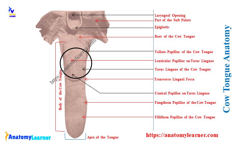
The cow tongue anatomy possesses three distinct parts that made of muscles, nerves, vessels, lingual glands, and tonsils. You will find different identifiable features on the various surfaces of the cow tongue. I will show you these important distinguishable anatomical facts from the cow tongue with the diagram. Quick overview: the cow tongue has a … Read more