The cow heart anatomy comprises both external and internal essential features. It occupies the more significant part of the middle mediastinum space (thoracic cavity) of a cow.
The shape of a cow heart is somewhat irregular and flattened cone (hollow muscular organ). For a veterinary student, it is essential to know the exact location of the cow heart and its different important anatomical facts.
In this article, I will show you (share) the exact location of the cow heart along with both external and internal anatomical features.
Quick summary: the cow heart anatomy consists of 2 receiving (atria) and 2 discharging (ventricles) chambers internally. Externally the base and apex are more visible and covered by the serous pericardium. In addition, there are 2 distinct surfaces and 2 borders in the anatomy of a cow’s heart.
I will share different labeled diagrams and actual pictures of the cow heart structure so that you can easily understand every fact. Make sure you read the session summary from each section of this article.
Cow heart anatomy
The cow heart is a hollow muscular organ of the cardiovascular system that acts as a central pump for systemic and pulmonary blood circulation. It locates at the ventral half of the middle mediastinum (extends from the third – sixth ribs) of the thoracic cavity of the cow.
First, let’s see what you should cover under the cow heart anatomy –
- External appearance – includes the location, attachments, surfaces, borders, and features of the pericardium,
- Internal structures – consists of the features both from the atrium and ventricle (both right and left), and
- Great vessels that arise from the cow heart – include the aorta (ascending and descending), pulmonary trunk,
Now, I will show you the different important external and internal anatomical features of the cow heart with the labeled diagram.
Cow heart identification
Let’s try to identify the below-mentioned external features from the cow heart structure –
- Pericardium – serous covering of the cow heart,
- Broad upper base and narrow pointed apex (ventrally) of the cow heart,
- The diaphragmatic surface of the cow heart (right atrial),
- The sternocostal surface of a cow heart (left auricular),
- Grooves on the external surface – longitudinal (2), transverse or coronary groove (encircled the heart),
- Right and left coronary arteries of the cow heart,
- Right ventricular border (cranial) of the cow heart, and
- Left ventricular or caudal border of cow heart,
You may also find all the great vessels (aorta, pulmonary trunk, pulmonary veins) from the external view of the cow heart. But I will show them all from the internal view of the cow heart structure.
Okay, let’s see the essential internal anatomical features from the 4 chambers of a cow heart (2 atria and 2 ventricles) –
First, let’s identify the 2 atria and 2 ventricles (both right and left) from the cow’s heart. Now, I will show you the specific features of each atrium and ventricle of the cow heart.
The right atrium and ventricle identification
- Sinus venarum cavarum – into which veins open (cranial and caudal venacava),
- The right auricle of the heart,
- Right atrioventricular orifice with the tricuspid valve, and
- Pectineus muscle in the right atrium’s wall,
There are some other features like vena Hemi azygos and fossa ovalis in the right atrium of a cow heart that I will describe in the details anatomical section of this article.
Again, from the right ventricle of the cow heart, you might identify the below-mentioned features –
- Conus arteriosus and pulmonary trunk,
- A small semilunar valve on the right atrium,
- Trabeculae carneae of the right atrium – ridge, papillary muscle, and septomarginal trabeculae (moderator band),
- Chordae tendineae on the right ventricle,
I hope you can identify all the above-mentioned anatomical facts from the right atrium and ventricle of the cow heart structure. Now, let’s find the exceptional and common features in the left atrium and ventricle of a cow heart compared to the right atrium and ventricle.
The left atrium and ventricle identification
Now, I will show you the anatomical facts from the left atrium and ventricle of the cow heart. Let’s try to identify the below-mentioned facts from the left atrium and ventricle of the cow heart –
- Opening of the pulmonary veins on the left atrium,
- Left auricle from the atrium,
- Pectinate muscle on the left atrium (less compared to the right),
- Left atrioventricular orifice with the bicuspid or mitral valve,
Again, in the left ventricle of a cow heart, you will see the below-mentioned anatomical features –
- A larger cavity in the left ventricle of a cow heart,
- Aortic orifice and origin of the aorta,
- Semilunar or aortic valve on the origin of the aorta,
- Few but larger chordae tendineae,
- Larger papillary muscles,
- 2 larger moderator bands on the left ventricle, and
- The interventricular septum of the cow heart,
Overall the trabeculae carneae (chordae tendineae, papillary muscle, and moderator bands) are more in the left ventricle of a cow compared to the right ventricle. If you want to learn (read) the details of these anatomical features from both atrium and ventricle of a cow heart, let’s continue this article till the end.
Unique facts of cow heart anatomy
I hope you may now enlist the unique features that represent the cow heart anatomy. Let’s see what the unique anatomical features of a cow heart are –
- The cow heart is a hollow muscular organ that lies at the more significant part of the middle of the mediastinum,
- It possesses a wider base and narrow apex and is covers with the serous pericardium,
- There are 2 surfaces (diaphragmatic and sternocostal) and 2 borders (right and left ventricular) in a cow heart,
- You will see a distinct transverse groove (posses the coronary arteries) and many longitudinal grooves in the external surface of a cow heart,
- There are 4 distinct chambers (2 atria and 2 ventricles) in the cow heart structure,
- The right atrium is a thin-walled smaller muscular chamber compared to the left one (thick-walled and larger) in the cow heart,
- The left ventricle of a cow heart is a thin-walled smaller chamber compared to the right ventricle,
- More significant veins like the cranial and caudal vena cava empty into the right atrium of a cow’s heart, whereas the pulmonary veins drain blood from the lung into the left atrium,
- Again, the pulmonary trunk arises from the base of the right ventricle of a cow hear, whereas the aorta arises from the base of the left ventricle,
- The trabeculae carneae are more in the wall of the cow’s left ventricle compared to the right ventricle,
Now, you may learn the details of anatomical facts from both the external appearance and internal structures (atrium and ventricle) of a cow heart with the labeled diagram. I will also compare the different structures from both the right and left atrium as well as the ventricle of the cow heart.
Cow heart location
Most of the part of a cow heart (5/7th) remains on the left side of the thoracic cavity (at the ventral half of the middle mediastinum). This is due to the larger right lung in the cow’s thorax, which extends from the 1st rib to the 10th rib.
You may learn more about the location and anatomical facts of the right and left lungs of animals (dog) from the below-mentioned article –
Here in this article, you will also find the unique features of the cow lung lobes compared to the dogs.
The direction of the cow heart – the heart is located median plane of the middle mediastinum, and its log axis is oblique (less than a horse),
If you want to identify the exact location of the cow heart, you should consider the location of its base and apex. You know the base of a cow heart is an upward wider part, whereas the apex is the downward directed narrow pointed part that closes to the diaphragm.
Now, let’s see how you will identify the location of the base and apex from the cow heart –
- The base of the cow heart – lies opposite the thoracic wall that extends from the 2nd intercostal space to the 5th intercostal space (3rd – 6th rib),
- The apex of the cow heart – lies opposite the 6th chondrosternal joint,
You may follow the article that I mentioned below, where I showed how you might practically identify the area of a cow heart –
Please see the section where I described the animal’s heart identification from the surface approach.
The external appearance of the cow heart
Under the external appearance of a cow heart anatomy, I will show you the followings –
- Attachment of the cow heart,
- Serous pericardium (covering of the cow heart),
- Surfaces of the cow heart, and
- Borders of the cow heart,
Now, let’s see the anatomical feature of these structures and parts of the cow heart. Let’s start with the attachment of the cow heart within the thoracic cavity of a cow.
How is the cow heart attached to the thoracic cavity?
The serous pericardium covers both the base and apex of the cow heart. At the broader base of the cow heart structure, you will find the following attachement –
- Aorta (descending and ascending) and pulmonary artery,
- Brachiocephalic trunk (have a different origin but continue from the aorta),
- Cranial and caudal vena cava (2), and
- 4 – 5 pulmonary veins (number may vary in different cows),
The narrower apex of the cow heart covers the pericardium, which attaches to the dorsal surface of the sternum by 2 ligaments. These are the right and left peri-cardiaco-sternal ligaments of the cow heart. They are the fibrous pericardium of the pericardial sac.
What is pericardium on cow heart?
The pericardium on a cow heart is a serous fibro sac that encloses the heart and great vessels like the aorta, pulmonary trunk, and cranial and caudal vena cava. You will find 2 parts in the structure of the pericardium or pericardial sac –
- Fibrous pericardium, and
- Serous pericardium,
Here, the fibrous pericardium of the pericardial sac is thin, strong, and inelastic in nature. It helps to form both the right and left peri-cardiaco-sternal ligament in the cow heart.
The serous pericardium is a close sac-like structure that also contains the following layers –
- Parietal layer of the serous pericardium – lines fibrous pericardium and remain closely attached, and
- The visceral layer of the serous pericardium – generally covers the heart (base) and great vessels,
The visceral layer of the serous pericardium is also known as the epicardium of the heart. You will find a small amount of fluid in between the parietal and visceral layers of the serous pericardium of the cow heart. This is the liquor pericardii of the pericardium of a cow heart.
Surfaces and grooves on cow heart anatomy
In the cow heart structure, you will find the below-mentioned surfaces –
- Diaphragmatic surface – considers as the right atrial surface, and
- Sternocostal surface – regarded as the left or auricular (ventricular) surface,
As the cow heart is cone-shaped, both surfaces are convex. You will find different grooves on the external surface of the cow heart –
- Transverse or coronary groove on the upper part, and
- Longitudinal or interventricular grooves (2),
These grooves (primarily transverse and longitudinal) indicate the division of the heart into 4 chambers. Here, the transverse or coronary groove encircles the cow heart at the base of the ventricle. But, you will find a little interruption at the origin of the pulmonary trunk.
This transverse groove indicates the division between the atrium and ventricle of the cow heart structure. Now, the longitudinal groove considers the septum between the right and left ventricles of the cow heart.
So, you will also find the right and left longitudinal grooves on the external surface of the cow heart –
- Right longitudinal groove – caudal in position and begins at the coronary groove and ends at the ventral to termination of the caudal vena cava,
- Left longitudinal groove – cranial in position and begins at coronary groove just caudal to the origin of the pulmonary trunk,
The left longitudinal groove of the cow heart descends almost parallel to the left ventricular border. On the other hand, the right longitudinal groove of the cow heart passes towards the apex. But, these two grooves do not meet at the apex of the cow heart.
You will also find some of the intermediate longitudinal grooves in the cow heart that descend from the transverse groove.
What structures pass within the grooves of a cow heart?
The left and right coronary arteries, along with their different branches, runs through these grooves (longitudinal, intermediate, and transverse) of the cow heart. I will show you the details course, of the coronary arteries in the heart’s blood supply section.
Borders of cow heart
Generally, the cow heart structure shows 2 important borders –
- Right ventricular or cranial border of the cow heart, and
- Left ventricular or caudal border of the cow heart,
The right cranial ventricular border of a cow heart is strongly convex and curves ventrally and caudally. Again, the more significant part of the right ventricular border runs parallel to the sternum.
The left ventricular or caudal border of the cow heart is nearly verticle. It is much shorter compared to the right ventricular border of the cow heart.
You will find the left ventricular or caudal border of the cow heart opposite the sixth rib and fifth intercostal space.
Cow heart chambers
In the internal cow heart anatomy, you will find the 4 distinct chambers –
- The right atrium of the cow heart – receives blood through the cranial and caudal vena cava,
- The right ventricle of the cow heart – discharge blood through the pulmonary trunk into the lungs of the cow,
- Left atrium of the cow heart – blood receiving chamber through the pulmonary veins (number of veins varies from 5 – 8),
- The left ventricle of the cow heart – blood discharging chamber through the aorta and brachiocephalic trunk into the all body and head regions,
I will show you the anatomical facts of these 2 blood receiving and 2 blood-discharging chambers with the labeled diagram. If you want to know – how they receive and discharge blood throughout the body, head, and heart, you may read the following article –
Here, I showed the general systemic and pulmonary blood circulation flow chart and the diagram.
Okay, let’s go back to the anatomical facts of the different chambers of a cow heart structure.
Right atrium of cow heart anatomy
The right atrium of a cow heart is a thin-walled muscular chamber. Its cavity is smaller than the left atrium of the heart.
The right atrium is from the right cranial part of the base of the heart and lies dorsal to the right ventricle. You will find the following structures in the right atrium of a cow heart –
- Sinus venarum cavarum (sinus venosus) – into which larger veins open,
- Right larger auricle,
- Muscles in the wall of the (cow’s heart) right atrium,
Here, the sinus venarum cavarum is the part of the cavity between the opening of the cranial and caudal vena cava and the atrioventricular orifice. Sometimes it is also known as the sinus venosus of the right atrium of the cow heart structure.
You will see the right auricle, which is the conical diverticulum. It curves around the right and cranial surface of the ascending aorta of the heart. The blind end of the right auricle appears on the left side, just cranial to the origin of the pulmonary trunk.
Again, the wall of the right atrium of a cow heart is almost smooth except on the right and in the auricle. You will find numerous muscular ridges that direct in various directions.
These are the comb-like structure and are considered the pectinate muscle of the right atrium of a cow heart.
5 chief opening of the right atrium of cow heart
You will find 5 chief openings in the right atrium of a cow heart structure. First, let’s see and try to identify these 5 chief openings from the right atrium of a cow heart –
- Opening of the cranial vena cava – in the dorsal part, chiefly opposite to the 4th rib,
- Opening of the caudal vena cava – at the caudal part, just opposite the 5th intercostal space,
- Coronary sinus – oepns ventral to the caudal venacava,
- Foramina venarum minimarum – opening of the small veins which empty directly into the cavity of the right atrium, and
- The right atrioventricular orifice – is the ventral part and leads into the right ventricle,
You will not find any valves in the cranial and caudal vena cava of the cow heart structure. But, you will find the valves in the structure of other veins or arteries.
The orifice of the coronary sinus is provided with a small semilunar valve. You will also find a small coronary vein that has a separate opening; just close the coronary sinus.
The right ventricular orifice is a larger oval guarded by the tricuspid valve. In addition to these 5 openings, you will also find the following structures in the right atrium of the cow heart –
- Vena Hemi azygos – opens below the opening of the caudal vena cava and close to the coronary sinus, and
- Fossa ovalis (foramen ovale in fetal life) – lies cranial to the opening of the caudal vena cava and at the upper aspect of the interatrial septum,
This fossa ovalis of the right atrium of the cow heart remains as a depression on the interatrial septum.
The right ventricle of cow heart
The right ventricle of a cow heart anatomy extends from the 4th intercostal space to the 5th intercostal space on the right. It is somewhat triangular in outline and crescentic in cross section.
This right ventricle of the cow heart forms the cranial part of the ventricular mass. It again forms almost all the cranial border of the cow heart. But, it does not reach the apex of the cow’s heart.
In the base of the right ventricle of a cow heart structure, you will find the followings –
- Right ventricular orifice (I already got an idea about it as I describe it in the right atrium), and
- Conus arteriosus – a projection on the left part of the right atrium,
You know, the base of the right ventricle essentially connects with the right atrium. It actually communicates through the right ventricular orifice, which is an oval opening located opposite the 4th or 5th ribs.
You already know there is a tricuspid valve (possessing 3 larger cusps) in the atrioventricular junction on the right aspect of a cow heart. You will find these 3 larger cusps in –
- Between the atrioventricular opening and conus arteriosus,
- One in the septum, and
- Another one in the right margin of the right ventricle,
Now, let’s see the structure of the conus arteriosus from the right ventricle of a cow heart. This structure is actually located at the left side and right atrioventricular orifice. From this conus arteriosus of the cow heart, the pulmonary trunk arises.
On the structure of the pulmonary trunk of the cow heart, you will also find the semilunar valve (pulmonary valve). The seminular or pulmonary valve is thin and convex at the ventricular surface.
This valve prevents the re-entry of blood when the ventricle relaxes.
Wall of the right ventricle of cow heart
You will find different muscular ridges and bands in the wall of the right ventricle of a cow heart. You know, these muscular ridges and bands are known as the trabeculae carneae.
There are three types of trabeculae carneae in the right ventricular wall of a cow heart structure –
- Simple ridges or columns in relief,
- Papillary muscle – somewhat conical and flattened projection, and
- Septomarginal trabeculae or moderator bands – larger muscular band in the right ventricular wall,
The papillary muscle continues at the base with the wall and gives off a special structure – chordae tendineae. Again, the septomarginal trabeculae (moderator bands) are the large muscular band that extends from the lateral wall to the interventricular septum.
Anatomically, this septomarginal trabeculae is the mixer of muscle and tendon (partly muscular and partly tendinous). These moderator bands (septomarginal trabeculae) prevent excess dilation of the cow heart.
Left atrium of cow heart anatomy
The left atrium of a cow heart structure is comparatively more extensive than the right atrium. It forms the caudal part of the base of the cow heart.
Again, the left atrium of the cow heart lies caudal to the pulmonary trunk and ascending aorta. The cavity of the left atrium of a cow heart receives oxygenated blood through the pulmonary veins. These pulmonary veins open in the dorsal wall of the sinus venous of the left atrium.
So, in the anatomy of the left atrium of a cow’s heart, you will find the below-mentioned features –
- Left sinus venosus,
- 5 – 8 pulmonary veins open into the left atrium,
- The left auricle of the cow heart,
- The cavity of the left atrium, and
- An atrioventricular orifice in the left aspect of cow heart,
The left auricle extends lateral and cranial on the left side. The blind end of the left auricle is caudal to the origin of the pulmonary trunk.
The 5 – 8 pulmonary veins open into the cavity of the atrium on its right caudal aspect (in sinus venosus). Again, the cavity of the left atrium of the cow heart is smooth except for the auricle.
You will find less pectinate muscle compared to the right atrium of the cow heart. There is an oval fossa (fossa ovalis) or depression on the interatrial septa of the cow heart.
The atrioventricular orifice is ventral and cranial in its location. I will discuss (details) the anatomical facts of the atrioventricular orifice of the cow heart in the next section of this article.
The left ventricle of a cow heart
The left ventricle of a cow heart anatomy forms the left caudal part of the ventricular mass. It is a more regular conical shape than the right ventricle of a cow heart.
You will find a much thicker wall (except in the apex) in the left ventricle of a cow heart structure. From the anatomy of the left ventricle of a cow’s heart, you might focus on the below-mentioned features –
- The base of the left ventricle,
- The cavity of the left ventricle,
- Left ventricular orifice, and
- Origin of the aorta,
The base of the left ventricle continues with the left atrium of the cow heart. It communicates through the left atrioventricular orifice of the cow heart.
The cranial part of the base of the left ventricle opens into the aorta. Again, the cavity of the left ventricle is smaller than that of the right ventricle of the cow heart. This is due to the more significant constriction of the left ventricle.
The left ventricular orifice of the cow heart lies opposite the 5th intercostal space. It is almost circular and guarded by the left atrioventricular valve.
This left atrioventricular valve is also known as the bicuspid or mitral valve. You will find more extensive and thicker cups in the mitral valve than on the right side.
The aorta of the cow heart leaves the left ventricle. It closely relates to the right and caudal aspects of the pulmonary artery.
You will find the aortic orifice, which directs dorsally and slightly cranially. This structure lies opposite the 4th rib and intercostal space.
In the aortic orifice, you will find an aortic valve that is much thicker and strong. The structure of the aortic valve is almost similar to the pulmonary valve. You will discover 3 semilunar cusps (right, left, and septal) in the structure of the aortic valve.
Wall of the left ventricle of a cow heart
In the structure of a wall from the left ventricle of a cow heart, you will find more trabeculae carneae. The features of this trabeculae carneae are similar to the right ventricular wall.
Let’s see the exceptional features of the 3 types of trabeculae carneae from the wall of the left ventricle of a cow heart –
- Chordae tendineae – you will find less number of chordae tendineae, but they are larger than these of the right ventricle,
- Papillary muscle – they are two large muscle structures in the left ventricle of the cow heart (one on each side), and
- Septomarginal trabeculae or moderator bands – usually 2 moderator bands may find in the structure of the left ventricle of a cow,
But the number of the moderator bands in the left ventricle of a cow heart may vary from 2 – 4. Here, the larger moderator bands extend from the papillary muscle to the septum.
The other trabeculae carneae are few and less prominent in the left ventricle of a cow heart. A thick interventricular septum (partition) separates the cavities of 2 ventricles.
This structure is placed obliquely in the long axis of the cow heart structure. The more significant part of the interventricular septum is thicker and more muscular. In contrast, the smaller part of the interventricular septa is thinner and membranous.
Cow heart anatomy labeled diagram
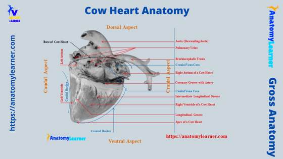
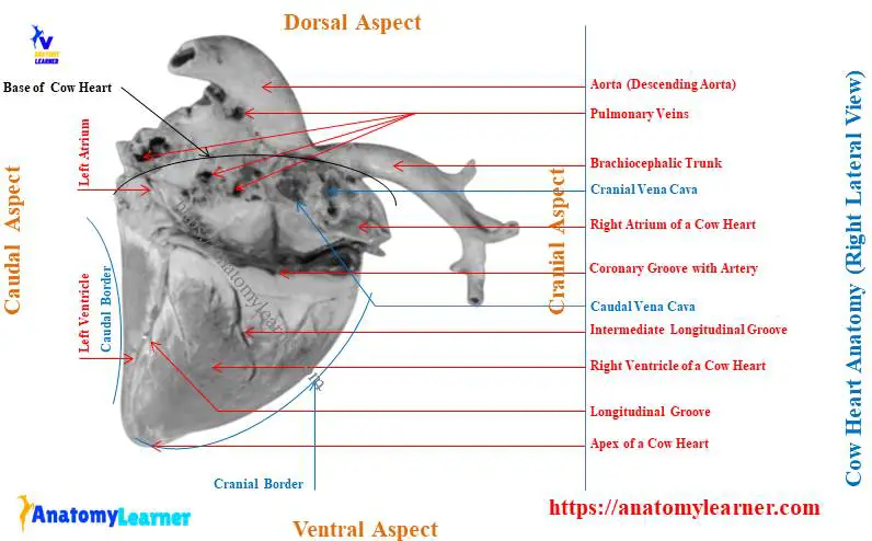
Now, I will show you the labeled diagrams on the cow heart. First, let’s see the anatomical facts from the external appearance (external view – both right and left) of a cow heart.
In the labeled diagram, I tried to show you the pericardium from the cow heart that covers the organ completely. The diagram also shows the diaphragmatic and sternocostal surfaces (identified) and right and left ventricular borders.
Here, the cow heart labeled diagram also shows the pulmonary trunk, aorta (descending and ascending parts), brachiocephalic trunk, and vena cava (cranial and caudal). The wider upper base and the narrow pointed lower apex of the cow heart are already identified in the labeled diagram.
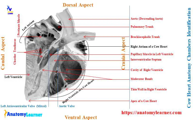
I will also show you the features from the internal surface of a cow heart structure (both in longitudinal and cross sections). The longitudinal section of the cow heart reveals all 4 chambers (right, left atrium, and ventricles).
Every single feature from the 4 chambers of a cow heart anatomy is identified in the labeled diagram. There is an interatrial septum between the right and left atrium of the cow heart. Again, there is an interventricular septum in between the right and left atrium of a cow heart.
The opening of the prominent veins and vena cava, like cranial and caudal vena cava, and pulmonary veins are also identified in the labeled diagram from the right and left atrium of a cow heart. Both the right and left atrioventricular orifices with their semilunar valves are also identified in the cow heart labeled diagram.
Finally, the diagram shows the different trabeculae carneae from both the right and left ventricles of the cow heart. Let’s find more diagrams on cow hearts from anatomy learners here.
Nerves and vessels of a cow heart
The cow heart innervates with the vagus and sympathetic nerves. You may learn more about the course of the vagus nerve from an animal from another article by anatomy learner.
The heart sends blood supply all over the animal’s body. But who supplies the heart itself? The cow heart receives blood from both the right and left coronary arteries.
Both the right and left coronary arteries arise from the ascending aorta. They are considered the vasa vasorum of the heart. This is because the heart itself is derived from blood vessels.
The left coronary artery of the cow heart is larger compared to the right coronary artery. It arises from the left side of the aortic sinus at the level of the semilunar valve.
The left coronary artery passes between the left atrium and pulmonary artery and reaches to atrioventricular groove. Then it divides into descending and circumflex branches.
Now the descending branch of the left coronary artery runs along the left longitudinal groove and reaches the apex. It anastomoses with the right coronary artery branches at the right longitudinal groove.
In addition, the circumflex branch of the left coronary artery runs within the transverse groove caudally and reaches the right longitudinal groove of the cow heart.
The right coronary artery is smaller and arises from the right side of the aortic sinus at the level of the semilunar valve. It runs (goes) along the right side of the transverse groove.
You will find descending and small caudal branches from the right coronary artery. The descending branch of the right coronary runs along the right longitudinal groove.
Cow heart vs. pig heart
Now I will show you a little difference between a cow heart and a pig heart. Compared to the cow heart; you will find the below-mentioned anatomical features in the pig heart –
- The size of the pig’s heart is comparatively smaller than these of the cows,
- You will see the broader base and narrow blunt apex in the pig heart,
- The auricles (right and left) are comparatively smaller than these of the cows,
- It is complicated to find out the intermediate grooves on the external surface of the pig heart,
- The arch of the aorta is strongly curved in the pig compared to that of the cow,
- You will find an extensive fossa ovalis in the interatrial septa of a pig heart anatomy,
In the internal features (atrium and ventricle) of the pig heart anatomy, you will find an almost similar feature to the cow’s heart.
Horse, dog, and rabbit heart anatomy compare to cows
Here, in this section of the article, I will show the comparative features of the hearts among different species, like horses, dogs, and rabbits. So that you may compare the anatomical features of their heart with the cow.
First, let’s see the notable anatomical facts from the horse heart anatomy –
Horse heart anatomy – unique features
- The horse heart extends from the 2nd intercostal space to the 6th intercostal space,
- You will find comparatively a flattened surface in the horse heart compared to the cow heart,
- The base is comparatively broader than these of the cow heart,
- The cranial border of the horse heart is more convex, whereas the caudal border is nearly verticle,
- You will see the more distinct right and left longitudinal grooves on both the right and left aspects of the horse heart compared to the cow,
The internal anatomical features of the horse heart are almost similar to these of the cow heart. You may know more about the horse heart anatomy and the great vessels from here.
Dog and rabbit heart anatomy
- The heart of the dog locate more obliquely compared to the cow’s heart,
- It extends from the third rib to the 6th intercostal space of the dog’s thoracic cavity,
- The base is wide, and the apex is blunt and narrow in the dog heart,
- You will find the strong Sterno-pericardiaco-ligaments (2) in the dog heart compared to the cow heart,
- The right and left longitudinal grooves of the dog heart structure meet together to the right of the apex,
The internal features from the atrium and ventricle (both right and left) show similar characteristics. Now, let’s see the exceptional anatomical features of the rabbit heart compared to the cow –
- It is comparatively smaller in size than these the cow heart,
- The rabbit heart shows the typical conical shape structure,
- You will find a strongly curved aortic arch in the rabbit heart anatomy,
- There is no well-marked sinus venosus and ductus arteriosus in the rabbit heart anatomy,
- The internal features of the rabbit heart are more or less similar to the cow heart,
But, if you want to learn the specific heart anatomy from these different animal species, you may go to the specific article published by anatomy learner.
Frequently asked questions on cow heart anatomy
Now, I will try to provide concise information on some of the essential questions that cow heart anatomy learners ask. But, it is recommended to go through (read) the whole article to learn the basics of the cow heart structure.
Okay, let’s see the questions and their concise answer that the cow heart learners frequently ask –
Is a cow heart similar to a human heart?
Yes, the anatomy of a cow’s heart is almost similar to the human heart. In both heart anatomy, you will find the 4 chambers (2 atria and 2 ventricles), aorta, pulmonary trunk, and right and left atrioventricular orifices along with their valves (bicuspid and tricuspid).
Again, in the human and cow heart, you will find the semilunar valve at the origin of both aorta and pulmonary trunk. In addition, the blood circulation from the heart to the lung, different parts of the body, neck, and head region is almost similar in humans and cows.
In the cow’s heart, it sends oxygenated blood to the different parts of the body and head region through the aorta. Similar functions are performed by the human heart.
Again, the cow’s heart sends the deoxygenated blood into the lungs through the pulmonary trunk. After purification of the deoxygenated blood, the pulmonary veins send it back (oxygenated blood) into the left atrium of the cow heart.
You will see similar blood circulation (general systemic and pulmonary) in humans like the cow heart perform.
Why do cows have 4 hearts?
Actually, there are no 4 hearts present in the cow. They are generally 4 chambers in the cow hearts – right atrium and ventricle, and left atrium and ventricle.
Number of chambers in the cow heart: 4
The atrium (both right and left) acts as the blood-receiving chamber through the vena cava or veins. Here, the cranial and caudal vena cava brings the deoxygenated blood into the right atrium of the cow heart.
Again, the pulmonary veins bring the oxygenated blood into the left atrium of the cow heart. Thus, this oxygenated blood is ready to transport into the body of the cow through the aorta (descending and ascending parts).
In addition, the ventricle of the cow heart anatomy discharges the blood from their cavities into the body and lungs. You will learn more about cow blood circulation from another article by anatomy learners.
Where is the heart located in the cow?
The cow heart is located in the thoracic cavity between two lungs. It locates at the level of the ventral half of the middle mediastinum, with the base directed upward and the apex downward.
Here, the base of the cow heart structure extends from the third rib to the sixth rib (3 – 6) just below the spine (a few centimeters – depending on the size of the cow). In contrast, the apex of the cow heart is very close to the diaphragm and opposite the sixth chondrosternal joint.
What is peri-cardiaco-sternal ligament?
The peri-cardiaco-sternal ligaments are the binding material between the apex of the cow’s heart and sternum. It is also known as the fibrous pericardium that attaches the heart’s apex to the dorsal surface of the sternum.
In the structure of the peri-cardiaco-sternal ligament, you will find the fibrous pericardium part of the pericardium of a cow heart. This labeled diagram will show you the details structure of the peri-cardiaco-sternal ligament from the cow heart.
Do cows have a bone in their heart?
Yes, cows have a bone in their heart. They are known as the os cordis and find in the heart of buffalo and the cow or ox.
This is one type of visceral bone in the animal. But where is this bone located in the cow’s heart? Well, you will find this bone in the junction between the interatrial and interventricular septum of the cow heart anatomy.
As it is very short and not so hard (like the real bone), sometime it may miss while exploring the anatomical facts of the cow heart. So, you need to keep patience while exploring the internal structure of the cow heart to find out the bone.
Conclusion
So from this article, you might learn the basics of the cow heart anatomy with the labeled diagram. Some basic features from the external and internal aspects of the cow heart should identify from the actual sample.
From the external appearance of the cow heart, you might have a good piece of knowledge on the base, apex, surfaces, and borders. Again, from the internal surface of the cow heart, you might know the anatomical facts of the atrium and ventricle from both the right and left aspects.
All the labeled diagrams on the cow heart anatomy might help you to get the proper idea of it. Again, these labeled diagrams and real pictures help you to identify all the features of the live cow heart.