
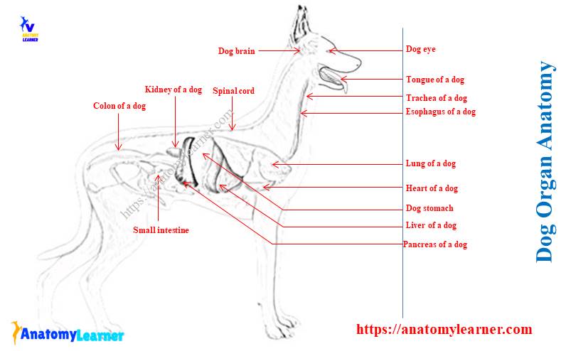
The dog organ anatomy consists of various organs from different body systems. As a veterinary student or a veterinarian, you might have a good concept of dog organs’ location from different systems. This article will help you know the location of a dog’s internal organs with different diagrams so that you may identify them so quickly from the real samples.
I will show you the small dog organ anatomy from the right and left sides. Again, you will also find a list of organs from male and female dogs with the labeled diagrams. You should emphasize the anatomical features of a dog’s thoracic, abdominal, and pelvic organs, as most of these are practically important.
Quick idea: in this article, you will learn the location of different organs from the different systems (like skeletal, digestive, respiratory, urinary, cardiovascular, endocrine, nervous, and special sense) of a dog with their important anatomical features. You will get different labeled diagrams on different organ systems of a dog that might help you gather the basic knowledge of the dog’s internal anatomy.
Dog organ anatomy
I hope you know the term ‘organ’ – the collection of specialized tissue to perform a specific important function. Various internal organs in the dog’s body are clinically valuable. So, the basic anatomy of dog organ is required for field practice.
Here, you will learn the different structures and internal organs of a dog from the following systems –
- Structures, organs, and parts from the locomotor apparatus,
- Organs from the digestive apparatus of a dog,
- Respiratory apparatus of a dog and its organs,
- Urinary organs of a dog,
- Endocrine glands of a dog,
- The cardiovascular system of a dog and its organs (heart and blood vessels),
- Organs, structures, and parts of the nervous system of a dog,
- Special sense organs of a dog (eye, ear, and nose), and
- Common integuments of a dog.
I hope you have a good piece of knowledge on the structures, organs, and parts of the locomotor apparatus of a dog. Again, you may read this article (locomotor apparatus of dog – features of dog skeleton) to memorize different bones from the dog anatomy.
Here, I would like to focus on the important internal organs of the dog’s digestive, respiratory, cardiovascular, endocrine, special sense, and urinary system. If possible, I will discuss the different important muscles of a dog at the end of this article.
But, before starting learning the internal dog organs anatomy, you might have a basic knowledge of the external features and the skeleton.
What are the important organs of a dog?
You should know the location of all the organs in dog anatomy. I will enlist all the organs of a dog from its different organs system in the next section of this article. But, let’s see some of the important organs that you should not avoid to learn their details and anatomical features. Fine, let’s see what they are –
Mouth and teeth – the mouth cavity of a dog consists of the lip, cheek, tongue, oral cavity, and different salivary glands. Again, the number of teeth may vary from the ruminant; usually, you will find forty-two teeth in a dog’s mouth.
Esophagus and trachea – both these two organs are clinically important as you may find partial or complete obstruction in different conditions like chock, chronic bronchitis, and others.
Lung, heart, and liver – the lung, heart, and liver are one of the vital organs in a dog’s internal anatomy. You will find four lobes in the right lung and two in the left lung of a dog. The dog heart is placed obliquely at the level between the third rib and to sixth intercostal space. Again, you will find five lobes in the dog’s liver – left lateral, left center, right lateral, right central, and caudate.
Stomach and intestine – dog, has a pyriform-shaped simple stomach that possesses glandular and non-glandular parts. You will find a strong ileocolic valve at the junction between the ileum and colon of a dog. The anatomy of the dog’s colon is also important as there you may find some difficulties practically.
Spleen – you will find a human foot-print-shaped spleen in a dog located below the proximal end of the last ribs.
Kidney – both the right and left kidneys are bean-shaped and possess a smooth surface.
Other organs from a dog’s internal anatomy
The list of organs I mentioned earlier is the most clinically important in a dog. But, that does not mean you will not learn another organ anatomy from the dog. You should also know the anatomical features of the other following organs from the dog’s internal anatomy –
Urinary bladder – the urinary bladder of a dog is covered by the peritoneum and extends considerably in distended conditions.
Pancreas – you will find two parts (right and left) in a dog’s pancreas. Both the parts meet together behind the pylorus and form a V-shaped structure that extends caudodorsally, mostly up to the corresponding kidneys.
Dog brain and spinal cord – a dog’s brain broadly consists of the brainstem, cerebrum, cerebellum, and medulla oblongata. Again, the structure of the spinal cord of a dog is almost similar to that of other animals, but the number of the spinal nerve that originated from it may vary.
Male and female organs – all the organs from the male and female dog are also clinically important for veterinary students and field practitioners. You will find the full list of all the organs of a male and female dog with its labeled diagrams here on social media.
Blood vessels – cephalic, saphenous, and external jugular veins are the most clinically important vessels in a dog. It is hard to show you all the blood vessels from the dog’s body in this article, but I will try to show some important vessels with their location. Again, this article might help you a little to understand the major vessels from the legs of a dog.
Dog leg anatomy with a labeled diagram
It will be better to learn about these organs according to the body systems.
Internal dog anatomy organs
I hope you got a basic idea of some clinically important organs from a dog with the diagram. But, I recommended learning dog organ anatomy separately with their details features from different systems of a dog’s body. Now, I will enlist and describe the organs from the different body systems of a dog.
So, what you should know from the every organ system of a dog? Here, you might know the location (surface anatomy and the topographic anatomy) with their little important features. So that you may identify the specific organs practically from the live dog.
Fine, let’s start with one of the important organ systems of a dog’s body: the digestive system.
Organs from dog digestive apparatus
The digestive apparatus of a dog consists of the oral cavity, pharynx, alimentary canal, and accessory organs. Again, the accessory organs of the dog digestive system consist of teeth, tongue, salivary glands, liver, gallbladder, pancreas, and paranasal sinus.
Okay, now enlist the organs from the dog digestive apparatus –
- Vestibule of dog mouth – includes the lip and cheeks,
- Oral cavity proper – includes palates, gums, teeth, tongue, and salivary glands (parotid, mandibular, sublingual, and zygomatic),
- The pharynx of a dog – nasal, oral, and laryngeal pharynx,
- The alimentary canal of a dog – includes the esophagus, stomach, small intestine (duodenum, jejunum, and ileum), large intestine,
- Liver, gallbladder, and pancreas of the dog.
If you are a veterinary student, you may write the accessory organs of the dog’s digestive system separately in your answer script. The dog’s digestive system diagram shows almost all the organs, parts, and structures.
Now, I will provide the surface and topographic anatomy of some organs from the dog digestive apparatus.
Dog tongue features
A dog’s tongue is a high mobile organ of its digestive system. The dog tongue is properly located in the oral cavity and possesses some special features from the other animals. You will find the dorsal median groove in the dog’s tongue.
There are exceptional features (lyssa body) present in the central ventral part of the dog tongue. This lyssa body is a thick muscular cord-like structure containing the fatty tissue, vessels, and nerves.
Again, a dog’s tongue possesses two to three vallet papillae on each side of the tongue. You will also find the foliate papillae in the tongue of a dog.
If you want to learn more anatomical features of the dog tongue, then the below-mentioned article might help you a great –
Dog tongue anatomy with special features and labeled diagrams.
Dog teeth anatomy
The teeth of a dog are the highly specialized structure in its mouth. You will find forty-two teeth in the mouth cavity of a dog. It is very important to know the eruption time of the dog’s permanent teeth a veterinarian.
In most dogs, the central, intermediate, and corner incisor teeth erupt at the time between two to five months. The canine teeth of the dog erupt between four to five months.
Again, the first premolar of a dog erupts between four to five months. The other premolar (second, third, and fourth) erupts within six months of age. All the molar teeth of a dog (first, second, and third) erupt between six to eight months of age.
You may learn more about the dog teeth anatomy from this article –
Anatomical features of the dog teeth with the labeled diagrams.
Salivary glands of a dog
Salivary glands are the accessory organ of the dog digestive system anatomy. In a dog, you will find the same salivary glands – parotid, sublingual, mandibular, and zygomatic as found in the other animals. These are the major salivary glands of the dog’s oral cavity. So, again, you will find some of the minor salivary glands like buccal, palatine, lingual, and molar in the oral cavity of a dog.
The parotid gland of a dog lies at the junction of the head and neck that overlies the buccal part of the auricular cartilage. This parotid gland of a dog is V-shaped or triangular, and its dorsal border is notched. You will find the opening ducts of the parotid gland just opposite the third upper premolar tooth.
The mandibular glands of the dog are larger than the parotid and lie between the ling facial and maxillary veins just caudal to the angle of the mandible. Again, the part of the sublingual glands of a dog lies adjacent to the ventral part of the rostral pole of the mandibular salivary gland.
The mandibular duct of a dog leaves the medial surface of the mandibular gland near the ventromedial portion. It closely relates to the major sublingual duct and opens with it.
You will find two sublingual glands (monochromatic and polychromatic), the smallest among salivary glands of a dog. They remain on the lateral surface of the styloglossus and lie medial to the body of the mandible.
The duct of the major sublingual glands lies between the genioglossal and mylohyoid muscles. It opens on a small sublingual caruncle that locates lateral to the rostral end of the frenulum.
The zygomatic gland of a dog is pyramidal in shape and lies against the ventral part of the periorbita.
The pharynx of a dog
The pharynx of a dog is a musculomembranous junction of respiratory and digestive tubes between the oral and nasal cavities rostrally and the esophagus and larynx caudally. You will find three different parts in the pharynx of a dog – nasal, oral, and laryngeal parts.
The nasal pharynx of a dog is the respiratory part dorsal to the soft palate. It extends from the choanae of the nasal cavity to the interpharyngeal opening of the pharynx.
You will find a pharyngeal opening of the auditory tube at the dorsolateral wall of the nasal pharynx. Again, there is an interphalangeal opening that opens the nasal pharynx into the laryngopharynx.
The oropharynx of a dog extends from the isthmus of the fauces to the base of the epiglottis. Do you know what the isthmus of fauces is? It is the orifice between the oral cavity and the oral part of the pharynx. You will find the palatine tonsil in the lateral wall of the oral part of the pharynx.
The laryngopharynx is that portion of the pharynx that lies dorsal to the larynx and extends from the intraphryngeal ostium and the nasal part of the pharynx rostrally to the beginning of the esophagus caudally.
The esophagus of a dog
The dog esophagus is the first part of the alimentary canal. It is a muscular tube connecting the laryngeal part of the pharynx and the stomach. Again, a dog’s esophagus passes most of the neck, all of the thorax, and ends on entering the abdomen.
So, you will find three different parts in a dog’s esophagus – cervical, thoracic, and abdominal. The dog has a small abdominal part of the esophagus compared to the other animals.
Dorsally, the cervical part of the dog esophagus relates to the left longus colli and longus capitis muscles. Again, this part of the dog esophagus relates to the trachea ventrally and the right.
On the left aspect, the left common carotid artery, vagosympathetic nerve trunk, internal jugular vein, and tracheal duct run at the angle between the esophagus and longus colli muscle.
The thoracic part of the dog esophagus extends from the thoracic inlet to the esophageal hiatus of the diaphragm. First, it lies to the left of the trachea and crosses the left face, then run over the dorsal surface.
It continues to run over the dorsal surface of the trachea up to the tracheal bifurcation (at the level of the fifth to sixth cervical vertebra). Again, it crosses the right face of the aortic arch and lies ventral to the right and left longus colli muscle.
There is a small abdominal part of the esophagus in a wedge-shaped dog. Dorsally, this small abdominal part immediately joins with the dog’s stomach.
Anatomy of a dog stomach
A dog’s stomach is one of the largest dilations of the alimentary canal that remains in between the esophagus and small intestine. It is pyriform in shape and possesses two surfaces (parietal and visceral), two curvatures (greater and lesser), and two orifices (cardiac and pyloric).
The anatomy of this organ of the dog differs from the ruminant. Ruminants possess a compound stomach, whereas dogs possess a simple stomach (glandular and non-glandular parts).
The size of the dog’s stomach varies from breed to breed. Again, the capacity and weight of the dog’s stomach may also vary within the same breeds.
The size of the dog’s stomach may increase during feeding; you will see the funds of the stomach increase first. Then it pushes caudodorsally on the left side. Do you know what the funds of a dog’s stomach are?
The dog’s stomach has three different regions – cardiac, fundus, and pyloric. The cardiac part of the dog’s stomach is that part that blends with the esophagus. Again, the fundus is the large blind out pocketing that locates to the left and dorsal to the cardia. The pyloric part of the dog’s stomach is the short distal part that continues with the small intestine.
You may learn more about the anatomical features of the dog stomach from the article mentioned below –
Dog stomach anatomy with the labeled diagrams
The visceral surface of the dog’s stomach presents a convex outer surface that faces dorsally. Again, the parietal surface of the dog’s stomach faces to the left and cranially and ventrally.
The greater curvature of the dog’s stomach forms the convex border, whereas the lesser curvature forms the concave border of the stomach.
The small intestine of a dog
The small intestine of a dog is the longest part of the alimentary canal. It extends from the pylorus of the stomach to the ileocecal orifice (junction between the small and large intestine). You will find three parts of a dog’s small intestine – the duodenum, jejunum, and ileum.
The duodenum of the dog is a relatively fixed part and possesses a short proximal loop. Again, the jejunum is the free movable long, middle, and distal part of the small intestine. The ileum is the short terminal part of a dog’s small intestine.
So, the duodenum is the first part of the small intestine that begins in the dorsal half of the right hypochondriac region opposite the nine intercostal spaces. It runs caudal to the tuber coxae and makes a U-shaped turn.
There are four portions and three flexures present in the duodenum of a dog. The portions of the duodenum of a dog include –
Cranial portion – short between the pylorus and cranial duodenal flexure,
Descending portion of duodenum – runs caudally from the cranial part near to the pelvic inlet,
Transverse portion – connect the ascending and descending portions from right to left. It lies ventral to the body and the right transverse process of the sixth lumbar vertebra.
Ascending portion – runs obliquely cranially and to the left from the transverse part. It obliquely crosses the uterus, sympathetic trunk, caudal vena cava, aorta, and lumbar lymphatic trunk.
There are cranial duodenal flexure, caudal duodenal flexure, and duodenojejunal flexure present in the duodenum of a dog.
Dog liver topographic anatomy
The dog liver is the largest gland in the body that possesses both exocrine and endocrine functions. The liver of a puppy is heavier than those of an aged dog. You will find red color on a dog’s liver in fresh condition.
The dog liver locates on the right side of the abdominal cavity in an oblique downward and forwards direction. It closely attaches to the diaphragm, and the major parts of the liver are located on the right side of the median plane. This gland extended from the lumbo costal angle to the seventh to eighth ribs level and was kept positioned with the abdominal cavity with ligaments.
In a dog’s liver, you will find two surfaces (diaphragmatic and visceral) and four borders (dorsal, ventral, right, and left). Again, you will see some impression of the organs on a dog’s liver. Four lobes, four sub loves, and two processes are present in a dog’s liver.
The dog liver’s diaphragmatic surface (parietal) is strongly convex in all directions. This surface of the dog liver lies mainly in contact with the diaphragm.
Again, the visceral surface of the dog liver is irregularly concave and faces mainly caudoventrally and to the left. You will find a close relationship between the visceral surface of the liver and with stomach, duodenum, pancreas, and right kidney.
Dorsal, ventral, left, and right borders are also present in a dog’s liver. All the borders of a dog liver possess sharp edges.
Lobes, process, and impression of a dog liver
The dog liver possesses four lobes, four sub-lobes, and two processes. You will find the below-mentioned lobes, sub-lobes, and impressions in a dog’s liver.
The left hepatic lobe – lies entirely to the left of the median plane,
Left lateral hepatic lobe – begins dorsally deep to the left crus of the diaphragm,
The quadrate lobe – is a deep liver wedge that lies in the median plane. You will see a fossa for the gallbladder at the middle surface of the quadrate lobe of the dog liver.
Left medial hepatic lobe – varies from being triangular to an oval shape,
Right hepatic lobe – smaller than the left hepatic lobe and lies completely to the right of the median plane,
Right medial hepatic lobe – fuses to the quadrate lobe medially. Again, the right half of this medial hepatic lobe forms the fossa with the quadrate lobe to host the gallbladder.
Caudate lobe – consists of caudate and papillary processes,
Right lateral hepatic lobes overlap the caudate process of the caudate lobe and fuse with it.
So, you will find eight lobes in the liver of a dog. Now, let’s see the processes present in the dog’s liver. You will find the following two processes in the dog liver –
Papillary process – pyramidal to tongue shape, and
Caudate process – form the caudal part of the liver.
Again, in the dog liver, you will find five important ligaments –
The coronary ligament of the dog liver,
A right triangular ligament of the dog liver,
The left triangular ligament of the dog liver,
A falciform ligament of the dog liver, and
The hepatorenal ligament of the liver.
In addition, you will find gastric, duodenal, and renal impressions in a dog’s liver.
The pancreas of a dog
The dog pancreas is the yellowish gray to reddish-gray structure in the digestive system of a dog. It locates in the dorsal part of the cranial and right lateral abdominal regions, caudal to the liver.
The pancreas also has exocrine and endocrine functions like the dog’s liver. The shape of the dog’s pancreas looks like a V shape. You will find a thin, slender right lobe and a short, thick, and wide left lobe in a dog’s pancreas.
The right lobe of a dog’s pancreas lies in the mesodudenum and extends from the transverse plane through the middle of the ninth intercostal space to the fourth lumbar vertebra. Again, the caudal extremity of the right lobe of a dog’s pancreas lies in the concavity of the duodenal loop.
The left lobe of the dog’s pancreas lies in the deep wall of the greater omentum. This lobe begins at the body and runs caudosinistrally.
The dorsal surface of the left lobe has a relationship with the caudate process of the liver, portal vein, caudal vena cava, and aorta. On the other hand, the ventral surface relates to the transverse colon ventrocaudally and the dorsal wall ventrocranially.
Dog respiratory organ anatomy
The respiratory system of a dog consists of the nose, nasal cavity, pharynx, larynx, trachea, bronchi, bronchus, and lungs. You might have a good anatomical knowledge of the trachea and lungs among these organs. Here, I will focus on the anatomy of the dog’s lung from the respiratory organ system. But, you will also get information on the anatomy of other organs from the dog’s respiratory apparatus.
So, first, let’s enlist the organs from the dog’s respiratory apparatus –
- The external nose of the dog – possesses a comma-shaped nostril and vomeronasal apparatus,
- Nasal cavity – possess nasal conchae, meatus, sinus, and glands,
- Larynx – consists of different cartilages,
- Trachea – do not possess any apical bronchi,
- Bronchi – consists of right and left bronchus,
- The lung of a dog – is the most vital organ of the respiratory system. It consists of right and left parts, which also contain different lobes.
Now, let’s know some of the important anatomical features of the respiratory organ of a dog. Then I will go for the details anatomical facts of the different organs separately with the diagrams.
Special anatomical features of a dog respiratory apparatus
The nostril of a dog is comma-shaped.
The size and shape of the nasal cavity vary according to the breed.
You will find a pair of extra cartilage – cuneiform cartilage that attaches to the arytenoid cartilage in the dog larynx.
There is no apical bronchus in the trachea of a dog.
The vocal cords are very prominent in the dog.
You will find four lobes in the right lung and two in the left lung of a dog. The lobes are very prominent, and fissures are deep in the dog’s lungs.
Now, let’s see the anatomy of the respiratory organs of a dog.
The external nose of a dog
The external nose of a dog is associated with the nasal cartilage and nasal cavity. You will find short hairs on the outer part of a dog’s nose. There are two nostrils present in the nasal plane separated by a groove.
The framework of the external nose of a dog consists of different nasal cartilage. You will find the following nasal cartilage in the dog’s nose –
Septal cartilage of the dog nose,
The dorsolateral nasal cartilage,
Ventrolateral nasal cartilage, and
The paired accessory nasal cartilage of a dog nose
You may find details about this nasal cartilage and the dog nose anatomy in the below-mentioned article by the anatomy learner.
Anatomical features of a dog nose with the labeled diagram
The most important structure of the dog nose is the paired vomeronasal organs (Jacobson’s organs) that locate in the rostral base of the nasal septum. You will find the dorsal and lateral nasal ligaments in the dog nose anatomy.
The nasal cavity of a dog’s respiratory anatomy
A dog’s nasal cavity extends from the nostril to the conchae and forms the facial part of the respiratory passageways. What are the main structures you should know from the nasal cavity of the dog nose? The most important part of the dog’s nasal cavity is the nasal conchae, meatus, sinus, and glands.
If you read the previously suggested article on dog nose, you will also get the details features of the nasal cavity.
You know the nasal conchae of a dog nose are the cartilaginous structures covering nasal mucosa. The dog’s nasal cavity shows dorsal, ventral, middle, and ethmoidal conchae. Again, you will find the following nasal meatus in the dog’s nasal cavity structure –
A dorsal nasal meatus of the dog nose,
The ventral nasal meatus of the dog’s nose,
A middle nasal meatus of the dog nose,
The common nasal meatus of the dog nose, and
Nasopharyngeal meatus of the dog nose.
You will find different paranasal sinuses in a dog’s nasal cavity, including maxillary sinus, frontal sinus, and sphenoid sinus. There are also serous lateral nasal glands present in the nasal cavity of a dog.
The larynx of a dog organ anatomy
The larynx is a musculocartilagenious organ in the dog’s respiratory anatomy that guards the trachea entrance. It serves as an air passageway, aids vocalization, and prevents the inspiration of foreign materials. The most important structures of the dog larynx are cartilages and muscles.
You will get the following laryngeal cartilages in the dog’s larynx –
- Epiglottis cartilage,
- Thyroid cartilage,
- Cricoid cartilage,
- Arytenoid cartilage,
- Sesamoid cartilage, and
- Interarytinoid or cuneiform cartilage of the dog larynx
The epiglottis cartilage of a dog resembles a sharply pointed spade that forms the base of the epiglottis. Again, the thyroid is the largest cartilage of the dog larynx that forms the middle part of the laryngeal skeleton.
The cricoid is the only cartilage that forms a complete ring in the dog’s larynx. In addition, the arytenoid cartilages are the irregular, paired cartilage that joins with the craniodorsal border of the cricoid cartilage.
You will find oval-shaped sesamoid cartilage in the dog’s larynx structure. Again, the inter arytenoid cartilage of the dog larynx is flat, small, and lies cranial to the cricoid lamina.
The muscles of the dog’s larynx include –
Cricothyroideus muscle,
Cricoarytenoideus lateralis and dorsalis,
Thyroarytenoideus muscle,
Vocalis muscle,
Ventricular muscle,
Arytenoideus transverse muscle, and
Hyoepiglotticus muscle of the dog larynx.
Again, you will find different ligaments in the dog larynx structure like vocal, vestibular, cricothyroid, and cricoarytenoid ligaments.
Dog trachea anatomy
A dog’s trachea is a muculocartilagenious structure that consists of C-shaped hyaline tracheal cartilages. You will find approximately thirty-five incomplete hyaline cartilaginous C-shaped rings that form the dog trachea’s skeleton.
The incomplete portion of the dog tracheal ring is faced dorsally and bridges with the smooth muscle fiber. Again, these tracheal rings unite in a longitudinal direction by the fibroelastic annular ligaments of the trachea.
This annular ligament of the trachea allows considerable intrinsic movement without collapse or breakage. Dorsal to the cranial part of the base of the heart, dog trachea bifurcated into left and right pulmonary bronchus.
Bronchi of the dog trachea
You will find the bronchial tree in the dog’s respiratory system that begins at the trachea’s bifurcation and forms the right and left principal bronchus. Each principal bronchus of the dog divides into lobar bronchi (secondary bronchi).
Again, the lobar bronchi of the dog divide into segmental bronchi within the lung tissue. You will also find the respiratory bronchioles that give rise to alveolar ducts, sacs, and pulmonary alveoli.
Dog lung anatomy
The lung is the main organ of the dog’s respiratory system that serves a passive function in the mechanical act of respiration. Before studying the lung anatomy, you might have a good knowledge of some terms like – mediastinum, pleura, and diaphragm.
Let’s start with the pleura – it is the thin transparent serous membrane that encloses the lung and lines the interior of the thoracic cavity of a dog. Again, you may say pleura is a sac-like structure that comprises parietal and visceral portions.
The pleura space contains the liquor pleurae that provide a frictionless environment during inspiration. Different parts like the dog pleura’s costal, diaphragmatic, and mediastinal parts.
The mediastinum is the intrapleural space that separates the right and left pleural sacs. It extends from the thoracic inlet to the diaphragm along the mid-axis of the thorax of a dog.
Again, the diaphragm is a musculoaponeurotic dome-shaped partition between the thoracic and abdominal cavities. The pleura covers the diaphragm’s thoracic surface, and the peritoneum covers the abdominal surface. The diaphragm contacts and move caudally to enlarge the pleural cavity.
The right and left lungs possess a base, an apex, costal surface, medial surface, cardiac impression, and cardiac notches. Each of these lungs has a concave diaphragmatic surface that lies against the convex surface of the diaphragm.
You may know more about the anatomical features of an animal lung from this article.
Left lung of a dog
The left lung of a dog only consists of two lobes – caudal and cranial lobes. Again, you will find the cranial and caudal parts in the cranial lobe of the left lung. The cranial part of the cranial lobe of the left lung is transversely compressed between the heart and the lateral thoracic wall.
This cranial part of the cranial lobe extends from the dorsal part of the fifth rib to the thoracic inlet. Again, the caudal part of the cranial lobe of the left lung (cardiac lobe) presents a thin dorsocranially convex border. It overlies the caudal thickened portion of the cranial part of the cranial lobe.
You will find a pyramidal caudal lobe (diaphragmatic lobe) in the left lung of a dog. It completely separates from the caudal part of the cranial lobe by the caudal interlobar fissure.
The right lung of a dog
The right lung of a dog possesses four lobes – cranial, caudal, middle, and accessory lobes. You will find two interlobar fissures in the right lung of the dog.
The cranial interlobar fissure separates the cranial and medial lobes of the right lung. At the same time, the caudal interlobar fissure separates the right middle and caudal lobes of the right lungs.
The right lung’s cranial lobe (apical lobe) extends from the dorsal part of the cranial interlobar fissure to the right of the median plane. Again, the medial lobe begins at the cranial interlobar fissure. You will find a cardiac impression at the medial surface of the medial lobe of the right lung.
The accessory lobe of the right lung (intermediate lobe) is the most irregular of all of the lobes of the lungs. Again, the caudal lobe of the right lung is also pyramidal and located at the right of the median plane.
Dog urinary organ anatomy
The urinary system of a dog consists of two kidneys (right and left), two ureters, a urinary bladder, and a urethra. From the dog’s urinary organ anatomy, I will show you the anatomical features of the kidney. But, you will also get a little information on the other urinary organs like the ureter and urinary bladder of a dog.
Fine, now, let’s see the anatomical features of the dog’s urinary organs –
- Kidney of the dog,
- Ureters of the dog,
- The urinary bladder of a dog, and
- The urethra of the dog.
First, let’s start with the kidney of dog anatomy.
Dog kidney anatomy
The kidneys are the paired retroperitoneal organ whose dorsal surface is in contact with the lumbar hypaxial muscle and is often surrounded by fat. Again, the ventral surface of the kidneys covers by the transparent parietal peritoneum.
Each dog’s kidney lies lateral to the aorta and caudal vena cava. You will find a less convex dorsal surface than the ventral surface in each kidney. There is a thin fibrous capsule that covers the surface of the kidney. The hilus of the kidney locates at the medial surface.
It is very important to know the surface anatomy of a dog’s both right and left kidneys. The right kidney of a dog locates opposite the body of the first three lumbar vertebrae.
Again, the left kidney of a dog lies ventral to the body of the third, fourth, and fifth lumbar vertebrae. You know both the right and left kidneys of a dog are bean-shaped and possess a smooth surface.
The cranial pole of the right kidney embeds in the fossa of the caudate process of the liver. You will find this extremity of the kidney at the level of thirteen ribs. It may be in contact with the diaphragm retractor costae muscle.
You will also find the right adrenal gland that relates to the cranial pole of the right kidney of the dog. The right kidney of a dog also has close contact with the caudal vena cava and pancreas.
Again, the left kidney of a dog has contact with the dorsal end of the medial surface of the spleen, greater omentum, and greater curvature of the stomach. The left kidney of a female dog has contact with the descending colon and mesovarium.
Nephrons and vessels in dog kidney
You will find millions of nephrons in the dog kidney. Each nephron begins at the double-layer glomerular capsule. Within the glomerular capsule, you will see glomerulus in the dog nephrons. Again, the glomerular capsule and the glomerulus from the renal corpuscle together.
It will be better to learn the nephron structure from the histology slide section. Because you will understand every single structure from the nephron so easily.
The kidney is a highly vascular organ where you will find the renal artery. Again, the renal artery of the dog kidney divides into dorsal and ventral branches. You will also find the interlobar arteries, arcuate arteries, and afferent and efferent arteries in the structure of a dog kidney.
Ureter of the dog
You will find two ureters in the dog’s urinary system. They (ureter) carry urine from the kidney to the urinary bladder. The length and size of the dog ureter depend on the age and breed. You will find the stronger ureter on the right side than the left.
There are two different parts in the ureter of a dog – one is the abdominal, and another is the pelvic part. Do you know where the abdominal part of the ureter starts?
The abdominal ureter begins from the renal pelvis and collects urine from the renal crest. You will see the right ureter of a dog lies in the close association with the caudal vena cava. Again, the uterus passes ventral to the deep circumflex iliac and external iliac arteries.
The pelvic ureter enters between the two layers of the peritoneum, forming the lateral ligament of the bladder. It reaches the dorsolateral surface of the urinary bladder just cranial to its neck.
If you see the structure of the ureter of a dog, you will find three different distinct layers of smooth muscles – outer longitudinal, middle circular, and inner longitudinal. Again, the mucosa layer of the ureter of a dog possesses the transitional epithelium.
The urinary bladder of a dog
The urinary bladder of a dog is a musculomembranous organ in the urinary system. This organ varies in form, size, and position, depending on the amount of urine it contains. The dog’s urinary bladder location also may vary with its condition.
When the urinary bladder is empty, you will find this organ on the pelvic floor of the dog. You may know more about the pelvic floor of an animal (how they form and what organs they contain). Again, the urinary bladder may extend into the abdominal cavity a little in urine.
You will find the same features in the urinary bladder of a dog as in other animals. The dog’s urinary bladder has a neck, body, and apex. The dog’s urinary bladder body connects with the urethra, and the apex is a cranial blind sac.
In the dog’s urinary bladder structure, you will also find three layers of smooth muscle that arranges in different patterns. Again, the mucosa also contains the transitional epithelium. You will also find the submucosa in the dog’s urinary bladder, consisting of dense connective tissue and other components.
Different ligaments fix the dog’s urinary bladder with the pelvic cavity. You will see the following important ligaments in the dog’s urinary bladder –
- A median ligament of the urinary bladder,
- The lateral ligament of the urinary bladder,
- A round ligament of the dog’s urinary bladder.
You will also find the cranial and caudal vesical arteries, pelvic nerve, pudendal nerve, hypogastric, and lumbar lymph nodes in a dog’s urinary bladder structure.
Male and female dog organs
These two systems (male and female dog organ systems) are very important for veterinary students and practitioners. The details guide on the male and female organs here in the anatomy learner in two separate articles. Or, you may go to the guide page of anatomy learner and find these two systems.
Again, I will share the list of male and female dog organs with a labeled diagram here. You will easily understand all the organs from both the male and female organs system as I provide the labeled diagrams.
These labeled diagrams might help you practically identify these male and female organs from the actual or live sample. Here, I will only show you the anatomical location and features of the ovary and uterus of a female dog.
Ovary of the female dog
The ovaries of a female dog are paired with elongated oval organs located below the third or fourth lumbar vertebrae. Each of these two ovaries is completely enclosed by a peritoneal pouch (ovarian bursa). You will find a small opening at the ventral part of the ovarian bursa.
Again, a dog’s ovaries are attached by a mesovarium to the body wall and the mesosalpinx. Besides this mesovarium, you will find some ligamentous attachment to the ovaries. There are suspensory and proper ligaments for the ovaries.
The suspensory ligament of the ovary attaches to the medial and ventral third of the last one or two ribs. Again, the proper ligament of the ovary attaches the uterine end of the ovary to the cranial end of the uterine horn.
In the structure of the dog ovary, you will find the medulla and cortex. The medulla of the dog ovaries contains nerves, blood vessels, lymphatics, and connective tissue fibers. Again, the cortex of the ovary contains connective tissue stroma, where you will find different types of growing follicles.
Uterine tube of a dog: it is located between the peritoneal layer of the mesosalpinx and connects the peritoneal cavity with the uterine cavity. It transports the oocytes from the ovary to the uterus. You will find the infundibulum, fimbriae, and uterine ostium in the dog’s uterine tube structure.
The ovarian extremity of the uterine tube is the infundibulum and locates near the edge of the opening of the ovarian bursa. Again, the fimbriae are usually visible, projecting out the opening of the ovarian bursa.
Dog uterus anatomy
The uterus is one of the most important organ in female dog anatomy. The whole structure of the dog’s uterus appears as a large V-shape. Again, the body is short, and the horn is very long in the dog’s uterus. The horns of this uterus are not tapered.
If you see a live uterus of a dog, you will find three different important parts – the neck, body, and two horns. The cranial end of the uterine horn connects with the ovary by the proper ligament of the ovary. You will find the body of the dog’s uterus in both pelvic and abdominal cavities.
The body of the uterus extends from the point of convergence of the uterine horns to the cervix. Again, the internal features of the dog’s uterus are described in the other article by the anatomy learner.
What are the ligaments that attach to the dog’s uterus? You will find a broad ligament, genital fold, and the round ligament of the uterus. The broad ligament contains fat and smooth muscle that attach the uterus and ovaries to the lateral body wall.
Cardiovascular organ of a dog anatomy
From the cardiovascular system of a dog, you might know the surface and topographic anatomy of the heart, pulmonary arteries and veins, aorta and its different parts, arteries of different limbs, and veins. But, the most important organs in the cardiovascular system of a dog are the heart, cephalic, and saphenous vein.
Let’s enlist the organs, parts, and different structures from the dog’s cardiovascular system –
- Heart of the dog,
- Brachiocephalic trunk and branches in the dog,
- Aorta and its branches – thoracic and abdominal parts,
- Arteries of pelvic limb – external and internal iliac artery, femoral artery, and popliteal artery,
- Median sacral artery of the dog,
- Veins of the thoracic and pelvic limbs of a dog.
Let’s start with the anatomical features of the dog heart.
Heart of a dog
First, let’s know where the dog’s heart is located. The dog heart is located obliquely at the level between the third and sixth intercostal spaces. You will find a blunt and rounded apex in the dog’s heart. The right and left longitudinal grooves meet together to the right of the apex.
The dog heart is the muscular organ in the cardiovascular system that covers the fibro serous envelops – pericardium. It is cone-shaped and obliquely placed into the thoracic and possesses a base and apex. The base of the dog heart faces dorsocranially, whereas the apex directs ventrocaudally and to the left side of the median plane.
There are many internal and external features in the dog heart that you should know as a veterinary student. Here, I will not provide all the anatomical facts about the dog heart as you will find a dedicated article on the dog heart anatomy. I request you to learn the full anatomical features of the dog heart from that article by the anatomy learner.
Anatomical features of the dog heart with the labeled diagrams
In the dog heart labeled diagram, I tried to show you the following structures, but you should learn the details of these structures perfectly.
- Blood vessels of the dog’s heart (coronary artery, cardiac vein, and others),
- The intraventricular septum of the dog’s heart,
- A thin intraarterial septum of the dog heart,
- Features of the left and right atrium,
- The right and left ventricles of the dog’s heart,
The left and right ventricles separate internally by the intraventricular septum. It extends from the cranioventral position on the left to the caudodorsal position on the right.
Atrium and ventricle of the dog heart
The right atrium of the dog heart receives the blood from the systemic veins and most of the blood from the heart itself. You know this right atrium lies dorsocranially to the heart’s right ventricle.
Again, the left atrium of the heart forms the left dorsocaudal part of the base of the heart. The right ventricle of the dog’s heart receives systemic blood from the right atrium and pumps it into the lungs.
You will find a conical shape left ventricle in the dog heart that forms the apex of the heart. There are different valves present in the dog heart structures like – right and left atrioventricular valves, aortic valve, and valve of the pulmonary trunk.
Aorta of a dog anatomy
The dog’s aorta leaves the left ventricle near the center of the base of the heart. You know this is a thick-walled vessel from where systemic blood passes. It is impossible to describe all the branches of the dog aorta; rather, I prefer to provide some important branches.
Brachiocephalic trunk: it is the first large artery from the aortic arch that passes obliquely to the right and cranially across the ventral surface of the trachea. The brachiocephalic trunk gives the first branch – the left common carotid artery.
Let’s see some of the branches of the common carotid artery, internal carotid, and external carotid artery –
Common carotid artery – caudal thyroid and cranial thyroid artery,
Internal carotid artery of the dog,
The external carotid artery of the dog – includes occipital, cranial laryngeal, ascending pharyngeal, lingual, facial, caudal auricular, parotid, superficial temporal, and maxillary arteries.
Please read the details of these arteries from the other articles from the anatomy learner.
The thoracic aorta of the dog
The thoracic part of the dog aorta continues from the aortic arch opposite to the fourth thoracic vertebrae. This thoracic aorta extends to the caudal border of the second lumbar vertebra. Then it enters the abdominal cavity and passes between the crura of the diaphragm.
You will find the visceral and parietal branches of the thoracic aorta in a dog. The visceral branches of the thoracic aorta include the bronchial and esophageal arteries. Again, the parietal branch of the thoracic aorta includes the dorsal intercostal, dorsal cost abdominal, and first two lumbar arteries.
The abdominal aorta of the dog
The thoracic aorta enters the diaphragm’s crura and continues as the abdominal aorta in a dog. This abdominal aorta ends at the level of the seventh lumbar vertebra and divides into right and left internal iliac arteries.
The dog’s abdominal aorta’s abnormal visceral branches, paired visceral branches, and parietal branches. The unpaired visceral branches of the abdominal aorta include celiac, cranial mesenteric, and caudal mesenteric arteries.
Again, the paired visceral branches of the abdominal aorta include adrenal, renal, and ovarian arteries. The caudal phrenic, cranial abdominal, lumbar, deep circumflex iliac, external iliac,
internal iliac and median sacral arteries are under the parietal branches of the abdominal aorta.
Arteries of the pelvic limb of a dog
The abdominal aorta of a dog divides into paired external iliac arteries that supply to the pelvic limb. These external iliac arteries of the dog ends at the internal iliac and median sacral arteries in a dog.
Here, I will provide a summary of the external iliac artery of the dog –
The external iliac artery is the largest parietal branch of the abdominal aorta. These arteries arise from the lateral surface of the aorta ventral to the intervertebral disc between the sixth and seventh lumbar vertebrae.
You will see a deep femoral artery that arises from the caudomedial surface of the external iliac artery. The pudendoepigastric trunk is short and extends to the deep abdominal inguinal ring.
You will see the caudal epigastric artery that runs cranially on the dorsal surface of the rectus abdominis muscle deep to the parietal peritoneum. The external pudendal artery arises from the ventral branch of the pudendoepigastric trunk.
You know the medial circumflex femoral artery continues the deep femoral artery in a dog. There is an obturator, ascending, acetabular, transverse, and deep branches found in the medial circumflex femoral artery of a dog.
The caudal abdominal artery arises from the cranial surface of the external iliac artery.
Femoral artery of a dog: it is the continuation of the external iliac artery from the vascular lacuna through the thing of a dog. The saphenous artery arises from the medial surface of the femoral artery just before the femoral artery disappears to the semimembranosus muscle.
The popliteal artery is the femoral artery’s continuation through the popliteal fossa. A small caudal tibial artery leaves the caudal surface of the popliteal artery at the interosseous space.
Internal iliac and median sacral artery of a dog
The internal iliac artery arises at the level of the seventh lumbar vertebra of a dog. You will see different branches of the internal iliac artery of a dog, like the umbilical artery, internal pudendal artery, prostatic artery, caudal vesical artery, and artery of the ductus deferens in a male dog.
You will also find other different branches of the internal iliac artery in both male and female dogs. The median sacral artery is the direct continuation of the aorta caudally after the internal iliac artery arises.
Cephalic and saphenous vein of a dog
In the dog’s cardiovascular organ anatomy, cephalic and saphenous veins are also important for practical uses. The cephalic vein of a dog is the only larger superficial vein of the thoracic limb. Do you know where the cephalic vein begins?
The cephalic vein of a dog begins on the medioplanter surface of the carpus, where it is a continuation of a radial vein. Again, distal to the radial vein, the superficial palmar arch arises that crosses the palmar side of the distal third of the second to fifth metacarpal bones.
You will find two branches of the saphenous vein – the lateral and medial saphenous vein in a dog leg. The lateral saphenous veins begin by collecting its cranial branch from the flexor surface of the tarsus and its caudal branch from the lateral surface.
Lymph nodes of a dog’s body
Lymph nodes are the structural and functional unit of the lymphatic system that locates in the course of the lymph vessels. You will find different lymph nodes and lymph centers in the dog’s body. Here, I will only enlist the lymph nodes from the different regions of a dog’s body.
Lymph nodes in the head and neck region
The parotid lymph center contains the parotid lymph node at the rostral base of the dog ear. They are the bean-shaped lymph nodes that remain deep to the posterodorsal border of the parotid salivary gland.
The mandibular lymph center posses the mandibular and buccal lymph nodes. At the angle of the mandible, you will see a group of two or three mandibular lymph nodes in a dog.
Again, the buccal lymph node locates at the dorsal, ventral, and rostral angles of facial and superficial veins. The medial retropharyngeal (Near pharynx) lymph node is the largest lymph node found in a dog’s head and neck region. It is an elongated, transversely compressed node with a pointed caudal end.
The lateral retropharyngeal lymph node is completely or partially covered by the caudal part of the parotid salivary gland. On the lateral surface (outer) of the serratus ventralis and scalenus muscle, you will find the superficial cervical lymph nodes.
The deep cervical lymph nodes are located along the cervical portion of the trachea. You may find the cranial deep cervical lymph node and middle deep cervical lymph node in a dog.
Lymph nodes in the thorax and thoracic limb
The axillary lymph center of a dog consists of the axillary lymph node and accessory lymph nodes. You will only find the axillary lymph node in the thoracic limb of a dog. The accessory lymph node lies caudal to the central node in the fascia between the deep pectoral and latissimus dorsi muscle.
In the thoracic region of a dog, you will find the ventral thoracic, dorsal thoracic, mediastinum, and bronchial lymph center. The cranial sternal lymph node locates either right or left of the median plane and serves both sides of the thoracic region.
A small spherical aortic thoracic lymph node lies in the vertebral end of either the fifth or sixth intercostal space. The number of the cranial mediastinal lymph node vary, and most of them are associated with the large vessels of the heart.
The bronchial lymph center of a dog consists of pulmonary, right, and left tracheobronchial and middle tracheobronchial lymph nodes.
Lymph nodes in abdominal and pelvic regions
The lumbar lymph center contains the lumbar aortic and renal lymph nodes. The lumbar aortic lymph node along the aorta and vena cava will find the lumbar aortic lymph node. Renal lymph nodes are very small and associated with the renal vessels of a dog.
The ileocecal lymph center of a dog consists of internal iliac lymph nodes and sacral lymph nodes. You will see the small internal iliac lymph node at the angle between the internal iliac artery and median sacral artery.
You will also find different visceral lymph nodes in the abdominal and pelvic regions of a dog. Please find the labeled diagrams on the visceral lymph nodes from the dog’s abdomen and pelvis here on social media.
Lymph nodes in the dog’s pelvic limb
There are popliteal lymph centers, iliofemoral lymph centers, and inguinofemoral lymph centers in a dog’s pelvic limb. The superficial popliteal lymph node is the largest in the pelvic limb of a dog. It is an oval lymph node that lies in the fat between the biceps femoris muscle’s medial border and the semitendinosus muscle’s lateral border.
You will find the distal femoral lymph node in the fat deep to the deep medial femoral fascia at the distal part of the femoral triangle. This is the very small lymph node in the pelvic limb of a dog. Another small external iliac lymph node lies on the ventral surface of the tendon of the psoas minor muscle and caudal to the external iliac vein.
There is two superficial inguinal lymph node in a dog that lies in the fat between the abdominal wall and the medial surface of the thigh.
Dog spleen organ anatomy
The spleen is another clinically important organ in dog internal anatomy. There is a roughly human foot-print-shaped structure spleen present in a dog. The ventral end is wider than the dorsal end of the dog’s spleen. Again, the dog’s spleen location is variable except for the upper end, which is below the proximal end of the last rib.
The parietal surface of the dog spleen is convex, and the visceral surface is concave. You will find the hilus in the ridge that locates along the organ’s length at its visceral surface.
Do you know why the position of the dog’s spleen varies? Well, it is due to the fullness of the dog’s stomach. In the empty condition of the stomach, the spleen is entirely cranial to the left costal arch. Again, in the distended condition of the stomach, the spleen displays fully into the flank and may reach the pelvic inlet.
You will find two extremities, two surfaces, and two borders in the dog’s spleen. The dorsal extremity of the dog spleen is rounded and wedged-shaped that lies ventral to the left crus of the diaphragm.
Again, the ventral extremity of the spleen is variable in both position and shape. You will see a convex parietal surface that faces the diaphragm and the lateral abdominal wall on the left side.
The visceral surface of the spleen is concave and faces medially where you will find the hilus. In addition, the cranial and caudal borders of the spleen are thin and irregular. Internally, you will find the white and red pulp in a dog’s spleen structure. The spleen histology slide will be the best option for understanding the white and red pulps of the dog’s spleen.
Canine thymus
The thymus is a light–gray, the distinctly lobulated pink organ in the dog. This organ is laterally compressed and lies in the cranial, ventral part of the thoracic cavity. The thymus grows rapidly during the first few postnatal months and reaches its maximum development between the fourth and fifth postnatal months.
The thymus of a dog locates almost entirely in the cranial mediastinum. Its maximum development may lie against the sternum ventrally with its cranial pole ventral to the trachea. Again, the caudal limit extends to the fifth or sixth costal cartilage.
You will find two lobes in the dog’s thymus – left and right lobes. The left lobe of the canine thymus extends further caudally than the right. It covers the right ventricle and its caudal border and lies between the left thoracic wall and the cranial aspect of the left ventricle.
The right lobe of the canine thymus lies on the cranial surface of the pericardial sac and expands laterally.
Dog nervous system organ anatomy
The most important organs of the canine nervous system are the brain and spinal cord. But, you will also find the importance of different nerves practically from dog anatomy. Here, in this part, I will show you only the specific features of a dog’s brain and spinal cord. In another article, I want to share information on the spinal nerves, brachial plexus nerves, and lumbosacral plexus nerves.
So, what you should learn from the canine nervous system as a veterinary student –
- Different parts of the brain (parts of the central nervous system),
- Anatomical features of the spinal cord (parts of the central nervous system),
- Cranial and spinal nerves (peripheral nervous system),
- Visceral peripheral nerves (peripheral nervous system).
As this is a large and important dog system, I recommend you read all the organs, parts, structures, and nerves (courses and distributions) separately with their details features. The anatomy learner describes most organs, parts, and structures separately.
Dog brain anatomy
The brain is the most important organ in dog nervous system anatomy. Here you will find a piece of concise information on the dog’s brain anatomy. In the dog brain, you might have learned the anatomical features of the different parts like – the brainstem, medulla oblongata, pons, midbrain, cerebellum, and cerebrum.
Twelve pairs of cranial nerves emerge from the brain and exit the cranial cavity to supply the head, neck muscle, and viscera of thoracic and abdominal cavities.
I’m not particularly eager to present the canine brain features in a complicated way; rather, I prefer to explain them simply. First, it will be better if you identify the different parts of the canine brain –
- Cerebral hemispheres of the dog brain,
- Cerebral peduncle and tectum,
- Ponds and medulla oblongata,
- The cerebellum of the dog brain,
- Transverse and longitudinal fissures of the canine brain,
- Meninges and their modifications (shown in the diagrams).
- Again, you might identify the other important structures from the canine brain –
- Thalamus in the forebrain,
- Hypothalamus in the forebrain,
- Lateral and third ventricles,
- Mesencephalic aqueduct, and
- The fourth ventricle of the canine brain.
The lateral and third ventricle locates in the forebrain prat of a canine brain. Again, the fourth ventricle finds in the area of the pons, cerebellum, and medulla oblongata.
In addition, you might also identify the origins of twelves pairs of cranial spinal nerves from the canine brain. I hope the below-mentioned canine brain labeled diagram might provide basic information on the different parts, the origin of nerves, and others perfectly.
You may also read the full guide on the canine brain anatomy from the other article published here in anatomy learner.
The cerebrum of the dog brain
The cerebrum of a dog consists of two cerebral hemispheres – right and left. You will find a cerebral transverse fissure that separates the cerebrum from the cerebellum. Again, a cerebral longitudinal fissure separates the right and left cerebrum hemispheres.
You will find some raised bands (cerebri gyri) separated by the groove (cerebri sulci) in the dog brain. There are also different lobes in the cerebrum of a canine brain – the frontal lobe, parietal lobe, occipital lobe, temporal lobe, and limbic area.
Now, let’s see the features from the dorsal view of a dog’s brain –
- Olfactory bulb of the brain,
- Longitudinal fissure of the brain,
- Prorean presylvian sulcus,
- Precruciate sulcus and gyrus,
- Postcruciate sulcus and gyrus,
- Coronal and ansate sulcus,
- Rostral suprasylvian sulcus and gyrus,
- Ectosylvian sulcus and gyrus,
- Ectomarginal sulcus and gyrus,
- Occipital gyrus of the dog brain.
Again, the ventral view of the dog brain shows the following important structures –
- Olfactory bulb,
- Olfactory tubercle of the dog brain,
- Diagonal gyrus,
- Rostral commissure,
- Column of the fornix,
- Body of the fornix,
- Parahippocampal gyrus,
- Hippocampal sulcus,
- Occipital lobe,
- A pyriform lobe of the dog brian.
In addition, you might have identified the different important features from the longitudinal and transverse sections of the dog brain.
The cerebellum of the dog brain
The cerebellum of a dog’s brain is the dorsocaudal part of the hindbrain. This cerebellum divides into lobes and lobules by the fissures. The cerebellum is attached to the brainstem by three cerebellar peduncles – caudal, middle, and rostral peduncles.
The caudal cerebellar peduncle contains both efferent and afferent axons. Again, the middle cerebellar peduncle is entirely afferent to the cerebellum. In addition, the rostral cerebellar peduncle comprises efferent axons from interpositus and lateral nuclei.
From the cerebellum of a dog’s brain, you might identify the following structures –
- Arbor vita of the cerebellum,
- Fasciculus cuneatus,
- Spinal tract of the trigeminal nerve,
- Caudal cerebellar peduncle,
- Ventral spinocerebellar tract,
- Transverse fibers of the pons,
- Rostral cerebellar peduncle,
- Trigeminal nerve root.
All other structures from the cerebellum are identified with a labeled diagram here.
The spinal cord of a dog
The dog’s spinal cord and the spinal nerve roots run through the vertebral canal and are covered by the three layers of the meninges. You will see the dura mater, arachnoid mater, and pia mater in the spinal covering (meninges).
The dura mater is the most superficial meningeal coat, thick and fibrous. Again, the arachnoid mater is thin and lines the inner surface of the dura mater. The pai mater is the most vascular part of the meningeal coat that lines the spinal cord.
In a cross-section of dog spinal cord, you will see the following structures –
Dorsal median, intermediate, and dorsolateral sulcus,
The dorsal intermediate septum,
Dorsal median septum,
Dorsal, lateral, and ventral horn of the spinal cord,
Lateral and central intermediate substances,
Ventrolateral sulcus,
Ventral medial fissure,
The central canal of the spinal cord,
Dura mater, arachnoid mater, and pia mater,
Subarachnoid spaces,
Gray and white mater of the spinal cord.
The spinal cord extends from the medulla oblongata to the sacrum. So, you will find cervical, thoracic, lumbar, and sacral segments concerning the vertebrae. At the last segment of the spinal cord form cauda equine.
Please read the cranial spinal nerves, nerves from the brachial plexus, and lumbosacral plexus from another article by an anatomy learner.
Endocrine glands of a dog
The endocrine system of a dog differs from the other body system as the component organs are not in direct continuity. Components of the dog endocrine system classify according to their activities. The primary endocrine glands of a dog are –
- Hypophysis or pituitary gland of a dog,
- Thyroid and parathyroid glands of a dog,
- The pineal gland of a dog, and
- Adrenal gland of a dog.
Again, dog ovaries and pancreas have both endocrine and exocrine functions. You will also find the endocrine functions in the kidney, liver, thymus, and fetal membrane.
Pituitary or hypophysis of a dog
The pituitary or hypophysis is a reddish appendage that attaches to the ventral midline of the diencephalon of the dog brain. The size of the hypophysis may greatly vary in different dog species and even in the individual breed.
This gland of the dog remains in the bony recess in the basisphenoid bone. This is the shallow, oval depression in the basisphenoid bone known as the hypophyseal fossa.
You will find two different main parts in the hypophysis of a dog – adenohypophysis and neurohypophysis. Again, the adenohypophysis shows four parts: pars distalis, pars intermedia, pars tuberalis, and cavum hypophysis.
Again, the neurohypophysis shows three major segments – infundibulum, pars cava, and pars nervous. You may learn more about the histological features of the different parts of the hypophysis of a dog.
Thyroid and parathyroid glands of a dog
The thyroid is another clinically important organ in dog anatomy. So, it is essential to know the exact location of the thyroid gland in a dog. The dog’s thyroid gland is an elongated, dark red structure that attaches to the external surface of the cranial part of the trachea. It may present laterally and somewhat ventrally on the trachea at the level of five to eight tracheal rings.
This is the largest ductless gland in a dog that performs only the endocrine function. The cranial pole of the dog’s right thyroid lobe lies at the level of the caudal border of the cricoid cartilage of the larynx. Sometimes, it may extend to the region of the fifth tracheal ring.
The left thyroid lobe of a dog is generally indistinguishable and differs in its location. You know the thyroid glands are intimately structurally related to the parathyroid glands.
Again, the parathyroid glands are (right and left) smaller endocrine glands located cranial to the thyroid glands.
Pineal and adrenal glands of a dog
The pineal gland (epiphysis) of a dog is an unpaired, cream-colored, and wedge-shaped structure located at the dorsal midline surface of the diencephalon. This pineal gland of a dog forms the caudal boundary point of the roof of the third ventricle.
It extends to the potential space between the cerebellum caudally and the two cerebral hemispheres dorsally and laterally. The parenchyma of the pineal gland comprises pinealocytes, which are acidophilic epithelioid cells, and astrocytes.
The adrenal glands of a dog locate near the craniomedial border of the respective kidneys. You will find an outer cortex (superficial) and inner medulla in a dog’s adrenal gland structure.
The left adrenal gland of a dog is larger than that of the right gland. Again, the cranial part of the left adrenal gland is somewhat flattened dorsoventrally and oval in outline.
The right adrenal gland of a dog is retroperitoneal in position and remains close to the hilus of the right kidney.
Small dog abdominal organ anatomy
For the convenience of description, the dog’s abdomen divides into three major regions – cranial, middle, and caudal regions. Again, these three regions of the dog’s abdomen sub-divides into three regions.
The small dog abdominal organ anatomy is essential for veterinary students and field practitioners to diagnose different conditions. I have already published a special article on the dog’s abdomen anatomy where I show the different abdomen regions with the clinically important organs and other structures.
So, here I will provide only a summary of the dog’s abdominal organs. You will find the following sub-regions in a dog’s abdomen –
The cranial region of the dog’s abdomen contains –
- Right hypochondriac region of the dog’s abdomen,
- Left hypochondriac region of the dog’s abdomen, and
- Xiphoid region of the dog’s abdomen.
Again, the middle region of the dog’s abdomen divides into –
- Umbilical regions of the dog’s abdomen,
- Right lateral region of the dog’s abdomen, and
- Left lateral region of the dog’s abdomen.
The caudal region of the dog’s abdomen again divides into the following sub-regions –
- Right inguinal region of the dog’s abdomen,
- Left inguinal region of the dog’s abdomen, and
- Pubic region of the dog’s abdomen.
Externally, you will find the most important features – the paralumbar fossa and fold of the flank at the left lateral region of the middle division of the abdomen.
Dog organ anatomy left side
Here, I will only enlist the organs from the left side of the dog’s abdomen. The location and surface anatomy of most organs are already described. But, you will find all the organ’s anatomy in the specific article from the anatomy learner.
Fine, let’s see the organs from the left side of a dog’s abdomen –
Left lung of the dog,
Heart of the dog,
Liver of the dog,
The stomach of the dog,
Left kidney of the dog,
Ureter of the dog,
The urinary bladder of the dog,
The urethra of the dog,
Greater omentum covering the small intestine of a dog,
The spleen of a dog,
Descending the colon of a dog,
Ductus deferens of a male dog,
Prostate gland of a male dog, and
The thymus of a dog.
As mentioned earlier, the labeled diagram on the left lateral abdominal organ of a dog shows all these organs.
Dog organ anatomy right side
Again, I will enlist the organs from the right side of a dog’s abdomen. But, you will get both the location and other anatomical features of the right side organs of a dog in the specific article of anatomy learner.
Nice, see the dog organs from the right side of its abdomen –
The right lung of the dog,
Heart of the dog (right part),
Stomach (right) of the dog,
Right kidney of the dog,
Ureter (right) of the dog,
The right part of the urinary bladder,
Part of the urethra (right),
Right greater omentum that covers the small intestine,
Uterine horn (right part),
Right ovary of the female dog.
You may identify all these right side organs from the live sample of a dog with the help of labeled diagrams.
Male dog anatomy organs
The canine male dog organs consist of the scroti, test, the epididymis, the ductus deferens, the spermatic cord, different accessory glands, pen, and urethra. As a veterinary student and practitioner, you might know the structure of the spermatic cord of a male dog.
You will find the following structures in the spermatic cord of a male dog –
Tunica propria layer, arteries, vein, lymphatic, nerves, cremaster muscles, and vas deferences
Only the vas deference remains in the caudal bundle of the spermatic cord of the dog. So, the other structures remain in the cranial bundle of the spermatic cord of the dogs.
Female dog organ anatomy
As I told you before, the organ of female dog anatomy is clinically important. So, you might have a piece of good knowledge on the anatomical features of all the female organs from a dog. The female organ system of a dog consists of ovaries, uterine tube, uterus, vestibule, pudendum femininum, and other accessory organs that show on the labeled diagram.
You will also find an aborad ligament in the male organ system that attaches the ovaries, uterine tube, and uterus to the abdomen’s dorsolateral wall to the pelvic inlet. The broad ligament of the female dog shows numerous blood vessels on it.
Morphologically, the broad ligament of the female dog divides into three major parts – mesovarium, mesosalpinx, and mesometrium. The mesovarium is that part of the dog’s broad ligament that attaches to the ovaries to the dorsolateral region of the abdominal wall.
Again, the mesosalpinx is the double fold of the peritoneum that extends laterally from the dorsal peritoneal layer of the mesovarium. This mesosalpinx curves around the dorsal and ventrolateral borders of the ovary to attach the medial surface of the broad ligament just dorsal to the ovary.
The mesometrium begins at the uterine tube’s cranial edge and continues with the mesovarium. This myometrium extends caudally to a point where the peritoneum of the broad ligament reflects onto the urinary bladder and colon.
Frequently asked questions on the dog organs anatomy
In this part, I will try to solve the frequently asked questions about the dog organs’ anatomy. As I regularly update this section, you will find more questions and answers on dog internal organs.
What organ is on the dog’s right side?
You will find the right lung, right part of the heart, stomach, right kidney, ureter, right part of the urinary bladder, greater omentum, ovaries, and the right uterine horn of a dog. For more organs from the dog’s right side, you may see the list that I provide earlier.
What are the main organs in a dog?
There are many internal organs in a dog, as there are many systems. I have already enlisted different main organs like heart, lung, liver, stomach, esophagus, trachea, small intestine, uterus, ureter, colon, and other organs from a different system of a dog.
But, you might know the surface anatomy of these organs from a dog – heart, esophagus, lung, uterus, urinary bladder, and ovary as they have clinical importance.
What organs are in a dog’s abdomen?
You will find different organs in the dog’s abdomen; for description purposes, you may divide the dog’s abdomen into three divisions – cranial, middle, and caudal. Please see the organs from the cranial, middle, and caudal parts of the dog’s abdomen (a list that I provided earlier).
What is the most sensitive part of a dog’s body?
The most sensitive part of a dog’s body is the muzzle and paws. You will find the details structures of the dog muzzle in another article from the anatomy learner. Again, I already describe the structure of a dog and cat’s paws in another article.
Conclusion
So, the dog organ anatomy consists of different important organs, structures, and parts from the different body systems of a dog. It includes the bones, muscles, digestive apparatus, respiratory apparatus, cardiovascular organs, brains, spinal cord, nerves, urinary organs, and male and female organs. You should always keep in mind the surface anatomy of some of the clinically important organs of a dog, like the heart, lung, liver, urinary bladder, uterus, and ovaries.
With the help of dog organ anatomy labeled diagrams, you might practice the surface anatomy of all the clinical organs from a live dog. I hope you like this guide to learn the dog organs anatomy perfectly.