The cow digestive system diagram helps you to identify all the organs with their location. Here, I will provide the surface and topographic location of the digestive organs of the cow or ruminant.
Quick overview: the cow digestive system diagram shows the organs or parts from the mouth to the last part of the large intestine. You will get an idea of the location of these main organs and other accessory organs (like the liver, salivary glands, and pancreas) of the ruminant digestive system.
So, to know and identify all the organs from the ruminant digestive system, let’slet’s continue this article till the end.
What is the digestive system organ of a cow?
A cow’scow’s digestive system or tract is a musculo-membranous tube that extends from the mouth to the last segment of the large intestine. This digestive tract passes throughout the cow or ruminant’sruminant’s body.
You will see (learn) the various parts of the digestive tract in a ruminant. They become modified according to the need of the animal body and has been named differently as mouth, pharynx, esophagus, stomach, and intestine.
You will also find some accessory organs (like the tongue, salivary glands, teeth, liver, and pancreas) in the cow digestive system.
Ruminant digestive system parts or organs
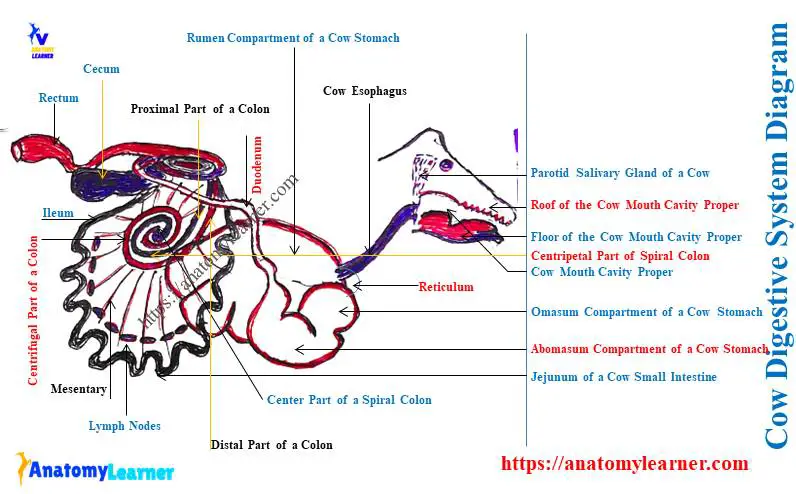
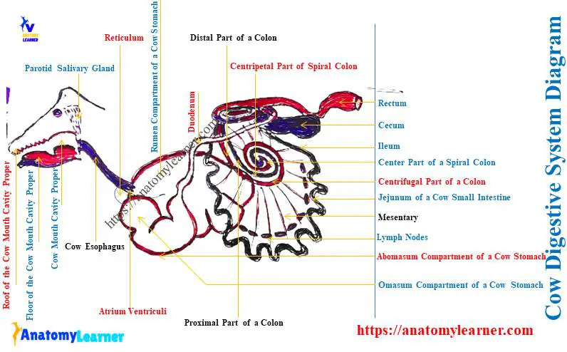
First, see the parts or organs of the cow or ruminant digestive system with the diagram. You will find the following organs or structures in the cow digestive system –
- The mouth of the cow – has 2 compartments (vestibule and mouth cavity proper),
- Pharynx – a space common both for the digestive and respiratory systems that continues with a caudal aspect of the mouth,
- Esophagus – a musculo membranous tube,
- Compound stomach – that possesses four compartments (rumen, reticulum, omasum, and abomasum),
- Small intestine – you will see the parts in the structure of the small intestine of a cow (duodenum, jejunum, and ileum),
- Large intestine – it consists of three major segments (cecum, various parts of colon, and last part of the large intestine),
You will find three parts in the structure of the cow colon – ascending, transverse, and descending. Again, the ascending part of the cow colon shows three parts – proximal, spiral, and distal.
The spiral segment of the ascending colon shows three other parts – centripetal, central, and centrifugal.
All the organs, as mentioned above or structures, are the main parts of the cow or ruminant digestive system. You will also see (notice) some of the other accessory organs or structures in the cow’s digestive system.
Let’sLet’s see the accessory organs from the cow digestive system –
- Tongue – a muscular organ,
- Teeth – includes incisors, premolar, and molar,
- Salivary glands – parotid, mandibular, and sublingual glands,
- Liver and gallbladder, and
- Pancrease of the cow,
The labeled diagram identifies all these main and accessory organs from the ruminant digestive system. Now, you will identify all the features of these digestive organs specifically.
Ruminant mouth cavity anatomy
This is an elongated cavity at the beginning of the ruminant digestive tract. It comprises two major compartments –
- Vestibules of the ruminant mouth, and
- Ruminant mouth cavity proper,
You will see similar structural features in the mouth cavity of horses or dogs. Let’sLet’s see the structural features of the dog mouth cavity from the below-mentioned article of anatomy learners –
Here, the vestibular part of the cow or ruminant mouth cavity is externally bounded by the lip and cheek. Again, in the internal boundary, you will find the gums and teeth in the vestibular part of the mouth.
The alveolar arches, gums, and teeth bound the cranial and lateral part of the cow mouth cavity proper. This structure of the cow mouth continues caudally, with the pharynx behind the oropharyngeal opening.
The roof of the cow mouth cavity proper consists of hard and soft palates. Again, the floor of the cow mouth cavity proper comprises mandibles, muscles, and mucous membranes. This roof and floor of the ruminant mouth cavity proper contain different structures.
I have already described these structures from the floor and roof of the ruminant mouth cavity in the previous article. Here, I will only show these features from the cow mouth cavity proper with the diagram.
The pharynx of the cow digestive system
The pharynx of a cow is the rough funnel shape space common for the digestive and respiratory systems. This space extends from the base of the cranial cavity to the caudal aspect of the nasal aperture.
The wall of this space is formed by the muscles, fibers, and mucous membranes. Many skull bones have formed their boundary, like temporal, pterygoid, vomer, sphenoid, palatine, and occipital.
You may get a basic idea (main) of the different bones of the cow skull from the below-mentioned article –
Other organs or structures like hyoid bone, mandibular salivary glands, carotid artery, and nerves (9th and 12th cranial nerves) are also related to the pharynx.
You will find the below-mentioned muscles in the structure of the cow pharynx anatomy –
- Palatopharyngus muscle,
- Thyropharyngeus muscles,
- Hyopharyngeus muscle,
- Criocopharyngeus muscle,
- Pterygopharyngeus and stylopharyngeus muscles of the cow pharynx,
The contraction of these pharyngeal muscle help to pass the feed from the pharynx to the esophagus of the cows.
You will commonly find three parts in the cow pharynx structure –
- Nasopharynx – dorsal part of the pharynx above the soft palate of the cows,
- Oropharynx – the part of the pharynx (PH) between the soft palate (SP) and root of the tongue, and
- Laryngopharynx – the part of the pharynx just dorsal and lateral to the larynx,
The pharynx of the cow gets blood supply from the branches of the carotid, occipital, and external maxillary arteries.
How many openings does the pharynx have?
Answer: the pharynx has 7 (seven) openings; 2 for eustachian tubes, 2 for nostrils, 1 for the oral cavity, 1 for the esophagus, and 1 for the larynx or trachea.
Esophagus of the cow digestive system diagram
An esophagus is a musculomembranous structure in the cow or the ruminant digestive system. The cow esophagus is divided into cervical and thoracic parts for description purposes. But, the abdominal part of the ruminant esophagus is practically absent.
The course of the cow esophagus is important for the veterinary student to understand. You may get the basic idea on the course of the cow or ruminant stomach from the below-mentioned article of anatomy learners –
In short, the ruminant stomach starts at the level of the cricoid cartilage of the larynx. Finally, this esophagus terminates in the shallow area between the rumen and reticulum (atrium ventriculi).
The compound stomach of the dog’s digestive system
The ruminant has a very extensive compound stomach which nearly fills three fourth of the abdominal cavity. You will typically find four compartments in the structure of the cow’s compound stomach.
I have tried to show (teach) you all the compartments of the cow stomach with the labeled diagram. But horses and dogs have a simple compartment, which usually possesses 2 parts – glandular and non-glandular.
So, you may have the question – how many stomachs does a ruminant have? For that, you may go through the below-mentioned article from anatomy leaner –
The ruminant (including large or small) has 4 compartments in its stomach – rumen, reticulum, omasum, and abomasum. Here, the rumen is the larger muscular sac of the stomach of a cow or ruminant.
Again, the reticulum part is the smallest compartment of a cow’s stomach. You may easily identify all the compartments of the cow compound stomach with their external and internal appearances.
Here, I will show you almost all external and internal anatomical features from the compound stomach of the ruminant.
Again, the rumen of the compound stomach has 4 compartments or sacs – dorsal, ventral, cauda-dorsal blind, and caudo-ventral blind sacs.
The surface and topographic location of the different compartments of the compound stomach is important for veterinarians. Let’s see the surface location of the rumen, reticulum, omasum, and abomasum of the compound stomach of the ruminant –
- Rumen – extends from the 7th ribs to the pelvic inlet,
- Reticulum – extends between 6th to 7th ribs,
- Omasum – lies right of the median plane and extends from the 7th to 11th ribs, and
- Abomasum – extends from the xiphoid cartilage to the abdominal floor,
You may understand the structure of the xiphoid cartilage of an ox sternum.
The small intestine of the cow digestive system
The small intestine is identified in the cow digestive system diagram. Here, you will see three major parts of the cow’s small intestine – duodenum, jejunum, and ileum.
The small intestine of the cow starts from the pylorus and ends at the ileocecal junction. Here, the diameter of the small intestine is smaller, but the length is more compared to the large intestine.
The average length of the small intestine of a medium-sized cow is about 40 – 45 meters. In contrast, small ruminants like sheep and goats are 20 – 25 meters long in their small intestines.
Again, the diameter of the small intestine in the large ruminant is 5 – 6 centimeters. And the goat and sheep show only 2 centimeters in diameter in their small intestine.
After the origin of the cow duodenum, it forms an S-shaped curve (known as the sigmoid flexure). Then it passes caudally, and this is the descending part of the cow duodenum.
You will also find a caudal flexure in the structure of the cow duodenum. The cranial part of the caudal flexure is continued as the ascending part of the duodenum.
The bile duct opens into the second bend of the S-shaped curved duodenum. Again, the pancreatic duct opens just behind the opening site of the bile ducts on the duodenum.
Jejunum and the ileum of the cow digestive tract
The length of the jejunum in the cow’s small intestine is larger compared to other parts. They form a good number of coils that forms the constricted and dilated portions.
Thus, you will see the U-shaped structure in the cow jejunum. You will see mesentery (modification of peritoneum) attached to the jejunum and several blood vessels (jejunal arteries).
The terminal part of the small intestine (after the jejunum) is the ileum. But how will you differentiate the ileum from the jejunum?
The last part of the jejunum becomes straight and becomes the ileum. So, the ileum is straight in structure and connects with the first part of the larger intestine.
The large intestine of the ruminant digestive system
The diameter of the cow’s large intestine is greater than these of the small intestine. You will find more diameter in the first part of the small intestine, and gradually it diminishes.
The major part of the cow’s large intestine locates on the dorsal aspect of the right side of the abdominal cavity. These parts of the large intestine also cover the modified peritoneum (mesentery).
The cow’s large intestine shows three different segments – the cecum, the colon, and the last segment. Here, the cecum of a cow is the blind sac-like structure that lies right off the median plane.
The length is about 65 – 75 centimeters with a 10 – 15 centimeter diameter. Again, the capacity of the cow cecum is about 7 – 10 liters.
The caudal blind is also known as the apex. You will see the connection of the cranial cecum with the colon of the cows.
The colon of the cow is longer and possesses 3 different segments –
- Ascending colon – consists of proximal, spiral, and distal loops,
- Transverse colon – passes left to the cranial mesenteric artery, and
- Descending colon – runs caudally and continues with the last segment of the large intestine,
Here, the spiral part of the ascending colon shows another 3 different coils –
- Centripetal – the spiral part that goes toward the center,
- Central flexure – a central part of the spiral colon, and
- Centrifugal coils – the part of the spiral colon that back from the center and continue with the distal part of the colon,
All the main organs or structures from the cow or ruminant digestive system are described. Now, I will provide information on the accessory organs or structures from the ruminant digestive system.
Accessory organs of the cow digestive system
First, let’s see the significant accessory organs from the ruminant digestive system. Here, the main accessory organs of the ruminant digestive system are – the tongue, teeth, liver, pancreas, gallbladder, and three major salivary glands.
You will find the description of the anatomical facts of these accessory organs of the cow digestive system individually.
The tongue of the ruminant digestive system
The tongue is considered the accessory organ in the ruminant digestive system. You will find the typical features of the tongue in a ruminant.
The ruminant tongue shows three major segments – root, body, and apex. Here, the dorsum surface of the ruminant tongue shows different anatomical features.
The torus linguae, transverse lingual fossa, and five various papillae on the dorsum surface of the ruminant tongue make them different from others. You will find the details features of these structures of the ruminant tongue from the below-mentioned article –
- Cow tongue anatomy – torus linguae, transverse lingual fossa, and varieties of papillae with diagram,
The fungiform, conical, and lenticular are the mechanical papillae of the ruminant or cow tongue structure. Again, the filiform and vallet are the gustatory papillae of the ruminant tongue that contain the taste buds.
Teeth of the ruminant
You will find a total of 32 teeth in the ruminant mouth cavity. The upper jaw doesn’t possess the incisor or canine teeth.
The cheek teeth (premolar and molar) are 6 in number on both sides of the upper and lower jaws. You may get a clear concept of the jaw bone structure of the ruminant from the below-mentioned article –
Again, I tried to show you the various teeth from the upper and lower jaws of the cow here.
Cow liver – accessory digestive organ
The cow liver is the solid and largest gland in the body. You will find the reddish brown color in a cow liver in its fresh condition.
The liver of a cow locates at the right of the median plane (MP) of the body. It directs obliquely downward and forward (cranioventrally) and extends from the last ribs to 6th ribs.
The cow or ruminant liver shows the followings –
- Two surfaces – diaphragmatic and visceral surfaces,
- Four lobes – right, left, caudate, and quadrate lobes of the cow liver, and
- Four borders – dorsal, ventral, right (caudal), and left (cranial) borders of the cow liver,
All of these surfaces, lobes, and borders from the cow or ruminant liver are identified in the labeled diagram. Here, the diaphragmatic surface has a contact with the diaphragm. The visceral surface connects or attaches to the reticulum, omasum, and abomasum.
So, you will find the impression or depression on the visceral surface of the ruminant liver for these organs mentioned above. All these impressions on the visceral surface of the liver are identified in the labeled diagram.
The caudate process of the ruminant liver possesses two processes – caudate and papillary process. Here, the caudate lobe of the ruminant liver forms the impression of the kidney with the right border of the right lobe.
There is another impression present on the visceral surface of the ruminant liver for the gallbladder. You will see the depression on the dorsal border of the cow’s (ruminant) liver for passing the vena cava.
If you like to learn the details facts of the ruminant (cow/goat/sheep) liver, you may read the below-mentioned article –
- Cow liver anatomy – lobes, surfaces, impression, and ligaments with diagram,
Salivary glands of the cow digestive system
You will see the three pairs of salivary glands in the cow digestive system (as the accessory glands) –
- Parotid glands – triangular and long reddish salivary glands in the cow,
- Mandibular glands – elongated and pale yellowish lobulated glands, and
- Sublingual glands – possess two separated parts (superior and inferior),
The parotid glands of a cow locate on the masseter muscle along the caudal border of the vertical part of the mandible. You will see two ends in the structure of a cow’s parotid glands – dorsal and ventral.
Here, the dorsal end of the cow parotid gland is broad and located near the base of the ear. In contrast, the ventral end of the parotid gland is narrow and located at the caudal aspect of the masseter muscle.
The parotid ducts (Stenson) open at the base of the papilla incisive just opposite the second molar tooth of the upper jaw.
The mandibular gland of a cow locates at the medial side (MS) of the angle of the mandible. This gland is longer than the parotid and extends from the wing of the atlas bone to the intermandibular space.
Let’s know the anatomical features of the cow atlas bone from the below-mentioned article of anatomy learner –
You will find two distinct surfaces (lateral and medial) in the structure of the cow mandible. Here, the lateral surface of the cow mandibular glands is related to the parotid gland, mandibular lymph nodes, sternomandibualr muscles, and facial vein.
The medial surface of this gland is related to the lymph node, pharynx, larynx, and carotid artery. You will find the opening of the mandibular ducts (Wharton’s duct) at the sublingual caruncles of the mouth cavity.
Sublingual salivary glands of a cow
The superior sublingual gland (also known as the polystomatic) is a lobulated and pale-colored structure. These superior sublingual glands of the cow locate under the mucous membrane of the mouth cavity properly.
The superior sublingual glands extend from the mandibular symphysis to the palatoglossal arch of the cows. Laterally, the superior sublingual glands of the cow are related to the myohyoideus, styloglossus, and genioglossus muscles.
You will see the numerous ducts of the superior sublingual glands in a cow. These ducts of the superior sublingual will open on either side of the frenulum linguae at the floor of the cow’s mouth.
The inferior sublingual glands (also known as the monostomatic) are the pink color salivary glands in cows. These glands are thicker and shorter than these of the superior sublingual glands.
The inferior sublingual salivary glands of the cows extend from the mandibular symphysis to the level of the last premolar tooth. You will see the opening of the inferior sublingual gland’s duct at the base of the sublingual caruncle.
Pancreas of cow digestive system
The pancreas is another accessory digestive gland in cows. This is another solid gland located at the right side of the median plane (MP) of the body.
You will see the pancreas attach to the medial aspect of the cow’s liver. The mesentery also covers the pancreas of the cows.
The color of the cow pancreas is reddish yellow. It is a flat and irregular triangular shape gland in the cow.
You will see two surfaces (dorsal and ventral) and four borders (cranial, caudal, lateral, and medial) in the structure of the cow pancreas.
The dorsal surface of the cow pancreas attaches to the diaphragm and liver. Again, the ventral surface of the ruminant pancreas is related to the dorsal sac of the rumen, colon, and hepatic artery.
The cranial border of the cow pancreas is more or less straight. You will see the notch at the caudal border of the ruminant pancreas for passing the portal vein.
The right border is long and parrel to the duodenum. Again, the left border of the cow pancreas is irregular and convex.
The dorsal surface of the cow pancreas is not only related to the diaphragm and liver. This surface is also related to the right kidney, vena cava, and mesenteric arteries.
Cow digestive system labeled diagram
Now, let’s see every single part or organ from the cow digestive system labeled diagram. First, I will show the main organs of the cow’s digestive system (ruminant).
The organs from the mouth to the last part of the large intestine are identified in the labeled diagram. Cow tongue labeled diagram also shows the different anatomical features.
The labeled diagram also identifies different compartments of the cow’s compound stomach. The segments from the small and large intestines of the cows are also shown in the labeled diagram.
The accessory organs, especially the liver, pancreas, gallbladder, and salivary glands, are also shown in the labeled diagram. Let’s find more labeled diagrams on the cow digestive system on the social media of anatomy learners.
Frequently asked questions on cow digestive system
Here, you will find the concise answer to the question related to the cow digestive system that the anatomy learners ask. But, it is suggested to go through the whole article to get the details facts of the ruminant digestive system organs.
Okay, let’s see the question on the ruminant digestive system’s organs with their concise answer –
Does a cow have 2 or 4 stomachs in their digestive system?
Quick answer: cows have only one (1) stomach in their digestive system. But, the cow possesses a compound stomach that has 4 (four) different compartments like rumen, reticulum, omasum, and abomasum.
How is the cow’s digestive system different from the horse’s?
Quick answer: you will find a significant variation in the organs of the digestive system between horses and cows. The cow possesses a compound stomach, whereas the horse has a simple stomach.
Again, you will see a great variation in the length and structure of the intestine between the cows and horses. The horse cecum is longer, and the apex is directed cranially.
The tongue of the horse is more or less different in structure compared to the cow. Again, the horse’s liver shows four distinct different lobes, whereas the quadrate lobes are indistinct in the cow’s liver.
Conclusion
The cow digestive system diagram might help you to understand the organs from the mouth to the last part of the large intestine. All the ruminant digestive system’s main organs possess unique anatomical facts that differ from other animals.
The surface and topographic location of the accessory organs of the cow’s digestive system are essential. Now, you should identify all of these organs from the cow digestive system with the help of the diagram.