The cow respiratory system includes the nasal cavity, pharynx, larynx, trachea, bronchi, and lungs. Here, the lungs are considered as the central organ of the animal respiratory system.
In this article, I will only focus on identifying different organs or structures from the ruminant respiratory system with a diagram. After completing this article, you may understand the topographic and surface anatomy of these respiratory organs or structures.
Quick overview: cow’s respiratory system comprises respiratory passages and lungs. The respiratory passages include the nasal cavity, pharynx, larynx, trachea, and bronchi. Two lungs (right and left) are the principal respiratory organs that exchange gas between the blood and air.
Let’s see the various features from the nasal cavity, pharynx, larynx, trachea, bronchi, and lungs of the cow respiratory apparatus. In the last part of this article, you will find some important differences among the respiratory organs of cows, horses, and dogs.
Cow respiratory system
The cow respiratory system starts from the nostrils and ends in the lungs. It connects with the oral cavity into the pharynx.
At the cranial end of the larynx, the respiratory passages subdivide into two parts –
- The upper respiratory passages and
- A lower respiratory passages,
Thus, the upper respiratory system includes the nostrils, pharynx, and
The lower part of the ruminant respiratory system includes the larynx, trachea, and bronchi.
And you already know that the lungs are the central organs of the respiratory system.
Now, let’s identify these main respiratory passages and organs from the cow or ruminant. You will find the below-mentioned passages and organs in the structure of a cow respiratory apparatus –
- Two nostrils (right and left),
- The nasal cavity, along with its conchae and meatus,
- Pharynx (a common space both for respiratory and digestive systems),
- Larynx (4 or 5 main cartilages on the structure of the cow larynx),
- The cow trachea (comprised of two main parts – cervical and thoracic parts),
- Apical and primary bronchus (you will find some other bronchus and bronchioles), and
- The lungs of the cow (right and left),
The ruminant respiratory system diagram identifies all the above-mentioned main structures and organs. But make sure you have a basic idea of the thoracic cavity of the cow while learning the details structure of the lungs from the cow’s respiratory apparatus.
You may read the below-mentioned article for anatomy learner to get a basic idea of the thoracic cavity of animals –
Cow respiratory system facts
Here, I will enlist the common facts from the ruminant respiratory system. Let’s see what they are in a cow –
- You will find the comma-shaped right and left nostrils on either end of the muzzle,
- The bilateral nasal cavity of the cow are irregular tubular passages that are enclosed by the facial bones and cartilage,
- The air passages through the nasal cavity and reaches the nasopharynx and oropharynx,
- You will find the musculo-cartilaginous elongated laryngeal compartment in the structure of a cow’s respiratory apparatus,
- The cow larynx comprises three unpaired (cricoid, thyroid, and epiglottis) and one paired (arytenoid) cartilages,
- The trachea of a cow is almost U-shaped structure and has cervical and thoracic parts,
- You will find 48 – 60 incomplete cartilaginous rings in the structure of a cow trachea,
- At the level of 3rd intercostal space, the cow trachea gives off the apical bronchus on its right side,
- You will find the right and left primary bronchus of the cow’s trachea at the level of the base of the heart,
- There are two lungs (right and left) in the thoracic cavity of the cow that separates from each other by the mediastinum,
You will also find a thin transparent serous membrane in the right and left lungs. This is the pleura of the cow lung and possesses two surfaces – parietal and visceral parts. You will find the pleural fluid within these two surfaces of the pleura.
Animal respiratory system parts and functions (ruminant)
The nasal cavity of the cow respiratory apparatus opens externally at the right and left nostrils. It communicates with the pharynx caudally through the nasal conchae or posterior nares.
The nasal cavity of the cow also possesses the olfactory apparatus that mediates the sense of smell.
You will see the space (pharynx), which is common for both air and food (respiratory and digestive systems of the animals. You may find the organs of the digestive organs of a cow in the below-mentioned article of anatomy learner –
The complex valvular apparatus (larynx) in the cow’s respiratory tract will also be seen. This larynx comprises the cartilage, and a regular volume of air passes through it.
The larynx of the cow is also considered the chief voice production organ. Again, the trachea is an important lower respiratory tract of the passages that contain several incomplete rings (48 – 60).
The bronchus that is formed by the bifurcation of the trachea is permanently open conducting tubes. All these upper and lower parts of the cow’s respiratory tracts help pass the air within and to the lungs.
Here, you might also understand the structure of the cow’s thorax, pleural sacs, and thoracic muscles (including the diaphragm).
Cow respiratory system organs identification
Now, let’s identify the various structures from the different organs or parts of the cow respiratory system. First, let’s start with the nasal cavity of the cow.
The nasal cavity of the cow
The right and left nostrils of the cow’s nose are relatively small and less dilatable than these of the horse. You will find the smooth and moist skin surrounding the nostrils of a cow.
The opening of the nasolacrimal duct is not visible on the lateral wall of the cow’s nostrils. Again, the nostrils are supported by two types of cartilage – parietal and alar cartilage.
On the other hand, the nasal cavity of a cow is short, wide cranially, and narrow caudally. The nasal cavity of a cow is complete divides into two compartments – right and left.
From the nasal cavity of a cow, you might identify the below-mentioned structures –
- Median nasal septum – perpendicular part separating the nasal cavity into right and left compartments,
- Dorsal nasal concha – extends from the cribriform plate of the ethmoid bone to the rostral part of the nasal cavity,
- Ventral nasal concha – an extensively folded structure that fills the middle lumen of the nasal cavity,
- Ethmo-turbinate or middle nasal concha – delicate, mucous-covered, bony scrolls that fills the caudal part of the nasal cavity,
Here, the ventral nasal concha is the separate and distinct bone (turbinate) of the cow’s skull. Again, the middle or ethmoturbinate is part of the ethmoid bone.
You may also know the other different bones of the cow skull (including the ethmoid and vomer) from the below-mentioned article of anatomy learner –
What is nasal concha in the cow nasal cavity?
Answer: these bony scrolls cover the nasal mucosa and fill each half of the nasal cavity. Here, the nasal concha and the nasal septum divide the cow’s nasal cavity into two complete compartments.
What is the meatus in the cow nasal cavity?
Meatus are the passageways between the nasal conchae of each half of the cow’s nasal cavity. You will find three main nasal meatuses in the structure of the cow nasal cavity.
Let’s identify the below-mentioned nasal meatus from the structure of the cow nasal cavity –
- Dorsal nasal meatus – narrow passages between the dorsal nasal concha and the nasal bone leading into the caudal nasal cavity,
- Middle nasal meatus – the passageways between the dorsal and ventral nasal conchae, and
- Ventral nasal meatus – largest nasal meatus between the ventral nasal concha and the hard plate,
You will also find (get) the common nasal meatus in the structure of the cow nasal cavity. It is a narrow verticle space between the median nasal septum and conchae.
Pharynx of the cow – identification
It is the funnel-shaped area at the base of the cranial cavity and behind the caudal nasal cavity. Here, you might identify the different parts of the pharynx and seven openings of the pharynx –
- Parts of the pharynx – nasopharynx, oropharynx, and laryngopharynx, and
- Seven openings of the pharynx – 2 for eustachian, 2 for caudal nares, 1 for esophagus, 1 for larynx, and 1 for mouth cavity proper,
All these structures from the cow pharynx are identified in the labeled diagram. But, you may know more details on these topics from the cow respiratory apparatus from other articles of anatomy learners.
Cow larynx anatomy
The cow larynx is the upper end of the windpipe that possesses a musculocartilaginous structure. This tube connects the laryngopharynx with the trachea and contains the vocal cords.
You will see a group of cartilage in the structure of the cow larynx. Both single and paired cartilages make the cow larynx.
Let’s identify the various cartilage from the cow larynx –
- Unpaired or single cartilages – cricoid, thyroid, and epiglottis, and
- Paired cartilage – arytenoid and corniculate,
All these cartilages from the cow larynx are identified in the labeled diagram.
Cricoid cartilage of the cow larynx:
It is signet-shaped cartilage that connects thyroid and arytenoid cartilage. The cricoid cartilage of the cow larynx is compressed laterally.
This cricoid cartilage locates rostral to the first tracheal ring. It possesses two parts –
- Dorsal quadrilateral lamina (not distinctly divides from the arches), and
- Ventral and lateral arches of the cricoid cartilage,
You will also find a median ridge on the lamina of the cricoid cartilage. This median ridge is the muscular process of the cricoid cartilage.
Thyroid cartilage of the cow larynx
The thyroid cartilage of the cow larynx is thin and broad U-shaped structure. You will see the ventral body and two lateral laminae in the structure of the thyroid cartilage of the cow larynx.
The lateral lamina possesses cornu on its cranial and caudal aspects (shown in the diagram). Again, the caudal third of the body of the thyroid cartilage possesses an eminence. This is the laryngeal prominence of the cow’s larynx.
The thyroid cartilage is relatively short in sheep and goats.
Cow epiglottis cartilage structure
The epiglottis is the most rostral cartilage in the cow larynx. It closes the laryngeal opening during deglutition and protects the lung from foreign bodies.
You will find the base, apex, two surfaces, and two lateral borders in the structure of the cow’s epiglottis. The two surfaces of the epiglottis are – the lingual and laryngeal surfaces.
The shape of the cow epiglottis is an obovate leaf shape. You will also find the same shaped epiglottis in the sheep larynx.
But, the goat has the cordate leaf-shaped epiglottis in their larynx. But, the epiglottis is comparatively short in cows and has a rounded apex.
The base of the epiglottis cartilage rest on the membranous structure of the thyroid cartilage. You will find the loose attachment between the base of the epiglottis and thyroid cartilage.
Arytenoid cartilage of the cow larynx
This is the only paired cartilage in the cow larynx, just rostral to the cricoid cartilage. It is three-sided pyramidal shape cartilage with a rostral apex and caudal base.
You will find a number of processes in the structure of the arytenoid cartilage. The processes of the arytenoid cartilages are –
- Vocal process,
- Muscular process,
- Corniculates process, and
- Cuneiform process,
But, you will only find the muscular and corniculate processes in the structure of the arytenoid cartilage of the cows.
You will find strong prominence in the junction between the lateral border and the base of the arytenoid cartilage. This strong prominence is the muscular process of the cow’s arytenoid cartilage.
Again, the corniculate process is the rostral horn-like projection that comes from the dorsal part of the laryngeal opening.
Here, in the structure of the cow larynx, you will find various laryngeal muscles and ligaments.
Cow trachea anatomy – identification
The cow’s trachea is comparatively short than the horse’s. A medium-sized cow possesses a trachea with a length of 25 inches. The caliber of the cow trachea is also small compared to the horse.
The cow trachea is a flexible, cartilaginous, and membranous structure. Dorsal to the base of the heart, the trachea gives the right and left primary bronchus.
Before that, you will find an apical bronchus on the right side of the trachea at the level of the third intercostal space. This apical bronchus ventilates the apical lobe of the right lung.
Now, let’s try to identify the below-mentioned features from the cow trachea –
- Tracheal rings (incomplete),
- Cervical parts of the cow’s trachea,
- The thoracic part of the cow’s trachea,
- Apical bronchus on the right side,
- Right and left primary bronchus of the trachea,
All these features from the cow trachea are identified in the labeled diagram. The tracheal ring of the cow trachea is U-shaped. And you will find an average of 50 tracheal rings (varies from 48 – 60) in the structure of the cow trachea.
You will find a great relationship in the course of the trachea and esophagus of the cows. Let’s get a basic idea of the relationship between the cow’s esophagus and trachea from the below-mentioned article –
The average length of the sheep trachea is 9 – 10 inches. Again, the caliber of the sheep trachea is less than one inch.
You will find three major bronchi that bifurcate from the trachea. An apical and two primary bronchi are found in the structure of the cow trachea.
Cow lung anatomy identification
Lungs are a pair of main respiratory organs that occupy most of the thoracic cavity. A cow’s right and left lungs are separated by the mediastinum and covered by the pleural sac.
In fresh condition, the lungs are pink, soft, and spongy. Here, the right lung of the cow is twice the time larger than the left lung.
But, the cow lungs are comparatively shorter than these of the goats and sheep. Why is the right lung larger in the same cows?
The possible causes of being larger lung on the right side –
- The right lung of a cow possesses an accessory lobe (on its medial aspect),
- You will find a larger apical lobe in the right lung structure in a cow, and
- The heart of the cow mostly occupies the left side of the median plane, and thus it also occupies the left lung,
What should you identify from the cow lungs?
You might identify the below-mentioned anatomical facts from the cow lung. Here, I will only enlist the most important features, but you might know the details of these features.
You will find the basic idea of these features from the cow lung in the next section of this article. Okay, let’s see the main anatomical facts of the cow lung anatomy –
- Lobes of the right and left lungs (4 lobes in right lung, and 2 lobes in left lung),
- The cranial apex of both lungs,
- Caudal base or diaphragmatic surface of both lungs,
- Costal and medial surfaces of both right and left lungs,
- Three borders of the lungs (dorsal, ventral, and basal),
- Costal impression on the costal or lateral surface of both right and left lungs,
- Cardiac impression (middle) on the medial surface of the lungs,
- Hilus of the right and left lungs (above the cardiac impression),
- Cardiac notch in the ventral border of the lungs,
- Fissures among the lobes of right and left lungs, and
- Groove for the caudal vena cava and phrenic nerve,
All the features mentioned earlier from the right and left lungs of a cow are identified in the labeled diagram. Now, let’s know more about these features of the right and left lungs of the cows.
How many lungs does a cow have?
Answer: the cow has 2 well-developed lungs – right and left. Here, the right lung is exceptionally larger than the left lung of the cow.
This is due to the larger apical lobe and a small accessory lobe on its medial surface. Let’s see the extension of the right and left lungs of the cows –
- Left lung of the cow – extends from the 1st rib to the 10th rib of the thoracic cavity, and
- The right lung of a cow – extends from the 2nd rib to the 11th rib of the thoracic cavity,
You will find a great variation in the number and structure of the lobes in both the right and left lungs.
How many lobes does the cow’s right lung have?
Quick answer: the cow’s right lung has 4 (four) lobes which are subdivided by the interlobar fissures. Let’s see the 4 lobes of the cow’s right lungs –
- Apical or cranial lobes of the right lung,
- Middle r cardiac lobe of the right lung,
- A diaphragmatic or caudal lobe of the cow’s right lung, and
- Accessory or intermediate lobe of the cow’s right lung,
Here, the apical lobe of the right lung is larger and from the larger portion of the cardiac impression. The middle or cardiac lobe is elongated and three-sided in appearance.
This middle lobe of the right lung also contributes to forming the cardiac impression on the right aspect. Again, the diaphragmatic lobe is the largest lobe in the right lung of a cow. This diaphragmatic lobe of the right lung is caudal in position.
The accessory or intermediate lobe is pyramidal in shape and located medial to the diaphragmatic lobe. The accessory and diaphragmatic lobes form a groove for the caudal vena cava and phrenic nerve.
How many lobes does the cow’s left lung have?
Quick answer: you will find only 2 (two) lobes in the structure of the cow left lung anatomy. Here, you will not find any intermediate or accessory lobes in the left lung of the cows.
Let’s see 2 lobes of the cow’s left lung –
- An apical or cranial lobe of the cow’s left lung, and
- The caudal or diaphragmatic lobe of the left lung,
Here, the apical lobe of the left lung shows 2 pats –
- The apical portion of the apical lobe, and
- Ventrally directed caudal portion of the apical lobe,
The apical part of the apical lobe is pointed, small, and cranially directed. On the other hand, the caudal part of the apical lobe of the left lung is larger and three-sided. You will find a small interlobar fissure that partially subdivides the apical and caudal parts of the apical lobe.
Common structure of right and left lungs of the cows
Each lung has an apex, base, two surfaces, and two borders. You identified these apex, base, surfaces, and borders from the cow lung labeled diagram.
Here, the right lung of a cow shows a larger apex than the left lung. The diaphragmatic surface of the lung is related to the convex thoracic (caudal) surface of the diaphragm. It is concave, more or less elliptical, and bounded by the basal border.
The costal surface of both lungs is larger and smooth, and convex. You will find a relationship between this costal surface with the inner surface of the ribs, costal cartilage, and intercostal muscles.
Thus, you will find the costal impression on the lateral or costal surface of the right and left lungs of the cows.
The medial surface of both lungs is extensive and possesses two parts –
- Vertebral part – related to the bodies of the thoracic vertebrae, and
- The larger mediastinum part – is concave and relates to the heart and pericardium,
You will find the concave and deep cardiac impression on the medial surface of the lungs. The right lung of a cow shows a more deep and more extensive cardiac impression.
The whole lungs cover with the pleura (a thin serous membrane). But above the cardiac impression, you will not find any pleural covering.
You will find the bronchi, vessels, lymphatic, and nerves in this area. This is known as the hilus of the cow’s lungs.
Borders and cardiac notch of the cow lungs
You will find dorsal, ventral, and basal borders in the structure of both right and left lungs. Here, the dorsal border of the cow lungs is thick and rounded.
Again, the basal border is smooth and rounded in the cows lungs. This basal border separates the diaphragmatic surface from the medial surface of the lungs.
The ventral border of each lung is sharp and irregular. You will find an identical cardiac notch in the ventral border of the cow’s lungs.
It is small and variable in size for the right lung of the cows. But the left lung of a cow shows an L-formed cardiac notch on its ventral border.
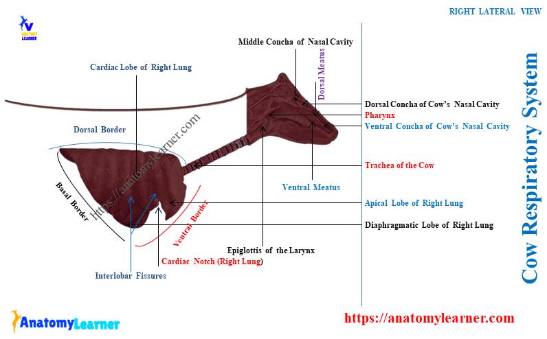
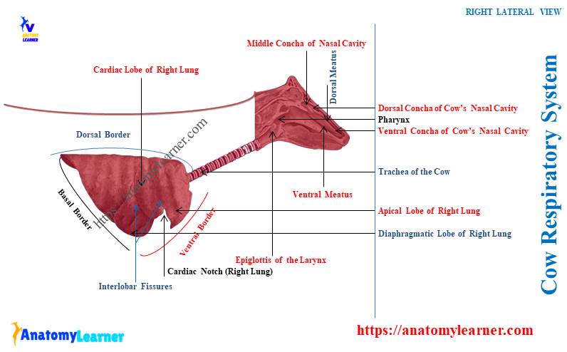
Cow respiratory system diagram
Now, let’s see the overview of the cow respiratory system labeled diagram. Here, I tried to show you every passage (both upper and lower) tracts and different lobes of both lungs.
From the nostril to the lungs from the ox respiratory system are identified in the labeled diagram. The conchae and meatus from the cow’s nasal cavity are also identified in the diagram.
I showed you the 4 different laryngeal cartilage from the cow larynx in the diagram. Let’s see the U-shaped tracheal cartilaginous rings on the provided diagram.
The diagram also shows the three bronchus – apical (on right side) and two primary (right and left) of the cow’s trachea.
Finally, 4 lobes from the right lung and 2 lobes from the left lung are also identified in the cow lung labeled diagram. The cow lung diagram also shows the base, apex, surfaces, and borders.
You may also find more diagrams and videos of the cow lung anatomy or other respiratory organs on social media of anatomy learners.
Frequently asked questions on cow respiratory apparatus
Let’s see the common questions on the cow respiratory apparatus that anatomy learners ask. Here, I will enlist the frequently asked questions on the respiratory system of animals with their concise answer.
What are some facts about the ruminant respiratory system?
Answer: the nasal cavity of the ruminant respiratory system is short and wide compared to other animals. Again, the larynx of the ruminant is more compact than the horse.
You will not find the cuneiform process in the structure of the arytenoid cartilage in the ruminant larynx. The trachea is shorter and possesses U-shaped (ox, goat) or C-shaped (sheep) structure.
Finally, the lobes of the ruminant lungs are prominent and more or less well-separated than the horses.
How to differentiate the horse’s respiratory organs from the cows?
Answer: the nasal cavity of the horse respiratory system is larger but narrower compared to the cows. You will not find the subdivision on the structure of the middle meatus of the horse.
The epiglottis of the horse larynx is pointed and long. You will find the prominent vocal cord in the structure of the horse larynx.
You will find three paired (cricoid, thyroid, and epiglottis) and 3 unpaired (arytenoid, corniculate, and cuneiform) cartilage in the horse larynx.
The trachea of the horse is comparatively long and has no apical bronchus like the cows. Lobes of both the right and left lungs of the horse do not show any well-separation or fissures.
Thus, the lobes of the horse lungs are not prominent, and the apical lobe of the right lung is not larger like the cows.
Conclusion
The cow respiratory system shows the typical features compared to other animals like horses or dogs. Various nasal conchae, meatus, and septum are important structures in the cow’s nasal cavity.
Different types of hyaline and elastic cartilages of the cow larynx should be identified perfectly by you. The lobes with their apex, base, surfaces, borders, and other features are also important for learning the cow lungs anatomy from its respiratory apparatus.