The dog nervous system is divided into the central and peripheral nervous systems. The brain and the spinal cord include the central nervous system (CNS). In contrast, the canine peripheral nervous system (PNS) consists of cranial nerves, spinal nerves, and visceral peripheral components.
The visceral peripheral components of the canine nervous system include the sympathetic and parasympathetic nerves. This article will shortly describe the different components and nerves of the dog nervous system.
So, you will learn about the different parts of the dog’s brain, spinal cord, and their covering with its modification. This article will also illustrate the origin and distribution of the cranial and spinal nerves from the canine nervous system.
In the end, you will also know how a dog’s sympathetic and parasympathetic nervous systems work. All these nerves and components from the dog will describe with possible and necessary labeled diagrams.
Dog nervous system
First, I would like to provide the overall idea of the different parts of the dog nervous system. The central nervous system of the dogs consists of a brain (encephalon) and a spinal cord (medulla spinalis). Again, the central nervous tissue divides into the white and gray matter based on the gross appearance.
In the gray matter, you will see the cell bodies of the neuron cells, neuroglia, intertwined dendrites, and axons (both myelinated and nonmyelinated). You know the basic, genetic, and functional unit of the nervous system is a neuron which is a highly specialized cell.
There are a cell body, an axon, and the dendrite in the structure of a neuron. You may find the details of form and different types of neurons in the below-mentioned article –
Structure of the animal neuron with the labeled diagram
Again, the white matter consists of the myelinated axons and the neuroglia associated with the white matter. The neuronal cell bodies of the gray matter of the spinal cord are arranged in longitudinal columns. Again, the white matter of the spinal cord (outer aspect) surrounds by the gray matter.
“In the dog brain, you will see the inner white and outer gray matters. But, in the spinal cord, you will see the inner gray and outer white matter.”
The peripheral nervous system subdivides into spinal, cranial, and visceral peripheral components. Again, the visceral peripheral element consists of the thoracolumbar (sympathetic system) and the craniosacral part (parasympathetic system).
You will see 36 pairs of spinal nerves (nerve spinales) in the dog. There are many essential nerves in a dog’s brachial and sacral plexus. Twelve (12) pairs of cranial nerves in a dog emerge or enter the brain through foramina.
Central nervous system
The dog’s brain is the first part of the central nervous system that divides into five major components – telencephalon, diencephalon, mesencephalon, metencephalon, and myelencephalon. But, in the embryonic state, the dog brain may divide into the forebrain, midbrain, and hindbrain.
- The forebrain of the dog – includes telencephalon and diencephalon,
- A midbrain of the dog – contains mesencephalon, and
- The hindbrain of the dog – includes metencephalon and myelencephalon.
But, here, I will describe the dog’s brain anatomy from three large regions – cerebrum, cerebellum, and brainstem. So, you may divide the dog brain into these three major parts for description purposes.
Here, the cerebrum is the telencephalon, whereas the cerebellum is the dorsal part of the metencephalon. The brainstem of the dog comprises the remaining portions, medulla oblongata, and pons. Caudally, the brainstem of the dog brain is continuous with the spinal cord.
Now, the spinal cord of a dog is enclosed within the vertebral canal, which also belongs to the central nervous system. But, dorsal and ventral spinal roots and spinal nerves belong to the peripheral nervous system.
The dorsal root carries the sensory fiber, whereas the ventral root conveys the motor fibers. They meet outside the intervertebral foramen, exchange their fibers, and form the spinal nerve.
Now, the spinal nerves of the dog that originated from the spinal cord innervate to the neck, trunk, tail, limbs, and caudal and dorsal surfaces of the head.
You will learn the anatomical facts about the brain and spinal cord of the dog in the next section of this article.
Dog peripheral nervous system
First, let’s discuss the spinal nerves from the dog peripheral nervous system. Each spinal nerve (total of 36 pairs) consists of 4 segments – root, main trunk, four primary branches, and numerous peripheral branches.
The root of the spinal nerve lies within the vertebral canal and consists of a dorsal root and a ventral root. Within the intervertebral foramen, the union of dorsal and ventral roots forms the main trunk of the spinal nerve.
You will see some small and variable meningeal branches of the spinal nerve within the intervertebral foramen. Again, the spinal nerve gives off a small dorsal communication and a large ventral branch. Finally, the dorsal and ventral branches subdivide into the lateral and medial branches, giving rise to numerous other small branches.
You will also see the spinal ganglion (also known as the dorsal root ganglia) within the corresponding intervertebral foramen. These are the aggregation of the pseudounipolar nerve cell bodies of the dorsal root.
Here, in the diagram, I tried to show you the structure of a spinal nerve of a dog. The dorsal branch of the spinal nerve extends dorsally and divides into lateral and medial branches. These branches of the dorsal part of the spinal nerve will supply to the muscle and skin near the dorsal midline.
The ventral branch is the largest of the four primary branches, also divided into medial and lateral parts. Again, the communicating branches of the spinal nerve differ from the dorsal and ventral branches. These communicating branches of the spinal nerve carry the general visceral afferent and efferent axons.
Division of the dog’s spinal nerves
The dog’s spinal nerves divide into 6 major groups. You will find a piece of short information on these spinal nerves from the different regions of a dog’s body in the next section of this article.
First, let’s see what the primary division of the dog’s spinal nerves is –
- A spinal nerve in the cervical region of a dog,
- The thoracic spinal nerve of the dog,
- Nerves of the dog brachial plexus (formed by the ventral branches of a few cervicals and thoracic spinal nerves),
- The spinal nerve in the lumbar region of a dog,
- Sacral plexus (spinal nerve in the sacral region of a dog), and
- The caudal spinal nerve of a dog,
There are eight pairs of cervical nerves in the dog. The dog brachial plexus nerves supply to the thoracic limb’s extrinsic and intrinsic muscles. Again, three major nerves from the dog’s brachial plexus (radial, median, and ulnar) supply up to the digits of the thoracic limb of a dog.
You will find the 13 pairs of thoracic spinal nerves in a dog. In comparison, the number of lumbar spinal nerves in a dog is 7 (seven). In the sacral plexus of a dog, you will see the largest ischiatic nerve that divides into common fibular, tibial, and caudal cutaneous sural nerves.
All the courses of the dog ischiatic nerve are shown in the labeled diagram in the dog nerve anatomy section. Finally, you may find four to seven (4 – 7) caudal spinal nerves in a dog.
Dog cranial nerves
As I told you before, 12 pairs of cranial nerves of a dog emerge or enter the brain through the different foramina of the skull. These cranial nerves of a dog possess sensory, motor, and mixed functions.
First, let’s see the 12 pairs of cranial nerves from a dog –
- Olfactory nerve (cranial nerve I),
- The optic nerve of a dog (cranial nerve II),
- The oculomotor nerve of the dog (cranial nerve III),
- Trochlear nerve (cranial nerve IV),
- Trigeminal nerve of the dog (cranial nerve V),
- Abducent nerve (cranial nerve VI),
- The facial nerve of the dog (cranial nerve VII),
- Vestibulocochlear nerve (cranial nerve VIII),
- The glossopharyngeal nerve of the dog (cranial nerve IX),
- Vagus nerve of the dog (cranial nerve X),
- The accessory nerve of the dog (cranial nerve XI), and
- The hypoglossal nerve of a dog (cranial nerve XII),
You might know the root of these cranial nerves and their supply to different organs and structures of the dog. The vagus and trigeminal are essential nerves from the cranial division of the dog peripheral nervous system. In this article, you will find the short course and distribution of the vagus and trigeminal nerves here with a labeled diagram.
Dog autonomic nervous system
The autonomic nervous system of a dog is divided into parasympathetic and sympathetic divisions. You will see the parasympathetic preganglionic axons that leave the brainstem as part of cranial nerves III, VII, IX, and X.
The dog’s autonomic nerves’ parasympathetic division (cranial part) is restricted to the eye, lacrimal, mandibular, sublingual, and parotid salivary gland. Again, they also control the function of the larynx, esophagus, heart, lung, and some of the abdominal viscera.
The sacral part of the parasympathetic division is restricted to the urogenital and different parts of a dog’s large intestine.
The preganglionic cell bodies of the dog sympathetic nerve fiber located in the lateral horn of the spinal cord gray matter from thoracic to lumbar segments. You will find the following division of the sympathetic autonomic nervous system –
- Sympathetic distribution in the dog’s neck and head,
- Sympathetic distribution in the thoracic region of a dog,
- The sympathetic distribution in the dog’s abdominal area,
- A sympathetic distribution in the dog’s pelvic area, and
- Sympathetic distribution in some somatic vasculature of the dog,
I will try to inform you of the anatomical facts of both the sympathetic and parasympathetic divisions of the autonomic nerve of a dog shortly. The labeled diagrams show the distribution of the parasympathetic and sympathetic nerve fibers in the different organs and structures.
Dog nervous system anatomy
In the dog nervous system anatomy, you will learn the anatomical facts of the brain, spinal cord, course, and distribution of the cranial and spinal nerves. Again, with different labeled diagrams, you will know the anatomy of a dog’s autonomic nervous division (parasympathetic and sympathetic).
- Anatomy of the dog brain – different structures from the cerebrum and cerebellum
- Anatomical facts of the brainstem – include the medulla oblongata and pons,
- Anatomy of dog spinal cord – different segments and features of the covering (meninges),
- Course and distribution of the cranial and spinal nerve of the dog, and
- Anatomical features of the autonomic nervous system (sympathetic and parasympathetic divisions),
From the dog brain (cerebrum, cerebellum, medulla oblongata, and pons) and spinal cord, I will try to show you all the essential structures and features with the labeled diagrams. Again, you will find all the spinal nerves from the dog with their significant supplies.
All 12 pairs of cranial nerve roots will identify from the dog brain with their distribution. Okay, first, let’s see the significant anatomical facts of the dog’s brain and the spinal cord.
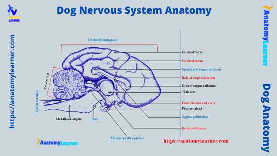
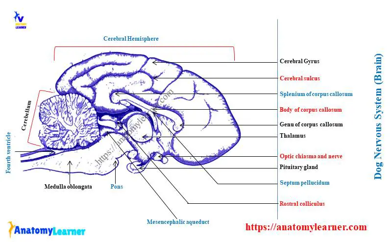
Dog brain anatomy – cerebrum part
In the dog brain anatomy, the cerebrum consists of paired cerebral hemispheres. There is a cerebral transverse fissure that separates the cerebrum from the cerebellum. Again, a cerebral longitudinal fissure separates the right and left cerebral hemispheres.
Each cerebral hemisphere of the dog brain consists of cerebral cortex or gray matter and cerebral white matter. You will see the lateral ventricle within the cerebral part of the dog’s brain.
The ventral region of the dog’s cerebral hemisphere is the rhinencephalon. You will find the limbic system structure within the rhinencephalon of the dog brain.
First, let’s see the different lobes of the dog’s brain anatomy. You will find the following lobes in the cerebral part of the dog brain –
- The frontal lobe of the dog brain (cerebral),
- The parietal lobe of the dog brain,
- A occipital lobe of the cerebral,
- The temporal lobe of the cerebral part of the dog brain, and
- Piriform lobe of the dog brain,
I tried to show you these different lobes in the dog brain labeled diagram. Now, let’s see the following critical anatomical features from the dorsal and ventral surfaces of the dog cerebrum.
From the dorsal surface of a dog’s brain –
- Longitudinal fissure of the cerebrum,
- Olfactory bulb of the cerebrum,
- Frontal, occipital, and parietal poles,
- Different types of the cerebral sulcus, and
- Different types of the cerebral gyrus (identified in the diagram),
From the ventral surface of a dog’s brain (cerebral part) –
- Olfactory tubercle,
- Olfactory peduncle,
- Diagonal gyrus,
- Rostral commissure,
- Columns and body of the fornix,
- Hippocampal sulcus,
- Optic tract and chiasma,
- Cerebral peduncle,
- Mammillary body,
- The occipital lobe, and
- Piriform lobe of cerebrum,
Again, the frontal section of the dog brain (cerebrum) clearly shows the caudate nucleus, choroid plexus, and hippocampus.
- Lateral ventricles of the dog cerebrum
You will see the lateral ventricle within each cerebral hemisphere of the dog brain. Two lateral ventricles from the cerebral hemispheres communicate with the third ventricle through an interventricular foramen.
Each lateral ventricles possess a choroid plexus that passes through the interventricular foramen. Again, they continue along the roof of the third ventricle.
Each choroid plexus arises from the tela choroidea and is the source of the cerebrospinal fluid. The lateral ventricle extends rostroventrally by a rostral horn and caudoventrally by the temporal horn.
Again, the roof of the lateral ventricle is formed by the corpus callosum. The septum pellucidum includes the medial wall of the ventricle. Finally, the floor of the dog’s lateral ventricle is formed by the caudate nucleus, thalamus, choroid plexus, and hippocampus.
“There are four ventricles in the dog brain ventricle system – two lateral, third, and fourth ventricle.”
The thalamus of the dog brain finds deep in the tissue of the posterior cerebrum. Its function is to process information from the sense organs and relay it to the cerebral cortex.
Again, the hypothalamus lies ventral to the thalamus and has several functions. It links the endocrine and nervous systems by secreting a series of hormones that are then stored in the pituitary gland.
The hypothalamus also helps control the autonomic nervous system by influencing various involuntary actions.
Transverse section of the dog cerebrum
Before reading the septum pellucidum, hippocampus formation, and limbic system from the dog nervous system, you may identify the following structures. Here, I tried to identify the basic structure from the transverse section of the dog cerebrum.
- Genu, splenium of the corpus callosum,
- Callosomarginal fissure,
- Septum pellucidum,
- Choroid plexus in the cerebrum,
- Thalamus and pituitary gland,
- Pineal body in the dog brain,
- Two lateral ventricles,
- Third ventricle in the dog brain,
- Superior and inferior colliculus,
- Aqueduct of the sylvius, and
- Pons and medulla oblongata of the dog brain,
The corpus callosum is the transverse commissure between the cerebral hemispheres. They lie at the bottom of the longitudinal fissure and are formed by the commissural fibers.
You will see two extremities and a central portion in the corpus callosum of the dog brain. The extremities are anterior thicker genu, posterior thicker splenium, and middle thinner truncus.
The fornix is the curved structure placed below the corpus callosum and above the tela choroidea of the third ventricle. You will see a bilaminar partition (septum pellucidum) extend from the undersurface of the corpus callosum to the upper surface of the body of the fornix.
It is thin and connects the thicker cellular septum to the corpus callosum. Then it caudally fills the space between the corpus callosum and fornix.
You will see a thin slit-like cavity between the two layers of the septum pellucidum of the dog’s brain anatomy.
Hippocampus of the dog nervous system
Hippocampus is the principal component of the limbic system of the dog. The hippocampus is the inner growth of the cerebral cortex at the lateral ventricle through the hippocampal sulcus.
Grossly, this part of the dog brain is the curved structure that occupies the posterior position of the lateral ventricles. It is made up of two curved horns which meet at the midline of the brain. The hippocampus is essential for spatial memory in the dog.
You will see the dentate gyrus, hippocampal proper, and subiculum in the dog hippocampal formation. These are the sequential regions joined together as the infolded cortex.
You know the dentate gyrus is a three-layered structure embedded into the concavity of the hippocampus proper. Again, the hippocampus proper is a gyrus folded concave medially. The subiculum consists of variable layers in a dog’s brain.
The hippocampal formation in a dog forms a rostrally concave structure. This structure includes the medial wall of the temporal part of the lateral ventricle. The ventral part of the hippocampus locates beside the amygdala, deep to the rostromedial surface of the caudal part of the piriform lobe.
Dorsally, you will find a prominent tubercle of the dentate. The hippocampal formation in a dog ends ventrally to the splenium of the corpus callosum of the brain.
The limbic system of the dog brain
The dog limbic system is very well developed and is the collection of the brain structures involved in affective behaviors and memory. A cerebral cortex has a significant function in controlling the dog’s limbic system.
In the dog limbic system, you will find the following structures –
- Olfactory pathway of the dog brain,
- Piriform lobe of the dog brain,
- Amygdaloid body of the brain,
- Hippocampal formation,
- The limbic lobe of the dog brain, and
- Hypothalamus of the brain,
All the above structures of the dog limbic system are associated with emotional integration. The dog’s limbic system is responsible for the outward expression of the internal state.
The olfactory pathway of the dog limbic system starts with the bipolar particular visceral afferent neuron that locates in the olfactory mucosa. A piriform lobe is concerned with the conscious olfaction that receives the mitral axon from the olfactory bulb.
The cerebellum of the dog brain anatomy
The dog cerebellum lies on the dorsal surface of the posterior part of the brain (hindbrain). It is the little brain or smaller version of the cerebrum of the dog brain. It has a spherical appearance and is covered in a deep fissure.
Anatomically, you will see a median vermis and bilateral cerebellum hemispheres in the structure of a dog cerebellum. Other fissures subdivide the dog cerebellum into different lobes and lobules.
You will see the narrow ridges on the dog cerebellum that are separated by the groove. The ridges on the cerebellum are the folium, whereas the grooves are the sulcus.
A uvulonodular fissure divides the cerebellum into a small floculonodular lobe and a cerebellar body. Again, the primary fissure divides the cerebellar body into a rostral and a caudal lobe.
The floculonodular lobe of the cerebellum possesses the function of the vestibular nucleus. You will see the connection of the rostral lobe with the spinal cord of the dog. This rostral lobe of the dog cerebellum regulates posture and gait.
The caudal lobe of the dog cerebellum connects with the forebrain and possesses the function of voluntary movement. Here, the transverse section of the dog cerebellum shows the medulla and white matter.
The white matter of the dog’s cerebellum appears as the branches of a tree commonly known as the arbor vitae. You will see a lamina in the folium and a central mass in the structure of the arbor vita of the dog cerebellum.
There are three cerebellar nuclei embedded deeply within the cerebellar white matter. From the medial to the lateral, these cerebellar nuclei are – the fastigial nucleus, interpositus nucleus, and lateral nucleus.
Cerebellar peduncle and cortex
The dog cerebellum attaches to the brainstem bilaterally by three cerebellar peduncles. These three cerebellar peduncles of the dog brain carry afferent and efferent cerebellar axons.
The caudal cerebellar peduncle (which contains both efferent and afferent fibers) attaches the cerebellum with the pons, medulla, and spinal cord. A middle cerebellar peduncle is formed by the projection to the lateral and ventral cerebellar zone from the cell bodies in contralateral pontine nuclei.
Again, the rostral cerebellar peduncle comprises the efferent fibers from the interpositus and lateral nuclei.
The cerebellar cortex of the dog brain shows three distinct layers – a superficial molecular layer, an intermediate Purkinje cell layer, and a deep granular cell layer. You may learn the details of these three layers of the cerebellar cortex of the dog brain from the below-mentioned article.
- Features of the three layers of the animal cerebellum with labeled diagram
The Purkinje cell layer of the cerebellum possesses a broad, flattened dendritic tree that extends up to the molecular layer. Again, the granular neuron of the cerebellum sends axons into the molecular layer. You will also see the mossy or climbing fibers (afferent fibers) in the structure of the cerebellar cortex of the dog brain.
Brainstems from the dog nervous system
You know the brainstem of the dog nervous system mainly consists of the pons and medulla oblongata. The pons lies ventral to the cerebellum and forms a nerve fibers bridge between the cerebellum hemispheres. Again, the medulla oblongata extends from the pons and enters the spinal cord.
Okay, let’s see the anatomical features of the pons and medulla oblongata from the dog brain anatomy –
- Anatomy of the dog pons and
- Anatomical features of the dog medulla oblongata,
Dog pons anatomy
The dog pons is a pair of transverse eminence located in front and ventral to the medulla oblongata. You will see a ventral bulging mass of the transverse fibers that separate from the dorsal cerebellum by the fourth ventricle.
Anatomically, you will find the dorsal tegmentum and ventral transverse pontine fibers in the dog pons. The pontine fibers of the dog pons run along the ventral surface of the pons. This structure helps to form the contralateral middle cerebellar peduncle.
The pontine fiber arises from the contralateral pontine nuclei deep to the transverse fibers. You will find the longitudinal axons in the corticopontine and the pyramidal tract.
The corticopontine axon and collateral branches of the pyramidal axon will synapse on neurons of the pontine nuclei. Again, the tegmentum of the dog pons resembles the medulla oblongata, where you will also see the fourth ventricle.
You know the rostral medullary velum forms the roof of the fourth ventricle. Again, the wall and floor of the fourth ventricle are lined by the gray matter layer.
In this periventricular gray matter, you will find the neuromodulatory cholinergic neuron. Locus ceruleus (neuromodulatory adrenergic neurons) are also located at the medial border of the rostral cerebellar peduncle.
In the rostral cerebellar peduncle, you will also find the parabrachial nuclei, which are essential visceral relay nuclei.
The trigeminal nerve originates from the anterior lateral aspect of the dog pons. Pons accommodates various nuclei and tracts that take part in the control of the different vital function of the body.
Dog medulla oblongata anatomy
The dog medulla oblongata is the cranial continuation of the spinal cord. This structure extends from the foramen magnum to the caudal margin of the pons.
The cranial part of the dog medulla oblongata is wide. This medulla oblongata is another vital part of the dog brainstem. It rests on the cranial face of the basioccipital bone.
The anterior two-thirds of the medulla oblongata is the open part that contains the posterior two-thirds of the fourth ventricle. Again, the posterior part of the medulla oblongata is the closed part that includes the central canal.
You will see a fragile membrane (known as the velum) on the roof of the fourth ventricle. There are superior and inferior median fissures of the spinal cord that continue cranially in the central line of the corresponding surface of the dog medulla oblongata.
You will see a transverse band at the ventral surface of the dog medulla oblongata just immediately behind the pons. This transverse band of the dog medulla oblongata is the corpus trapezoidum.
There are also a pair of elongated rounded masses on this surface of the corpus trapezoidum. These are the pyramid of the dog medulla oblongata that places on either side of the median groove.
Each pyramid consists of a myelinated axon that originates from the neuronal cell bodies in the cerebral cortex. Again, the axons within the pyramid go to the medulla oblongata or the spinal cord.
You will see a prominent olivary nucleus at the caudal part of the dog medulla oblongata. This structure locates dorsolateral to the pyramid and lateral to the medial lemniscus.
The motor nucleus of the hypoglossal nerve is evident dorsally beside the midline of the medulla oblongata.
Ventricular system of the dog nervous system
You already know that there are four ventricles in the dog nervous system – two lateral ventricles (right and left), a third ventricle, and a fourth ventricle. The two lateral ventricles and third ventricles locate in the forebrain of a dog. Again, the fourth ventricle locates in the hindbrain of the dog.
Two lateral ventricles of the dog brain communicate with the third ventricle by the foramen Monro. This foramen Monro is also located in the rostral part of the dog brain (forebrain).
The third ventricle communicates with the fourth ventricle by the cerebral aqueduct or aqueduct of Silvius. Finally, the fourth ventricle opens into the central canal, which is located in the dog’s spinal cord.
Let’s know a little about these four ventricles of the dog brain. The lateral ventricles are two irregular elongated cavities within each cerebral hemisphere. These two ventricles remain separate from each other by the thin septum pellucidum.
The corpus callosum forms the roof and lateral wall of the lateral ventricles. Again, the medial wall is formed by the septum pellucidum.
The third ventricle is a fissure-like space in the midline between the thalami. You will find the tela choroidea in the roof of the third ventricle.
There are two choroid plexus of the third ventricle that project downward.
The lateral wall of the third ventricle will form with the thalami and hypothalami. Again, the floor is created by the infundibulum, optic chiasma, and tuber cinerarium. Anteriorly, you will find the lamina cinerea and, posteriorly, the pineal gland.
The tela choroidea (dog brain) forms the roof of the fourth ventricle. On the floor, you will find the median sulcus. Bilaterally, a sulcus limitans marks the transition from the bottom to the wall of the fourth ventricle.
Dog spinal cord anatomy
The dog spinal cord is a thick cylindrical structure in the vertebral column. It extends from the foramen magnum to the middle of the sacrum.
The thickness of the dog’s spinal cord is not uniform. At the beginning of the spinal cord (caudal to the medulla oblongata), it becomes dilatation. Again, you will also see two other dilatation in the dog’s spinal cord – origin for the nerve of the brachial and lumbosacral plexus.
The thickness gradually decreases from the lumbar region. It terminates in a tapering and pointed structure at a second sacral vertebra level.
The tapered terminal portion of the dog’s spinal cord is called the conus medullaries. A fragile fiber that extends from the conus medularies to the first caudal vertebra is the filum terminale.
Again, you may see a lash of long nerve root of some sacral and caudal spinal nerves at the terminal part, which looks like the tail of a horse. This structure is the cauda equine of the spinal cord.
You will see a protective layer (meninges) that covers the dog’s spinal cord. These meninges have three layers – the most superficial meningeal layer (dura mater), a thin arachnoid mater, and the most profound vascular pia matter. You may learn these three layers’ anatomy from the brain and spinal cord in the next section of this article.
The paimater thickens bilaterally along the lateral margin of the dog’s spinal cord and forms the denticulate ligament. Each of these ligaments possesses periodic lateral extensions that attach to the dura mater of the meninges.
Structure of the dog’s spinal cord
In the center of the spinal cord, you will see the center canal that fills with the cerebrospinal fluid. This canal is slightly enlarged at the caudal termination of the spinal cord that forms the terminal ventricle.
Again, the gray matter surrounds the central canal that forms the core of the spinal cord. The white matter covers the gray matter and is positioned at the superficial part of the dog’s spinal cord.
The dog’s spinal cord possesses the following structures –
- Dorsal median sulcus,
- Dorsal intermediate sulcus of the spinal cord,
- Dorsolateral sulcus of the cord,
- Dorsal intermediate and median septum,
- Lateral and central intermediate substances,
- Dorsal, lateral, and ventral horns,
- Dorsal, lateral, and ventral funiculus,
- Ventrolateral sulcus,
- Ventral median fissure,
- The white commissure of the cord, and
- Three layers of meninges and ligament,
The gray matter of the dog’s spinal cord possesses cell bodies and neuron processes. They also contain glial cells. You will find numerous capillaries in the gray matter of the dog’s spinal cord. But the myelinated axons are sparse in the gray matter of the spinal cord.
You will see the butterfly-shaped gray matter in the transverse section of the dog’s spinal cord. It possesses bilateral wings that connect across the midline by a central intermediate substance.
The central intermediate substance surrounds the central canal and contains the gray commissure. Again, the lateral intermediate substance is the bilateral extension of the major substance.
There are three (dorsal, lateral, and ventral) horns that extend from the lateral intermediate substance of the dog’s spinal cord into the white matter. The dorsal horn extends dorsally to the lateral intermediate substance. In comparison, the ventral horn extends ventrally from the lateral intermediate substance into the white matter of the spinal cord.
The white matter of the dog’s spinal cord
The white matter of the dog’s spinal cord contains the packed myelinated axons. You may also find the nonmyelinated axons in the structure of the white matter of the dog’s spinal cord.
The white matter of each half of the dog’s spinal cord divides into three funiculi – dorsal, ventral, and lateral. Medial to the dorsolateral sulcus, you will see the dorsal funiculus. From the dorsal funiculus, the dorsal root of the spinal nerve enters the spinal cord.
The ventral funiculus is medial to the area from where the ventral roots exist from the spinal cord. Again, the lateral funiculus locates between the dorsal and ventral root attachments.
A white commissure ventral to the gray commissure connects the right and left ventral funiculi. A ventral median fissure divides the spinal cord into symmetric right and left halves.
There are dorsal median sulcus and median dorsal septum that extends from the sulcus to the gray commissure of the spinal cord. Again, a dorsolateral sulcus, dorsal intermediate sulcus, and dorsal intermediate septum are also found in the structure of a spinal cord.
According to the dog’s body region, the spinal cord also divides into the cervical, thoracic, lumbar, sacral, and caudal regions. A cervical enlargement gives rise to the spinal nerve that forms the brachial plexus. You know the nerves from the dog brachial plexus innervate to the skin and muscles of the thoracic limbs.
Again, the lumbosacral enlargement innervates the pelvic cavity and pelvic limbs. Caudal to the lumbar region, the spinal cord tapers into the elongated cone (conus medullaries). Finally, caudal to the last segment, the dog’s spinal cord reduces to form a uniform strand of glial and ependymal cells (filum terminali).
Anatomy of dog meninges
To protect the dog’s brain and spinal cord, they enclose three protective layers – dura matter, arachnoid mater, and pia matter. These three layers (dura, arachnoid, and pia mater – outside to inner side) are collectively known as the meninges.
The duramatter is the outermost solid and tough fibrous covering. The duramater that covers the dog’s brain is the cranial duramater, whereas the duramater that surrounds the spinal cord is the spinal dura mater.
You will find three significant modifications of the cranial duramater of the dog brain –
- Flax cerebri,
- Tentorirum cereberii, and
- Diaphragm sellae,
Falx cerebri is the sickle-shaped vertical fold at the midline between two cerebral hemispheres. Tentorium cereberii is the infolding of the duramater that forms the partial transverse partition between the cerebral hemisphere and cerebellum.
Diaphragm sellae is another fold of the duramater that extends transversely across the sella turcica and makes the roof over the pituitary gland. The flax cerebri and tentorium cereberii contain large venous channels (cranial venous sinuses).
Spinal duramater (spinal cord covering) starts from the foramen magnum and ends in the middle of the sacrum. There is no modification of the spinal duramater like the cranial duramater. But, you will find the epidural and subdural spaces in the structure of the spinal duramater of the dog.
The subdural space is between the duramater and arachnoid mater and contains a small amount of fluid.
Arachnoid and pia mater of meninges
The arachnoid mater is a fragile, delicate membrane that covers the brain and spinal cord. Its remains between the duramater and piamater of the meninges. There is a subarachnoid space between the arachnoid and piamater of the meninges.
In this subarachnoid space, you will find cerebrospinal fluid. The arachnoid mater of the cranial meninges is thick than these of the spinal part.
Finally, the piamater is the more delicate membrane that closely attaches to the brain and spinal cord of the dog. You will see the minute blood vessels and soft areolar tissue in the structure of the piamater of the meninges.
The spinal part of the paimater is thicker and less vascular. A lamina splendens lies along the inferior median line of the spinal paimater. The spinal paimater produces a longitudinal band (denticulate ligament) in the spinal cord.
Spinal nerves from the dog nervous system
Spinal nerves are essential elements of the dog nervous system. I have already described the formation of the dog’s spinal nerve earlier. You know there are 36 pairs of spinal nerves in the dog.
The dog skeleton has seven cervical vertebrae, but you will find 8 pairs of the cervical spinal nerve. Many ventral branches of the cervical nerves communicate with one another and run variable distances. They form the variable cervical plexus that can include axons of all cervical nerves.
The first cervical nerve of the dog arises from the first segment of the spinal cord. It locates just caudal to the foramen magnum and surrounds the cranial portion of the atlas.
The first cervical spinal nerve exits from the lateral vertebral foramen of the atlas vertebra and divides into the dorsal and ventral branches. You will not find any medial, lateral, or cutaneous branches of the first dorsal cervical spinal nerve.
The dorsal branch of the first cervical spinal nerve lies initially deep to the cranial part of the large obliquus capitis caudalis muscle.
Again, the ventral branch of the first cervical spinal nerve lies in the osseous groove of the atlas vertebra. Then it runs transversely to the alar notch from the lateral vertebral foramen.
The medial retropharyngeal lymph node covers this ventral branch of the first cervical spinal nerve. It continues its course caudally in the neck of the dog in close relation to the vagosympathetic nerve trunk.
Again, this nerve communicates with the smaller descending branch of the hypoglossal nerve to form the cervical loop. This cervical loop may extend up to the third cervical vertebrae of the dog skeleton.
Other cervical spinal nerves of a dog
The second cervical spinal nerve of a dog is somewhat different from the typical spinal nerve. Here, the dorsal and ventral roots of the second cervical spinal nerve fuse peripheral to the second intervertebral foramen.
Again, you will see a sizeable spinal ganglion in the second cervical spinal nerve that lies outside the vertebral canal. There are also dorsal and ventral branches in the second cervical spinal nerve of the dog.
The dorsal branch of the second cervical spinal nerve runs caudodorsally and locates deep in the obliquus capitis caudalis muscle. This dorsal branch gives off the muscular and cutaneous branches.
The muscular branch innervates to the semispinalis capitis and splenius muscle of the dog’s neck. Again, the cutaneous branch innervates the skin and dorsal aspect of the temporal muscle.
The ventral branch of the dog’s second cervical spinal nerve runs caudoventral on the lateral surface (both side) of the mastoid part of the cleidocephalicus muscle. It gives two ventral cutaneous branches – the transverse cervical and great auricular nerve.
The transverse cervical nerve of the dog runs deep to the platysma and crosses the maxillary and linguoficial veins. Again, the great auricular nerve runs dorsocranially to the base of the pinna of the ear and again subdivides into two branches.
The dorsal branches of the other cervical spinal nerves (third to eighth) vary in their course and formation. Each dorsal branch sends a small branch medially to the multifidus cervicis muscle.
The dorsal branch of the third to eighth cervical spinal nerve divides into the medial and lateral branches. Again, the ventral branch of the third to eighth cervical spinal nerves passes between the muscle bundle of intertransversarius cervicis.
The phrenic nerve of the dog
The phrenic nerve arises from the fifth, sixth, and seventh cervical spinal nerve that supplies to the diaphragm of the dog. At the origin, the phrenic nerve runs caudally and dorsomedial to the brachial plexus.
The fifth to seventh cervical spinal nerve branches run in the fascia adjacent to the external jugular vein and unit to form the phrenic nerve. Then the nerve of each side passes through the thoracic inlet ventral to the subclavian artery.
The right phrenic nerve passes from the cranial and middle mediastinum and plica of vena cavae. Again, the left phrenic nerve also departs from the same area of the mediastinum.
Now each phrenic nerve of the dog innervates on the respective surface of the diaphragm. You will see the three divisions (dorsal, ventral, and lateral) of the phrenic nerve on the dog diaphragm.
Thoracic nerves of the dog
You know there are 13 pairs of thoracic spinal nerves in a dog. Each of these thoracic nerves possesses the typical features of the spinal nerve. You will find the dorsal and ventral branches from each thoracic spinal nerve of the dog.
Again, the dorsal branch of each thoracic spinal nerve gives off the lateral and medial branches. The medial branch runs dorsolaterally, parallel and caudal to the thoracic vertebral spine.
Here, the medial branch supplies the multifidus thoracic, longissimus dorsi, and semispinalis thoracic muscles. You will not find any cutaneous branches from the medial thoracic spinal nerves.
The lateral part of the dorsal branch of the thoracic spinal nerves runs caudolaterally to a sagittal plane. Laterally, these lateral branches may course between the medial longissimus dorsi and iliocostal muscle. Finally, they reach the medial surface of the serratus dorsalis muscle of the dog.
The lateral branch of the dorsal thoracic spinal nerve again divides into a short medial cutaneous and a more extended lateral cutaneous branch. These two nerves of the lateral branch supply the dorsal skin third of the thorax.
The ventral branches of the dog’s thoracic spinal nerves are the intercostal nerves (except the first and thirteen nerves). An intercostal nerve possesses two branches – communicating and ventral branches and is embedded into the dorsal border of the internal intercostal muscles.
You will not find any ventral branch in the eleventh and twelfth intercostal nerves. The lateral cutaneous branches of these two nerves are much longer.
The ventral branch of the thirteen thoracic spinal nerves is the costoabdominal nerve. This muscle supply to the costal arch and the abdominal wall that locates on the caudal border of the last rib.
Brachial plexus from dog nervous system
The brachial plexus is the extensive somatic nerve plexus in a dog nervous system. This plexus is formed by the ventral branches of the sixth, seventh, and eighth cervical spinal nerves and first and second thoracic spinal nerves. The nerves that originate from the dog brachial plexus innervate to the skin and muscles of the thoracic limb and a minor part of the thorax.
First, let’s see the nerves in the dog’s brachial plexus –
- The suprascapular nerve of the dog,
- Subscapular nerve (usually single but double in ruminant),
- Cranial lateral cutaneous brachial nerve,
- The axillary nerve of the dog,
- The musculocutaneous nerve of the dog,
- Radial, median, and ulnar nerves of the dog brachial plexus – supply up to the digits,
- A caudal cutaneous antebrachial nerve of the dog,
- The brachiocephalic nerve of the dog,
- The cranial pectoral nerve of the dog,
- A thoracodorsal nerve of the dog brachial plexus,
- The long thoracic nerve of the dog,
- The lateral thoracic nerve of the dog’s brachial plexus, and
- A caudal pectoral nerve of the dog’s brachial plexus,
All the nerves, as mentioned earlier, are identified in the dog brachial plexus labeled diagram. You may learn the anatomy of the dog brachial plexus from another article by anatomy learner.
But, here, I will also provide a little information on the different nerves from the dog’s brachial plexus.
Nerves of the dog’s brachial plexus
The suprascapular nerve of a dog arises from the ventral branch of the sixth cervical spinal nerve. This nerve enters the distal end of the intermuscular space between supraspinatus and subscapularis muscles. It passes over the scapular notch and innervates the supraspinatus muscle.
The single subscapular nerve arises from the union of ventral branches of the sixth and seventh cervical spinal nerves. It may divide into the caudal and cranial parts that enter the medial surface of the subscapular muscle.
A cranial lateral cutaneous brachial nerve leaves the axillary nerve before entering the deltoid muscle. This muscle runs distally on the lateral head of the triceps muscle.
The axillary nerve arises from the ventral branch of the seventh and eighth cervical spinal nerve. It supplies mainly the muscle of the shoulder joint and the caudoventral border of the subscapular muscle.
The musculocutaneous nerve of a dog arises from the seventh cervical spinal nerve and supplies to the coracobrachialis, biceps brachii, and brachialis muscles. You will see the different branches of the dog’s musculocutaneous nerve like the proximal, distal muscular branch, and communicating branch.
You know the radial, median, and ulnar nerve of the dog brachial plexus supply to the different regions of the thoracic limb. They also innervate to the digits of the dog’s thoracic limb. You may learn details of the supply of these three nerves from another article by anatomy learner.
The cranial pectoral nerve supply to the superficial pectoral muscle of the dog. Whereas the caudal, pectoral supply to the deep pectoral muscle.
The long thoracic nerve supply to the serratus ventralis muscle. A branch from the lateral thoracic nerve supply to the deep pectoral and cutaneous tunci muscles. Dog thoracodorsal nerve supply to the latissimus dorsi.
Dog lumbar nerves
There are seven pairs of lumbar spinal nerves in the dog. The dog’s lumbar spinal nerve structure follows the typical structure of a spinal nerve.
The dorsal branch of the dog’s lumbar spinal nerves is similar throughout the regions. They are also divided into the lateral and medial branches like the thoracic spinal nerves.
The lateral branches of the dorsal branch of the lumbar spinal nerve are separate from the medial branch. Here, you will also find the dorsal cutaneous branches of the lateral extension of the dorsal lumbar spinal nerve.
You will see the medial and lateral cutaneous branches of the lateral branch of the dorsal lumbar spinal nerve. Again, there is a cranial clunial nerve branch in the lateral section of the lumbar nerve. The lateral branch of the dorsal lumbar nerve supply to the longissimus dorsi and iliocostalis muscles.
The medial branch of the dorsal lumbar spinal nerve gives branches to the multifidus lumborum and interspinalis lumborum. You will not find any cutaneous branches from the medial branch of the dorsal lumbar spinal nerve of the dog.
The ventral branches of the lumbar nerves are variable in a dog. Again, the ventral branch of the last lumbar and first two sacral spinal nerves fuses to form the lumbosacral plexus in a dog.
The ventral branches of the dog’s first and second lumbar nerves are the cranial and caudal iliohypogastric nerves, respectively. Both the cranial and caudal iliohypogastric nerves give off the medial branch. These medial branches supply the quadratus lumborum and psoas minor muscles.
You will find the lateral and medial branches in the cranial iliohypogastric nerve of the dog. The lateral branch passes through the obliquus internus abdominal muscle. Again, the medial branch lies close to the lateral surface of the transverse abdominis.
Lumbosacral plexus of dog
The ventral branch of the last five lumbar spinal nerves and the first three sacral spinal nerves communicate to form the lumbosacral plexus in a dog. So, in the lumbosacral plexus of a dog, you will find two parts – the lumbar plexus and sacral plexus.
A lumbar plexus of a dog provide the nerves that supply the cranial and medial muscles of the pelvic limb. Again, the sacral plexus provides the nerves that supply to caudal muscle and skin of the thigh.
An ilioinguinal nerve is the ventrolateral continuation of the significant part of the ventral branch of the third lumbar nerve. Here, this ilioinguinal nerve provides the lateral and medial branches.
The medial branch of the ilioinguinal nerve will supply to the psoas major, psoas minor, and ilicus muscles. Again, the lateral cutaneous branch of the ilioinguinal nerve supplies the skin of the cranolateral surface of the thigh.
The ventral branch of the fourth and fifth lumbar spinal nerve helps form the lateral cutaneous femoral nerve. This nerve passes from the abdominal wall, lumbar and inguinal part of the obliquus internus abdominal muscle.
The lateral cutaneous femoral nerve gives a branch to the tuber coxae and cranial part of the pelvic region. It also gives off another branch that supplies to the skin over the cranial part of the thigh and the lateral surface of the stifle joint.
The genitofemoral nerve of the dog arises from the ventral branch of the third and fourth lumbar nerves. Generally, this genitofemoral is the single, small, and long nerve in the lumbar plexus of a dog.
Other nerves from dog lumbosacral plexus
The femoral nerve is another important structure in the dog nervous system. It arises from the ventral branch of the fifth and sixth lumbar spinal nerves. This nerve continues caudally in the substance of the psoas muscle.
The dog femoral nerve sends the branches to the psoas and iliopsoas muscles. It supplies the four heads of the quadriceps and articular coxae.
There is no cutaneous nerve branch from the dog femoral nerve.
When the dog femoral nerve exits from the iliopsoas muscle, at that point, the saphenous nerve arises. This is only the superficial branch of the femoral nerve that relates to the medial surface of the tensor fascia latae.
The dog femoral nerve again divides into the muscular and cutaneous branches. A muscular branch of the dog femoral nerve bifurcates and supplies to the cranial and caudal belly of the Sartorius’s muscle.
The cutaneous branch of the dogs femoral nerve is long and slender and appears on the cranial surface of the femoral artery. It (cutaneous branch) sends branches to the skin of the medial and middle surfaces of the dog’s thigh.
The saphenous nerve of the dog continues with the cranial branch of the saphenous artery distal to the stifle joint. A cutaneous area of the saphenous nerve is extensive and lies mainly on the medial aspect of the pelvic limb.
The obturator nerve of the dog arises from the fourth, fifth, and sixth lumbar spinal nerves. This nerve forms within the caudomedial part of the psoas major muscle of the dog.
The obturator nerve exists from the psoas muscle, lies ventral to the iliac vein, and enters into the pelvis of the dog. There is no cutaneous branch found in the dog obturator nerve.
Anatomy of dog sacral nerves
You know the ventral branches of the sacral nerve forms the sacral plexus in a dog. The more prominent lumbosacral plexus continue with the dog pelvis as the sciatic nerve. In the sacral plexus of a dog, you will find the following nerves –
- A cranial gluteal nerve of the dog,
- The caudal gluteal nerve of the dog,
- A caudal cutaneous femoral nerve,
- The pelvic nerve of a dog,
- The pudendal nerve of a dog’s sacral plexus,
- A perineal nerve of a dog,
- A sciatic or ischiatic nerve of the dog,
- The lateral cutaneous sural nerve of the dog,
- Distal caudal cutaneous sural nerve, and
- A fibular and tibial nerve of the dog sacral plexus (branches of the sciatic nerve),
A cranial gluteal nerve in the dog’s lumbosacral plexus arises from the ventral branch of the sixth and seventh lumbar spinal nerve and the first sacral spinal nerve. It exists in the dog’s pelvis through the greater ischiatic foramen and enters into the lateral muscles of the pelvis.
The cranial gluteal nerve of the dog continues cranioventral between the middle and deep gluteus muscle. Two caudal gluteal nerves in the dog sacral plexus arise from the caudal margin of the lumbosacral trunk.
It runs parallel to the ventrocaudal border of the lumbosacral trunk and crosses the caudal border of the piriform muscle. You will see a branch that comes from the caudal gluteal to the middle gluteus muscle of the dog.
The caudal cutaneous femoral nerve of the dog arises from the ventral branch of the first and second sacral spinal nerves. This nerve courses caudally on the caudal attachment of the sacrotuberous ligament and divides into cranial and caudal branches.
Other sacral nerves of a dog
The pelvic nerve arises from the ventral branches of the first and second sacral spinal nerve. It also appears from the pudendal nerve of the dog. This pelvis nerve supply to the viscera of the pelvic cavity and urinary bladder of a dog.
The pudendal is another clinically significant nerve in a dog that arises from the ventral branch of the first, second, and third sacral spinal nerves. It runs obliquely caudoventral to the pelvic outlet and lies lateral to the coccygeal muscle.
Then the pudendal nerve appears superficially medial to the superficial gluteal muscle. The nerve has a close association with the pudendal vessel.
A perineal nerve in a dog’s sacral plexus has two major divisions – superficial and deep perineal. The deep branch of the perineal has several long branches from the pudendal nerve at the pelvic outlet.
Again, the superficial branch of the perineal nerve also sends several branches into the skin of the perineum. The superficial branch is also supplied to the dog gracilis muscle.
Sciatic nerve from dog nervous system
The sciatic or ischiatic nerve of a dog is the largest peripheral nerve in its body. It arises mainly from the ventral branch of the sixth, seventh lumbar spinal nerve, and first and second sacral spinal nerve.
The division of the sciatic nerve of a dog is variable. First, it lies on the Gemelli muscle and the tendon of the obturator caudomedial to the hip joint. It passes distal to the thigh and lies on the quadratus femoris, adductor, and semimembranosus muscle.
The sciatic nerve of a dog covers by the superficial gluteal and biceps femoris muscle (gluteobiceps). There are three major muscular branches of the dog’s sciatic nerve – thin, thick, and small muscular branches.
These muscular branches of the dog’s sciatic or ischiatic nerve remain between the medial gluteal and cranial gemellus muscles. Again, the main ischiatic nerve lies cranial and parallel to the sacrotuberous ligament, caudal gluteal artery, and vein.
The more caudal branch of the muscular ischiatic nerve enters the middle part of the semitendinosus muscle. Again, the more cranial branch runs distally and bifurcates into different branches. One branch enters the cranial belly, and another enters the caudal belly of the semimembranosus muscle.
You will see two other branches of the dog’s ischiatic or sciatic nerve – lateral cutaneous sural nerve and distal caudal cutaneous sural nerve. The lateral cutaneous sural nerve arises from the lateral surface of the common fibular part of the dog ischiatic nerve.
The lateral cutaneous sural nerve of the dog passes through the biceps femoris muscle and supplies distal to the thigh, stifle, and crus. Again, the distal caudal cutaneous sural nerve is a slender nerve that arises from the caudal border of the tibial part of the sciatic nerve.
Fibular and tibial nerves of a dog
The course and distribution of a dog’s fibular and tibial nerves are very complicated. Here, I will show you the main branches of a dog’s fibular and tibial nerves. But, you may learn anatomy and the innervation of the dog fibular and tibial nerves from the following articles.
- Dog leg anatomy with the labeled diagrams,
- Dog sciatic or ischiatic nerve anatomy with a labeled diagram,
The fibular nerve lies deep in the thin terminal part of the biceps femoris muscle. This nerve runs directly distal, obliquely crossing the lateral head of the gastrocnemius muscle. It sends an auricular branch to the lateral collateral ligament at the level of the stifle joint.
The fibular nerve also crosses the head of the fibula and reaches the thin lateral border of the flexor digitorum lateralis muscle. A small branch from the fibular nerve will be sent into the fibular longus muscle.
Then it divides into the superficial and deep fibular nerves. The superficial fibular nerve is the most caudal terminal branch of the common fibular nerve of the dog.
This superficial branch of the dog fibular nerve will supply to the fibularis brevis and lateral digital extensor muscles. It also innervates the deep and distal part of the fibularis longus muscle of the dog.
The branches of the dog’s superficial fibular nerve are identified in the labeled diagram. You may find a more labeled diagram where I show the different courses of the dog’s ischiatic nerve.
The deep fibular nerve arises as the cranial terminal branch of the common fibular nerve on the lateral surface of the gastrocnemius muscle. Its proximal part lies along the superficial aspect and passes between the lateral digital extensor, flexor cranially, and the fibularis longus caudally.
Dog tibial nerve
The dog tibial nerve lies between the medial caudal part of the semimembranosus and the biceps femoris laterally. It is a more significant nerve than the fibular and is flattened transversely.
The dog tibial nerve arises from the ventral branches of the first two sacral spinal nerves. It separates from the sciatic nerve in the proximal two-thirds of the dog’s thigh.
Again, this nerve enters between the two heads of the gastrocnemius muscles. The nerve supplies all the muscles on the caudal aspect of the tibia and fibular bones of the dog.
It also innervates the stifle, tarsal, and digital joints of the dog leg. The terminal part of the nerve also innervates the plantar muscles, skin, and foot pads.
You may learn the details anatomical facts (origins and distribution) of the dog tibial nerve from another article by an anatomy learner. There are numerous muscular branches in the dog’s tibial nerve. Again, you will see one or more cutaneous branches of the tibial nerve that innervate the skin around and slightly distal to the medial aspect of the calcaneus tuber.
You will also see the four to seven pairs of caudal nerves in a dog. Like the other spinal nerves, the dog caudal nerves immediately leave the intervertebral foramina and divide into dorsal and ventral segments.
The ventral branch of the dog’s caudal nerves is larger than the dorsal nerves. Again, both dorsal and ventral branches of the dog’s caudal nerves possess muscular and cutaneous segments.
Cranial nerves from the dog nervous system
You already know 12 pairs of cranial nerves are in the dog nervous system. I have already enlisted these 12 pairs of dog cranial nerves. You may also see these nerves from the table or picture below.
So, you see, there are sensory, motor, and mixed-function in these cranial nerves of the dog. I will describe the origin, types of function, and the main distribution of the dog cranial nerves with a labeled diagram.
But, it is your first duty to identify all the roots of these 12 pairs of cranial nerves from the dog brain. Here, I tried to show you the roots of the dog’s cranial nerves with the labeled diagram.
- Sensory cranial nerves – olfactory, optic, and vestibule cochlea,
- Motor cranial nerves – oculomotor, trochlear, accessory spinal, hypoglossal, and
- Mixed cranial nerves – trigeminal, abducent, facial, glossopharyngeal, and vagus,
Let’s start with the first cranial nerve of the dog (olfactory nerve).
Olfactory nerve: this is the first cranial nerve of the dog that consists of numerous nonmyelinated axons, which cell bodies present in the olfactory epithelium. Axon from these olfactory cells enters the skull through the cribriform plates of the ethmoid bone.
They also enter deep into the caudal part of the dog’s nasal cavity. Finally, these axons reach the olfactory bulb of the brain. But, these axons do not form any trunk as they enter the olfactory bulb through numerous cribriform foramina.
Quick summary of the olfactory nerve: the sensory nerve attaches to the olfactory bulb. They are distributed in the olfactory areas of the pyriform cortex and hippocampal gyrus.
Olfactory cells of the nasal mucosa are the cells of origin. The afferent fibers of the olfactory are for the sensation of smell.
Dog optic and oculomotor nerves
The dog optic nerve is sensory, whereas the oculomotor is a motor type of nerve. You will see the attachment of the optic nerve to the optic chiasma of the brain. It passes from the optic foramen of the dog.
The optic tract diverges from the optic chiasma, and each winds around the concerned cerebri. Finally, these tracts of the optic nerve enter the lateral geniculate body.
The component of the dog optic nerve is the afferent fibers responsible for the sensation of the vision. Again, some of the afferent fibers of the optic nerve are for the pupillary light reflex.
The axon of the ganglionic cells continues the fibers of the optic nerve that exits the eyeball through the blind spot. Then it locates a little medial to the posterior pole of the eyeball.
The optic nerve distributes at the visual cortex of the occipital pole of the dog brain.
Oculomotor nerve: the dog’s oculomotor nerve originates from the medial aspect of the cerebral peduncle. It leaves the cranial cavity through the foramen orbitorotundum.
The dog’s oculomotor nerve supplies all the extraocular muscles of the eye except superior oblique and lateral muscles. Again, the functional component of the oculomotor nerve is the efferent fibers responsible for the eyeball’s movement.
The branch of the dog’s oculomotor nerve will course rostrally through the middle cranial fossa, lateral to the hypophysis, and dorsal to the carnivorous sinuses. The dorsal branch supplies the levator palpebrae superior muscle.
The ventral branch of the oculomotor nerve continues rostrally, lateral, and slightly ventral to the optic nerve. It provides different branches to the medial, ventral, and ventral oblique muscles of the eyeball.
Dog trochlear nerve
The dog trochlear nerve is the slender most cranial nerve. It is the unique cranial nerve in a dog for the following two reasons –
It is the only cranial nerve in a dog that emerges from the brainstem dorsally, and
This is the only cranial nerve that crosses entirely to innervate the muscle of the lateral aspect of the eye,
The cell bodies of the dog trochlear nerve lie in the trochlear nucleus of the caudal mesencephalon. Again, the trochlear nucleus is caudal to the oculomotor nucleus in the caudal part of the mesencephalon.
An axon from the dog trochlear nerve will course dorsally around the mesencephalic aqueduct. It enters the rostral medullary velum caudal to the colliculi.
The nerve then exists from the velum caudal to the lateral colliculi and passes rostroventrally over the side of the mesencephalon. Finally, it leaves the cranial cavity through the orbital fissure and enters the periorbital area.
Then the nerve fiber innervates the dorsal oblique muscles of the eyeball.
Summary of the dog trochlear nerve: the trochlear nerve is a motor type in which afferent fibers are responsible for the movement of the eyeball. You will see the attachment of the dog trochlear nerve to the superior colliculus (posterior aspect).
It passes through the orbital fissure and distributes to the superior oblique muscles of the dog eyeball.
Trigeminal from dog nervous system
The trigeminal nerve from the dog peripheral nervous system possesses both motor and sensory functions. This nerve penetrates the pons just caudal and ventral area that continue dorsally with the middle cerebellar peduncle. You know this middle cerebellar peduncle is rostral to the lateral end of the trapezoid body.
The dog trigeminal nerve enters the trigeminal canal on the rostromedial aspect of the temporal bone. You will see a large trigeminal ganglion within the canal that contain sensory cell bodies.
There are three divisions of the dog trigeminal nerves that exit from the trigeminal canal –
- The ophthalmic division of the dog’s trigeminal (sensory),
- The maxillary division of the dog’s trigeminal (motor), and
- Mandibular part of the trigeminal (both sensory and motor),
Quick information on the dog trigeminal nerve: this is the largest cranial nerve that has an attachment to the anterolateral aspect of the pons by a large sensory and a small motor root. There are three branches where the ophthalmic and maxillary branch passes through the foramen orbitorotundum.
Again, the mandibular branch of the dog trigeminal passes through the foramen ovale. You will find a trigeminal ganglion in the dog’s trigeminal nerve.
This trigeminal nerve distributes to the face, teeth, gum, muscles of mastication, and face skin. You will also see the distribution of the dog trigeminal to the lacrimal gland, nasal cavity, tongue, palates, and eyelids.
The afferent fibers of the dogs trigeminal nerve are responsible for the pain, touch, and temperature sensation of the skin, face, teeth, and gum. Again, the efferent fibers of the trigeminal nerve (for muscles) are responsible for the movement of the lower jaws.
Now, let’s see the anatomical facts of the different branches of the dog’s trigeminal nerves.
The ophthalmic branch of dog trigeminal
The ophthalmic branch of the dog trigeminal nerve consists of the general somatic fibers from the eyelids, eyeball, nasal mucosa, and nose skin. This branch of the dog trigeminal passes through the orbital fissure of the skull.
You will see three major subdivisions of the ophthalmic branch of the dog trigeminal within the periorbita –
- The frontal branch of the ophthalmic,
- Lacrimal branch of the ophthalmic, and
- Nasocilliary branch of the ophthalmic,
The frontal branch is the most dorsal that passes rostrally in the periorbita dorsal to the dorsal oblique muscles of the eyeball. Again, this frontal branch divides into supraorbital and supratrochlear nerves just caudal to a periorbital ligament.
The lacrimal branch of the ophthalmic nerve is small than these two branches. It runs along the lateral straight muscle of the eyeball to supply the lacrimal gland.
The nasociliary is the most medial and most prominent branch of the frontal nerve of a dog. It runs medially deep in orbit across the dorsal surface of the retractor muscle of the eyeball.
You will see a communicating branch to the ciliary ganglion from the nasociliary branch. Again, the nasociliary branch of the frontal nerve gives off numerous long ciliary nerves. These nerves enter the eyeball dorsal and medial to the optic nerve.
The nasociliary branch again divides into ethmoid and infratrochlear branches. Here, the ethmoid nerve turns medially to pass through the rostral ethmoidal foramina and cribriform plate.
The ethmoidal branch enters the nasal cavity and divides into medial, lateral, and external nasal branches. Medial nasal nerve supply to the nasal septum and parts of the wall of the nasal cavity.
The infratrochlear nerve runs along the medial aspect of the orbit. It supplies the surrounding area of the medial canthus.
Maxillary branch of the dog trigeminal nerve
The maxillary branch of the dog trigeminal nerve exists from the cranial cavity through the round foramen, alar canal, and rostral alar foramen. It courses rostrally on the dorsal surface of the medial pterygoid muscle to the maxillary foramen.
You will see the significant three branches of the maxillary dog nerve at the orbit –
- Zygomatic branch of the maxillary,
- The pterygopalatine nerve of the maxillary, and
- Infraorbital branch of maxillary,
The zygomatic nerve arises from the maxillary nerve proximal to the round foramen. It divides into the zygomaticofacial and zygomaticotemporal within the periorbita.
The zygomaticotemporal branch is more dorsal that runs rostrally in orbit deep to periorbital. Again, the zygomaticofacial is the more ventral branch that emerges medial to the orbital ligament.
The pterygopalatine arises from the deep surface of the maxillary nerve on the dorsal surface of the medial pterygoid muscle. You will see different branches of pterygopalatine join with the branch of zygomaticotemporal and ophthalmic nerves.
The major, minor, and accessory palatine are the major branches of the pterygopalatine nerve. Again, the major palatine nerve gives a branch to the caudal nasal that continues rostrally and enters the palatine canal.
The caudal nasal nerve continues the pterygopalatine nerve that lies dorsal to the major palatine nerve. You will see some small branches from the caudal nasal nerve of the dog.
The infraorbital is the direct continuation of the maxillary nerve after the caudal nasal nerve. An infraorbital nerve gives the following branches at a different level –
- Caudal superior alveolar branch,
- Middle superior alveolar branch, and
- Rostral superior alveolar branch,
Again, the dog’s infraorbital nerve divides into external, internal, and superior nasal branches external to the infraorbital foramen.
The mandibular branch of the dog trigeminal nerve
The mandibular branch of the dog trigeminal nerve possesses both sensory and motor functions. This nerve passes the cranial cavity through the foramen ovale.
You will find the following primary branches of the mandibular nerve –
- Masticator nerve,
- Lateral and medial pterygoid nerves,
- Tensor tympani nerve,
- Tensor veli palatine nerve,
- Buccal nerve,
- Auriculotempral nerve,
- Lingual nerve, and
- Inferior alveolar nerve,
A masticator nerve attaches to the caudal belly of the digastricus muscle of the dog that helps to open the mouth. Again, a masseteric and deep temporal nerve to the temporalis muscle helps to close the mouth.
The lateral and medial pterygoid nerves innervate small lateral and larger medial pterygoid muscles. A tensor tympani nerve has an active role in modulating the movement of the malleus of the ear.
The buccal nerve is sensory to the mucosa and skin of the dog’s neck. It supplies to the cutaneous area of the ventral zygomatic arch, dorsal and caudal commissure of the dog’s mouth.
An auriculotemporal nerve exists from the mandibular branch at the foramen ovale. It passes medial and caudal to the retroarticular process of the temporal bone.
There are different branches of the auriculotemporal nerve of a dog –
- External acoustic meatus nerve,
- Rostral auricular nerve, and
- Transverse facial branch,
The external acoustic meatus nerve is sensory to the skin of the external acoustic meatus near the tympanic membrane. Again, the rostral auricular branch supplies the skin over the lateral aspect of the tragus, the medioventral part of the pinna.
The transverse facial branch of the mandibular nerve supplies to skin extending dorsal and ventral from the zygomatic arch. A cutaneous area of the transverse facial nerve overlaps with the cutaneous area of the rostral auricular and communicating branch.
Other branches of dog mandibular nerve
The inferior alveolar nerve arises from the mandibular nerve on the lateral aspect of the medial pterygoid muscle. This nerve (inferior alveolar) enters the mandibular foramen on the medial aspect of the ramus of the mandible.
It passes through the mandibular canal and supplies caudal, medial, and rostral alveolar sensory nerves to the teeth. Finally, the inferior alveolar nerve exists from the mental foramen as the mental nerve.
A mylohyoid nerve may arise inferior to the alveolar nerve in different species of dogs. It runs distally medial to the ramus of the mandible. You will see a muscular branch to the rostral belly of the digastricus muscle from the mylohyoid nerve.
The lingual nerve is sensory to the rostral two-thirds of the tongue. This is the large branch of the mandibular nerve that crosses the pterygoid muscle. It gives a branch to the buccal mucosa and passes between the styloglossus and mylohyoideus muscles.
At the base of the dog tongue, you will see the sublingual nerve that innervates the sublingual mucosa. Again, the sublingual nerve gives off the communicating branches that reach the lingual through the chorda tympani of the facial nerve.
The abducent nerve of the dog
The dog abducent is the sixth nerve that originates from the rostral medulla at the level of the trapezoid body. It passes ventrally through the foramen orbitorotumdum and reaches the dog orbital cavity.
The general somatic efferent fibers from the abducent nerve innervate the eyeball’s lateral straight and retractor bulbi muscles. Again, the genu of the facial nerve passes dorsal to the abducent nerve.
The small abducent nerve of the dog will course rostrally through the medial cranial fossa beside the hypophysis. This small abducent nerve exists from the cranial cavity through the orbital fissure.
Then the tiny abducent also enters into the periorbital and innervates the lateral straight and retractor bulbi muscles of the dog eyeball.
Facial nerve from dog nervous system
The facial nerve is another vital nerve of the dog peripheral nervous system. It provides the general somatic innervation for all the superficial muscles of the head, face, and external ear. You will also find the innervation of the facial nerve in the caudal belly of the digastricus, stapedius, stylohyoideus, and platysma of the dog neck.
The dog facial nerve originates from the lateral aspect of the medulla oblongata in front of the auditory nerve. Both the facial and auditory nerves enter into the internal auditory meatus of the dog.
The dog’s facial nerves enter the facial canal and form the geniculate ganglion. The facial canal is an irregular space between the internal auditory meatus and stylomastoid foramen. The facial nerve leaves the cranial cavity through the internal acoustic meatus accompanied by the vestibular and cochlear nerves.
The dog’s facial nerve gives superior and inferior branches within the acoustic meatus. The superior division remains in contact with the facial nerve and enters the facial canal.
You will see the major petrosal nerve from the facial nerve in the rostral wall of the petrous part of the temporal bone. Again, the major petrosal nerve emerges from the petrosal canal and joins with the deep petrosal nerve.
The union of the significant petrosal and deep petrosal nerves will form the nerve of the petrosal canal. You will see this canal in the dorsomedial to the alar canal in the pterygoid process of the basisphenoid bone.
The facial nerve continues with the canal and forms the stapedial nerve to the stapedius muscle. Again, the dog facial nerve also gives off the chorda tympani branch that runs in the canaliculus chordae tympani.
After emerging from the stylomastoid foramen, the dogs facial nerve increases its size and joins with the vagus nerve.
Other branches of the dog’s facial nerve
The dog facial nerve also sends the muscular branch to the caudal auricular muscle and forms the caudal auricular nerve. It curves ventrally and rostrally around the annular cartilage and gives off the digastricus and stylohyoideus branches.
The facial nerve curve around the caudal border of the mandible and divides into three main branches –
- The cervical branch of the facial nerve,
- Buccal branch of the dog’s facial nerve, and
- Auriculopalphebral branch of the dog facial nerve,
The cervical branch of the dog’s facial nerve passes superficially to the mandibular salivary gland. It innervates to the parotidoauricularis and sphincter colli muscles.
There are two buccal branches of the dog facial nerve that runs rostral over the masseter muscle. It innervates to the buccinators, orbicularis oris, and lateral surface of the nose.
Finally, the auriculopalphebral branch of the dog’s facial nerve is coursing dorsally from the base of the ear. Here, this auriculopalphebral nerve divides into the rostral auricular and palpebral nerves.
The palpebral branch forms a rostral auricular plexus between the eye and ear. Again, the rostral auricular branch of the auriculopalphebral nerve innervates the superficial and deep scutuloauriculairs muscles.
The vestibulocochlear nerve of the dog
The dog vestibulocochlear nerve comprises two roots – vestibular root and cochlear roots. Here, the vestibular root of the vestibulocochlear nerve innervates to the hair cells in the cristae ampullae and macula utriculi.
Again, the cochlear root locates in the spiral ganglion within the osseous modiolus of the cochlea. It innervates the hair cells of the spiral organ in the cochlear duct.
The nerve fibers of the vestibulocochlear originate from the inner ear of the petrosal part temporal bone. It leaves the skull through the internal acoustic meatus and the facial nerve and courses to the adjacent brainstem medulla.
Vestibular part: The vestibular ganglionic cell axons are located within the petrous part and pass through the internal auditory meatus. These neurons carry the impulse from the semicircular canal of the internal ear.
Again, they end at the vestibular nucleus in the medulla oblongata, deep at the floor of the fourth ventricle.
Cochlear part of the vestibulocochlear nerve: the axon from the cell bodies of the spiral ganglia organ of Corti comes out of the petrous bone through the internal acoustic meatus. Then, it joins with the vestibular division from the cochlear nerve.
Again, it enters the brainstem at the caudal border of the pons, and the fibers end at the cochlear nuclei. From the medial geniculate body of the axon, pass below the lenticular nucleus to the hearing center of the temporal bone.
Summary of vestibulocochlear nerve: it is a sensory nerve that has an attachment with the brain at the junction between the (dogs) pons and medulla oblongata. It passes through the internal auditory meatus and innervates to the cochlear vestibule and semicircular canal.
Dog glossopharyngeal nerve
The dog glossopharyngeal is the ninth cranial spinal nerve containing sensory and motor functions. This nerve carries the general visceral afferent fibers from the caudal part of the tongue, pharyngeal mucosa, and carotid sinuses.
Again, this nerve also contains the special visceral afferent axon for taste from the buds in the foliate and vallate papilla of the dog tongue. The dog glossopharyngeal nerve emerges from the lateral aspect of the medulla oblongata just caudal to the vestibule cochlear nerve.
Again, the axon of the glossopharyngeal nerve leaves the cranial cavity through the jugular foramen and tympniooccipital fissure. Through these foramen and fissures, the vagus and accessory spinal nerves also exist.
Between the jugular foramen and tympanioocipital fissure, the glossopharyngeal nerve gives the tympanic nerve. This tympanic nerve enters the adjacent tympanic cavity and forms the tympanic plexus.
You will also see a minor petrous nerve originating from the glossopharyngeal nerve. This minor petrous nerve synapse with the otic ganglia adjacent to the foramen ovale.
Again, the glossopharyngeal nerve crosses the medial side of the cranial cervical sympathetic ganglia at the level of the tympaniooccipital fissure. After passing, this fissure provides the branch to the carotid sinus that supplies the carotid body.
Again, the glossopharyngeal nerve gives off a small stylopharyngeal branch that innervates the stylopharyngeus muscle. You may also see a small tonsilar branch from the glossopharyngeal nerve that supplies to the palatine tonsil.
The terminal branch of the dog glossopharyngeal nerve
At the terminal part of the glossopharyngeal nerve, you will find two main branches – the lingual and pharyngeal branches. The lingual branch of the glossopharyngeal runs rostroventral and enters the base of the tongue medially.
It supplies to the mucosa of the caudal third of the tongue. Again, the lingual branch of the glossopharyngeal nerve supplies the parasympathetic axon to the glands of the dog tongue.
The pharyngeal branch of the glossopharyngeal nerve joins with the pharyngeal branch of the vagus nerve. Then they form the pharyngeal plexus, which also receives the hypoglossal nerve.
So, the pharyngeal muscles of the dog are jointly supplied by the vagus and glossopharyngeal nerves. Again, the glossopharyngeal and vagus nerves supply sensory fibers to the dog pharynx.
Vagus nerve from dog nervous system
This is the longest of all the cranial nerves of the dog nervous system. This nerve attaches to the lateral aspect of the medulla oblongata with the fibers of hypoglossal and accessory spinal nerves. Again, the vagus nerve leaves the cranial cavity through the jugular foramen.
The jugular ganglion in the jugular foramen communicates with the accessory spinal, hypoglossal nerves, and ganglion of the glossopharyngeal nerve. The vagus nerve supply to the viscera of the cervical, thoracic, and abdominal regions.
Again, the vagus nerve leaves the lateral surface of the medulla oblongata and passes through the jugular foramen and tympaniooccipital fissure along with the glossopharyngeal and accessory spinal nerve. There are two sensory ganglia in the vagus nerve – jugular ganglion (told earlier) or proximal ganglion and distal ganglion.
The distal ganglion lies external to the tympaniooccipital fissure ventral and medial to the tympanic bulla. Again, caudal to the distal ganglion vagus nerve bound with the sympathetic trunk in the neck without the carotid covering.
Thus, it forms the vago-sympathetic trunk, where the sympathetic fibers come from the cervical cranial ganglion. A common trunk passes through the carotid covering, descend deeply to the jugular furrow, and enters the thorax.
In the thoracic cavity, the right vagus nerve passes under the right axillary artery and right aspect of the trachea. Again, the left vagus also passes under the left axillary artery and reaches the root of the lung.
The right and left vagus are divided into major superior and inferior branches and pass the caudal mediastinum. Finally, the dog vagus nerve enters the abdominal cavity through the esophageal aperture.
Now, let’s see the significant branches of the vagus nerve in the dog’s cervical, thoracic, and abdominal regions.
Cervical branches of the dog vagus nerve
In the cervical region of a dog, the vagus nerve gives off different branches like – auricular, pharyngeal, esophageal, cranial laryngeal, and aortic nerves. Let’s see the origins and distribution of these cervical branches of the dog vagus nerve.
The auricular branch of the dog vagus arises from the jugular ganglion and joins with the facial nerve. It supplies to the skin of the back of the ear and external auditory meatus.
The pharyngeal branch of the dog vagus nerve originates from the distal ganglion and joins with the communicating rams of the glossopharyngeal nerve. Both the pharyngeal and communication branch of glossopharyngeal form the pharyngeal plexus.
This pharyngeal plexus innervates to the pharyngeal muscle and cranial part of the esophagus. Again, the pharyngeal branch of the vagus continues into the pharynx and innervates to the cricopharyngeal muscle and cervical part of the esophagus.
The cranial laryngeal also originates at the distal ganglion and passes ventral to the larynx. Here, this cranial laryngeal nerve divides into an external and internal ramus.
The external ramus supplies the cricothyroid muscle, whereas the inner ramus supplies the laryngeal mucosa. You may also see a depressor nerve that also arises from the cervical part of the vagus
just adjacent to the origin of the cranial laryngeal nerve.
In front of the thoracic inlet, the left vagus detaches from the vagus that supplies to the aorta. This is the aortic nerve of the dog.
Thoracic branches of the dog vagus nerve
In the thoracic region (learn here) of a dog, you may see the branches of the vagus nerve that supply to the different visceral organs like the heart, lung, and trachea. The major branches of the thoracic part of the dog vagus nerve are cardiac, recurrent laryngeal, caudal laryngeal, ventral, and dorsal vagal nerves.
The cardiac branch of the dog vagus nerve originates near the thoracic inlet and innervates to the heart. Again, the left vagus nerve provides the left recurrent laryngeal nerve that arches around the ligamentum arteriosum and aorta.
Then it crosses cranially along the trachea and gives branches to the trachea and esophagus. Finally, it terminates as the left recurrent laryngeal nerve. These branches of the nerves supply to the muscles of the larynx.
The right vagus nerve also gives off the right recurrent laryngeal nerve that also courses along the trachea and terminates as the right caudal laryngeal nerve. The dorsal and ventral vagal branches are on each side of the trachea (within the thorax). These branches supply the bronchi of the dog’s lungs.
Just caudal to the dog’s heart, the right and left ventral vagal branches to join to form the ventral vagal trunk. Again, at the diaphragm level, the dorsal branches of the right and left vagal nerve to join to form the dorsal vagal trunk.
Abdominal branches of the dog vagus nerve
In the abdominal region, the dog vagus nerve also supplies to the different organs or structures like the stomach, mesentery, and intestine. The dorsal vagal trunk courses through the celiacomesenteric plexus at the stomach level.
The axon will synapse with a cell body of the postganglionic axon in the wall of the gastrointestinal tract. There are different abdominal sympathetic ganglions present in the dog’s abdomen. The dorsal vagal trunk accompanies the postganglionic sympathetic axon from these abdominal ganglions.
The ventral vagal trunk innervates the lesser curvature of the stomach and the liver. Again, the vagal branches supply to the other different parts of the digestive tract and flexure the colon. The descending colon receives its parasympathetic innervation from the sacral region of the spinal cord.
Summary of the dog vagus nerve
The dog vagus nerve is the mixed (possesses sensory and motor functions) and longest cranial nerve. This nerve attaches to the lateral aspect of the medulla oblongata and leaves the cranial cavity through the jugular foramen.
You will see two major ganglions in the vagus nerve. It supplies to the pharynx, larynx, esophagus, stomach, intestine, trachea, lungs, liver, spleen, kidney, and heart.
The accessory cranial nerve of the dog
This is the eleventh cranial nerve, also known as the spinal accessory nerve. You will find two origins of this nerve – in the medulla oblongata and the spinal cord.
The cranial root possesses the cell bodies in the caudal part of the nucleus of the medulla oblongata. Lateral to the medulla oblongata, the cranial root joins with the axon of the spinal root.
The accessory cranial nerve of the dog leaves the cranial cavity through the foramen magnum. You will see the two significant branches of the dog’s spinal accessory nerve – internal and external branches.
You know the internal branch of the spinal accessory nerve joins with the vagus nerve within the space between the jugular foramen and tympanooccipital fissure. Here, the internal branch carries the somatic efferent axon that innervates the muscles of the larynx and esophagus.
The spinal root of the dogs accessory cranial nerve emerges laterally from the spinal cord midway between the dorsal (upper) and ventral root of the cervical spinal nerves. Within the cranial cavity, the spinal part of the accessory nerve lies on the ventrolateral surface of the medulla.
The external branch of the accessory nerve courses caudally in the dog’s neck. First, it courses ventral to the neck, then dorsal to the transverse process of the cervical vertebrae.
The dorsal branch of the accessory nerve of the dog supplies the trapezius and cleidocephalicus muscles. Again, a ventral branch of the accessory nerve innervates to the sternocephalicus muscle.
Summary of the spinal accessory
It is a motor type of nerve that possesses two major roots. The cranial root attaches to the dorsolateral aspect of the medulla oblongata, whereas the spinal root attaches to the first cervical segment of the spinal cord.
This nerve passes through the foramen magnum and supplies to the muscles of the neck and shoulder, larynx, and soft palate muscles.
Dog hypoglossal nerve
The dog hypoglossal nerve is the twelfth cranial nerve that possesses the somatic efferent fibers. It originates from several roots in the ventrolateral aspect of the medulla oblongata.
The dog hypoglossal exists from the hypoglossal foramen and gives a twig to the first cervical nerve. Again, it crosses the external carotid artery and swings forward.
The hypoglossal nerve innervates both the intrinsic and extrinsic muscles of the dog tongue. You know these muscles of the dog tongue are hyoglossus, genioglossus, and styloglossus.
The nerve appears larger after emerging from the dog’s skull. This hypoglossal nerve’s expansion is caused by myelination and a connective tissue covering.
As I told you before, the hypoglossal nerve courses the external carotid artery ventrolaterally. Ventral to the external carotid artery, the dog hypoglossal nerve, possesses two communication with the ventral branch of the first cervical spinal nerve.
These two communications (branch of the hypoglossal) and the first cervical spinal nerve form the cervical loop. The branches of the cervical loop majorly supply the sternothyroid and sternohyoid muscles.
Dog autonomic nervous system
This is the visceral component of the dog nervous system. The nerve fibers are distributed here to the viscera, blood vessels, glands, and muscles.
The autonomic nervous system work at a subconscious or unconscious level. The autonomic nervous system is not absolute autonomous as the central nervous system partially regulates it. They also use the somatic system as the afferent path.
The structural and functional view of the autonomic nervous system is divided into two major parts –
- Parasympathetic nervous system and
- The sympathetic nervous system,
The parasympathetic nervous system uses some cranial and spinal nerves as its pathway. In contrast, the sympathetic nervous system comprises the ganglia and their branches of distribution.
But, most of the viscera receive nerve supply from both sympathetic and parasympathetic systems. These two systems (sympathetic and parasympathetic) are antagonistic in their functions.
The preganglionic efferent fibers emerge through some cranial and sacral spinal nerves in the parasympathetic system. Thus, it forms the craniosacral outflow in the parasympathetic system.
Again, the fibers come from the lumbar and thoracic spinal nerves in the sympathetic system. Thus, this sympathetic system forms the thoracolumbar outflow.
The peripheral efferent pathway has two principal sets of neurons – preganglionic and postganglionic neurons.
The parasympathetic nervous system in dogs
Parasympathetic preganglionic axons of dogs leave the brainstem as part of the cranial nerves – oculomotor, facial, glossopharyngeal, and vagus. There are four peripheral ganglia in this parasympathetic part of the dog autonomic nervous system.
First, let’s know what these four peripheral ganglia of the parasympathetic nervous system are –
- Ciliary ganglia of the oculomotor nerve,
- Pterygopalatine ganglia of the facial nerve,
- Submandibular ganglia of facial nerve,
- Otic ganglia of dog glossopharyngeal nerve, and
- Ganglia of the dog vagus nerve,
The vagus nerve ganglia are present in the heart, around the root of the lungs, and at the wall of the intestine.
Parasympathetic division in oculomotor
The preganglionic parasympathetic neuron of the oculomotor nerve is unpaired in a dog. It locates on the medial plane ventral to the central gray matter of the rostral mesencephalon.
The axon runs as part of the oculomotor cranial nerve through the middle cranial fossa. Then it leaves the cranial cavity through the orbital fissure.
They also leave the oculomotor nerve by a short root and terminate at the ciliary ganglia. You will see a synapse of the ciliary ganglion and the postganglionic axons. Finally, they leave the synapse as the ciliary nerve.
The preganglionic parasympathetic axon supplies to the ciliary muscle and sphincter of the iris. Again, cholinergic and adrenergic innervation also occur in the dog’s eye. You may see an accessory ciliary ganglion in the oculomotor nerve of the dog.
Parasympathetic division in facial nerve
The preganglionic parasympathetic efferent fibers are associated with the dog facial nerve. It forms two small nuclei in the medulla – the facial and intermediate nerve parasympathetic nuclei.
A parasympathetic nucleus for the facial nerve is closely associated with the somatic motor facial nucleus. The preganglionic axons (dogs) of these small cell bodies emerge from the medulla, which immediately follows the facial nerve.
Within the facial canal of the temporal bone, these preganglionic axons leave the facial nerve in the significant petrosal nerve. And this continues to orbit in the nerve of pterygoid nerve.
Finally, the preganglionic axons terminate in the pterygopalatine ganglion. This ganglion locates medial to the sphenopalatine nerve on the medial pterygoid muscle.
Some axons may go directly to the gland and smooth muscles of the nasal and oral cavities.
The parasympathetic nucleus of the intermediate nerve locates in the medulla oblongata dorsomedial to the somatic motor nucleus. Here, these axons leave the brainstem with the pars intermedia of the facial nerve.
The preganglionic parasympathetic axons reach the lingual nerve and enter the submandibular ganglion. You may find a synspse in mandibular galnglion and sublingual ganglion.
Now, the postganglionic axons innervate the sublingual and mandibular glands of the dog. Again, the parasympathetic nucleus of the intermediate nerve also supplies to the gland of the dog tongue.
Parasympathetic division in the glossopharyngeal nerve
The cell bodies of the preganglionic neurons travel with the glossopharyngeal nerve. It locates in the medulla in the parasympathetic nucleus of the glossopharyngeal nerve.
You know, this nucleus locates at the rostral end of the parasympathetic nucleus of the vagus nerve. Again, it locates lateral to the hypoglossal nucleus and adjacent to the floor of the fourth ventricle.
The preganglionic axons of the glossopharyngeal nerve leave the medulla and enter the jugular foramen. These axons join with the tympanic plexus on the ventral surface of the temporal bone.
Again, these axons exist in the minor petrosal nerve and synapse with the otic ganglion. This structure locates adjacent to the external opening of the foramen ovale and the origin of the mandibular nerve.
The postganglionic axons of the glossopharyngeal nerve are supplied to the parotid salivary gland. You may also see the zygomatic salivary gland also receive the postganglionic parasympathetic axons from the otic ganglion.
The parasympathetic nervous system in a dog (vagus division)
The preganglionic parasympathetic efferent fibers of the vagus nerve of the dog nervous system derive from the dorsal nucleus. These are the parasympathetic nucleus of the vagus nerve that locates in the dorsal part of the medulla oblongata lateral to the hypoglossal nucleus.
The vagus root leaves the brain along the dorsolateral sulcus of the medulla oblongata with the cranial nerve accessory spinal and hypoglossal. There is a small vagal ganglion located distal to the origin of the vagus.
The proximal vagal ganglion locates at the level of tympaniooccipital fissure. It contains the unipolar general somatic afferent neurons that axons are distributed with the auricular branch.
Again, the pharyngeal branch of the vagus provides branches in between the proximal and distal vagal ganglion. The dog’s cranial laryngeal nerve leaves the vagus at the level of the distal ganglion. You will see the sympathetic cranial cervical ganglion in the rostral and medial to the distal vagal ganglions.
Distal to the dog’s distal vagal ganglion, the vagus nerve joins with the sympathetic trunk. Again, the right recurrent laryngeal nerve leaves the right vagus at the level of a middle cervical ganglion.
The right recurrent laryngeal nerve provides the large cervical cardiac nerve that receives a contribution from the middle cervical ganglion. Here, the cervical cardiac nerve possesses postganglionic sympathetic, preganglionic parasympathetic, and visceral afferent axons for the dog heart.
The main trunk of the dogs right vagus continues caudally dorsal to the root of the lung. Here, it provides several prominent branches along the bronchi of the lung.
Caudal to the root of the lung, both the right and left vagi, divides into dorsal and ventral branches. Here, you will see the ventral vagal and dorsal vagal trunks. The dorsal vagal trunk form near the diaphragm of the dog.
Importances of the parasympathetic system in a dog
One preganglionic parasympathetic fiber synapses with the one postganglionic parasympathetic fiber. Thus it formed the accurate and localized action in the parasympathetic nervous system.
On stimulation, the parasympathetic nervous system produces different significant effects –
- Inhibition of the dog heart rate,
- Constriction of the dog pupil,
- Fall the blood pressure,
- Constriction of the bronchi and bronchioles,
- Relaxation of the sphincters of the intestine of the dog, and
- Promotion of secretomotor and peristaltic activities in the dog intestine.
Now, I will discuss the parasympathetic system of the dog shortly. You may learn the details of the parasympathetic system of a dog from another article by an anatomy learner.
The parasympathetic nervous system of the dog
The parasympathetic nervous system of the dog comprises two ganglionated sympathetic trunks extending along each side of the vertebral column. You will also find some ganglionated plexus and several communicating and distributing fibers in the structure of the parasympathetic nervous system.
The ganglia of the sympathetic trunk connect with the central nervous system by white and gray rami communicans. Sometimes sympathetic preganglionic axons may appear in the ventral root of the more caudal cervical segment.
A preganglionic axon exists in the caudal thoracic and lumbar sympathetic trunk in the splanchnic nerve. It synapses in autonomic plexus ganglia (celiac, cranial and caudal mesenteric ganglia).
The postganglionic axons of the dog parasympathetic system may be distributed in many different ways –
- The axons leave the sympathetic trunk ganglia through the communicating rami to return to the spinal nerve at the same level,
- Again, the axon runs either rostrally in the sympathetic trunk and attaches to the adjacent spinal nerve,
- The axon leaves the sympathetic trunk ganglia such as the cervico thoracic to go directly to the visceral structure,
- Again, the axons leave the cervical cranial ganglia for distribution as plexus along the head regions arteries.
- The postganglionic fibers synapse with the ganglia cells at the same level. Some postganglionic fibers go cranially through the chain and synapse with the cells in cranially placed ganglia.
- Again, some other fibers pass down without synapsing in any ganglia of the paravertebral chain. Few postganglionic fibers terminate directly to the visceral organs.
Sympathetic distribution in dog head and neck
The preganglionic cell bodies from the cervical to thoracic spinal segment provide axons. They run as a part of the vagosympathetic trunk to synapse in the cervical cranial ganglia.
The cervical cranial ganglia lie deep to the tympanic bulla and proximal part of some cranial nerves (glossopharyngeal, vagus, accessory, and hypoglossal). Again, a large part of the postganglionic axons leave the ganglion and continue as the plexus along the arteries of the head region.
The external carotid nerve forms a plexus around the external carotid artery. In contrast, the internal carotid nerve forms a plexus around the internal carotid artery to supply postganglionic axons to the cerebral arteries and the structure of the orbit. Again, the sympathetic postganglionic axons also follow the common carotid artery.
Axons from the cervical cranial ganglia may join with the glossopharyngeal, vagus, accessory, and hypoglossal nerves. The sympathetic axons also supply the adventitia of the cervical lymphatic tracheal duct.
Sympathetic distribution in dog thorax
The sympathetic trunk of the caudal cervical and cranial thorax form the cervicothoracic and middle cervical ganglia. You know the cervicothoracic ganglia is the largest autonomic ganglion in the dog. It (autonomic ganglion) is located on the lateral surface of the longus Colli muscle at the level of the first intercostal space.
These cervicothoracic ganglia receive mixed rami from the thoracic (first three) spinal nerves. The preganglionic axons that end in the cervicothoracic ganglion originate from the fifth thoracic spinal nerve.
Again, the preganglionic axons in the communicating rami connect to the eight and ninth cervical spinal nerves. The ramus of the seventh cervical fuses with the vertebral nerve, and both may again fuse with the third cervical nerve.
The sympathetic trunk divides and passes around the subclavian artery as the ansa subclavia, just cranioventral to the cervicothoracic ganglion. At the junction of the vagosympathetic trunk with the ansa subclavia lies the middle cervical ganglia of the sympathetic division.
The ansa subclavia contains the preganglionic sympathetic axons and medium visceral axons. The sympathetic axons may synapse in the middle cervical ganglion as part of the cervical sympathetic trunk.
A cervical cardiac nerve arises from the middle cervical ganglia and cervicothoracic ganglia. The cervical cardiac ganglia contain the postganglionic sympathetic axons and postganglionic parasympathetic axons.
Sympathetic distribution in dog abdomen
The thoracic and abdominal sympathetic trunk branches supply preganglionic axons to the dog’s abdominal and pelvic regions. Again, these branches also carry a few postganglionic axons and some visceral afferent axons.
You will see a significant splanchnic nerve that leaves the sympathetic thoracic trunk at the level of thirteen thoracic ganglions. Again, the minor splanchnic nerve leaves the sympathetic trunk just caudal to the major splanchnic nerve.
The primary splanchnic nerve sends axons to the thoracic aorta and adrenal glands. You will find the termination of this nerve into several branches of the celiomesenteric plexus.
The minor splanchnic nerve branches join the major splanchnic nerve to the first lumbar splanchnic nerve. First, you will see the smaller lumbar plexus, which becomes larger caudally.
The first four lumbar splanchnic nerves distribute to the aorticorenal, cranial mesentery, intermesentery, renal, and gonadal plexus. Again, from the fifth to seventh lumbar splanchnic nerves go to the caudal mesenteric ganglion.
A celiacomesenteric plexus consists of paired celiac ganglia and unpaired cranial mesenteric ganglia. The aorticorenal ganglion gives the axons on the glomerulus to the macula densa of the dog kidney.
The medulla of the dog kidney innervates with the preganglionic axons that originated from the fourth lumbar to the first sacral spinal nerves. The external and internal iliac arteries arise from the lumbar splanchnic nerve.
The right and left hypogastric nerves possess the postganglionic fibers. These left and right hypogastric nerves connect the caudal mesenteric ganglion and pelvic plexus.
The caudal part of the female dog’s uterus innervates with hypogastric and pelvic nerves. Again, the vagus nerve and sympathetic axons supply the cranial part of the female dog’s uterus.
Importance of dog sympathetic nervous system
Here, I will tell you the importance of the dog sympathetic nervous system in short. One preganglionic neuron of the sympathetic system makes a synapse with more postganglionic sympathetic neurons. Thus, this system produces a widespread response and different effects on the dog’s body.
Let’s see some of the effects of the sympathetic system in the dog body –
- It helps to vasoconstrict the cutaneous vessels,
- Vaso dilatation of the coronary and skeletal muscles,
- Dilatation of the pupil, bronchi, and bronchiole,
- Stimulates the sphincter muscles of the intestine,
- Decrease the peristalsis, glandular secretion, and absorption of the intestine,
- Stimulate the sweat glands of the dog’s body,
These are concise information on the effect of the sympathetic system on the dog’s body.
Dog nervous system anatomy diagram
Throughout this article, you already have labeled diagrams on the dog nervous system. I will again provide almost all the diagrams from the dog’s nervous system for your easy study. If you are a veterinary student and want to learn the anatomy of the different parts of the nervous system, then you may follow this.
So, what are the principal labeled diagrams on the nervous system –
- Different parts of the brain with its covering 9modification),
- Roots of the different cranial nerves from the dog brain,
- Segments of the dogs’ spinal cord,
- Formation of the spinal nerve in a dog,
- Covering and different spaces (like epidural, subdural, subarachnoid spaces) in the spinal meninges,
- Distribution of the parasympathetic system in a different region of the dog body,
- Formation of the sympathetic system in a dog with their distribution,
Most of these diagrams are provided here in this article (in the different sections). But, you may get all these labeled diagrams on the nervous system anatomy here.
You may also find more updated nervous system labeled diagrams on the social media of anatomy learners.
Dog nervous system problems
I will discuss different problems in the dog nervous system in another article. But, here, I will enlist some of the common problems in the nervous system of dogs.
First, I will enlist some of the dog’s primary and common neurological problems. Then, I will discuss the problem with the dog’s cranial nerves.
Let’s see the common problems of the nervous system of the dogs –
- The intervertebral disc problems (most common),
- Neoplasia of the basal nuclei, thalamus, and hypothalamus,
- Neurodegenerative condition on the pudendal nerve,
- Upper and lower motor neuron problems,
These conditions may lead to dog urogenital problems. But, some other neurological conditions may also lead to urogenital problems in the dog.
Let’s see some other different problems in the nervous system –
- Brain trauma and hypoxia,
- Different overgrowth in the temporal lobe, limbic system, and hypothalamus,
- Age-related brain lesion,
These conditions may change the behaviors of the dogs. There are also different problems in the nervous system of the dogs (find from another article by anatomy learner).
Problems in the dog’s cranial nerves
Sometimes you may find different problems in your pet dog due to dysfunction of the cranial nerves. In this part of the article, I will describe some common problems in the dogs’ nervous system due to cranial nerve dysfunctions.
You may find three conditions in your dog due to problems in the olfactory nerves –
- Anosmia – loss of sensation of the smell,
- Hyperosmia – excessive sensitiveness to the smell, and
- Caeosmia – this is the condition when a dog imagines no existence smell.
You may find ptosis, proptosis, and diplopia if the oculomotor nerve does not work correctly. The ptosis is the drooping of the dog’s upper eyelid. Again, the abnormal protrusion of the eyeball is known as proptosis. Finally, diplopia means double vision in the dog’s eye.
Sometimes the dog feels sudden severe pain in the distribution of the trigeminal nerve area. This condition of the dog is known as trigeminal neuralgia.
If the paralysis of the abducent nerve has occurred, your dog may show double visison (diplopia). You may see three different problems when the dysfunction occurs in the vestibule cochlear nerve of the dog.
- Vertigo- a sensation of the whirling motion,
- Ataxia – this is the condition when the dog loses muscular coordination, and
- Nystagmus – this is the condition when the spasmodic movement of the dog’s eyeballs occurs.
The dog vagus nerve also shows dysfunction like dysphagia and change of voice. Dysphagia is the condition when a dog faces difficulty in swallowing. Again, you may see the absence of particular palate movement due to disfunction of the dog vagus nerve.
Common inquiries in dog nervous system anatomy
You may skip this part if you read the information mentioned earlier in this article. In this part, I will try to solve the common inquiries on the dog nervous system anatomy that the anatomy learners ask.
How does the nervous system work in a dog?
The nervous system informs the dog about its environment (both external and internal) and initiates the responses to that environment. All animal body systems are regulated and coordinated by the nervous system.
You know there are three major parts in the nervous system of dogs – the central nervous system (CNS), peripheral nervous system (PNS), and autonomic nervous system. All these parts of the nervous system are responsible for the stimulus.
Then the response interaction occurs between the environment and the organism. Some parts of the system analyze and integrate the stimulus and process for different coordinated and appropriate responses.
Thus, the nervous system works and regulates and coordinates other different body systems.
What type of nervous system does a dog have?
A dog has two types of nervous systems: the central and peripheral. The central nervous system of dogs includes mainly two essential parts – the brain and the spinal cord.
Again, the peripheral nervous system of the dogs comprises two main parts – the craniospinal part and the visceral peripheral part. The craniospinal part of the peripheral nervous system includes the cranial and spinal nerves.
Again, the visceral peripheral part of the nervous system includes the sympathetic (thoracolumbar) and parasympathetic parts (cranio sacral).
What are the symptoms of neurological problems in dogs?
There are different symptoms of neurological problems in dogs. I have already listed some of these neurological problems earlier in this article like – intervertebral disc disease, neurodegenerative conditions in basal nuclei, thalamus, and hypothalamus, different trauma, and brain hypoxia.
You may find different problems in the dogs due to the disfunction of their different cranial nerve like olfactory, oculomotor, trigeminal, abducent, and vagus. Let’s see the information on the different problems in the dog cranial nerves from the above section of this article.
What causes nervous system issues in dogs?
In a dog, nervous issues may cause due to different factors. Probably, the most common cause of dog nervous issues is due to genetic disfunction. Again, some infections and trauma may also lead the nervous issues in dogs.
Finally, chronic systemic problems also may cause nervous system issues in dogs.
What are the 7 parts of the dog nervous system?
In the dogs’ central nervous system, you may find the 7 different essential parts. The central nervous system of the dogs includes the cerebral hemispheres (two), cerebellum hemispheres (two), diencephalon (thalamus, hypothalamus), brainstem (contains pons and medulla oblongata), and spinal cord.
All these 7 parts of the dogs’ central nervous system are shown on the labeled diagrams.
What are the 5 main parts of the dog’s nervous system?
The main 5 parts of the dogs’ nervous system are – the central nervous system, cranial nerves, spinal nerves, sympathetic nervous system, and parasympathetic nervous system. The sympathetic and parasympathetic systems are under the visceral peripheral or autonomic nervous system.
Conclusion
I think you got the basic idea of the different parts of the dog nervous system from this article. The dog’s brain and spinal cord play an essential role in the nervous system. There are two major segments in the dogs’ nervous system – the central and peripheral nervous system.
Again, the dog nervous system anatomy includes the features of different parts of the brain, spinal cord, and formation of the spinal nerves. The major part of the dog’s spinal nerve is the formation of the dog’s brachial plexus and lumbosacral plexus.