
Cerebrum of animal’s normal brain is composed of paired cerebral hemispheres. In the cerebrum histology you will find the outer gray matter (cerebral cortex) and inner white matter (known as cerebral medulla).
Hi there, do you want to learn layers of cerebrum histology with slide images and labeled diagram? Fine, I am here to help you, I will show you the two different layers of cerebrum with their histological features.
If you want to identify cerebrum histology slide then this article is for you as I am going to enlist the most important identification points for cerebrum slide.
At the end of this article I will share the simple cerebrum histology drawing images with you that will help you to understand the every single structure and cells from the cerebrum. So, if you really interest to learn the histological features of cerebrum cortex and medulla of animal then you may continue this article till end.
Cerebrum histology
You know, each of the cerebral hemispheres of cerebrum contains gyri and sulci (groove). The surface of cerebrum is coated by the gray matter which is known as the cerebral cortex and the inner white matter known as cerebral medulla.
Before to going details discussion on cerebrum histology I would like to enlist the important structures from cerebral cortex that you might identify under the light microscope at laboratory.
#1. Pia matter
#2. External molecular layer of cerebrum
#3. External granular layer of cerebrum structure
#4. External pyramidal layer of cerebral cortex
#5. Internal granular layer of cerebral cortex
#6. Internal pyramidal layer or ganglionic layer of cerebral cortex
#7. Polymorphic layer of cerebral cortex
#8. Different types of cell in different layers of cerebral cortex
It is so difficult to identify all of the layers separately and all of the cells from the cerebral cortex under light microscope.
Cerebrum histology identification points
Do you want to identify the cerebrum histology slide under light microscope? These are the possible identification points for cerebral cortex that you may write –
#1. Presence of different types of cell in the gray matter of cerebral cortex
#2. There are six different layers in the provided tissue section (cerebral cortex)
#3. Presence of lightly stained molecular layer (superficially)
#4. There are different types of neuroglial cells present in the molecular and external granular layer
#5. Presence of pyramidal and granule cells in the deep layer of cerebral cortex
So, this is a slide of cerebral cortex of animal brain.
Layers of cerebrum histology with slide images and labeled diagram
Fine, do you want to know details about the different layers of cerebrum histology with real slide images? Okay, I am going to discuss on the two main parts of cerebrum structure separately.
Histological features of cerebral cortex
You know the cortex of cerebral consists of gray matter that covers the cerebral hemispheres. The cortex of cerebral made with the mixture of nerve cells, fibers, neuroglia cells and blood vessels.
You will find the five different types of nerve cells in the gray matter of cerebral cortex –
#1. Pyrimidal cells of cerebral cortex
#2. Stellate or granule cells in cortex
#3. Fusiform cell of cerebral cortex
#4. Horizontal cells of cerebral cortex and
#5. Cells of martinotti in cortex
The pyramidal cells are the most common type of cell in the cerebral cortex and pyramidal in shaped. These pyramidal cells located in the different layers of cerebral cortex and progressively increase their size.
Stellate or granule cells are small, star shaped cells and have short axon. The fusiform cell is the spindle shaped cell and located at the right angle to the surface of deep layers. Horizontal cells are also the spindle shaped but oriented horizontally in the superficial molecular layer of cerebral cortex. Cells of martinotti are the small multipolar cells that found in the deep layers of cerebrum cortex histology.
Layers of cerebral cortex
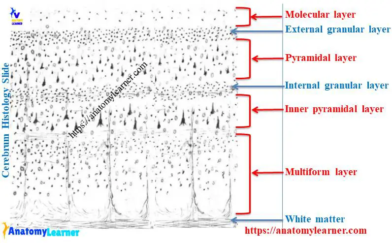
In the cerebral cortex histology, you will find the six different layers which contains different nerve cells and association fibers. But it (six layers of cerebral cortex) is poorly distinguished under light microscope.
#1. Layer 1: Molecular layer of plexiform layer of cerebral cortex
#2. Layer 2: External granular layer of cerebral cortex
#3. Layer 3: External pyramidal layer of cortex
#4. Layer 4: Inner granular layer of cerebral cortex
#5. Layer 5: Inner pyramidal layer of cortex and
#6. Layer 6: Multiform layer or layer of polymorphic cells of cerebral cortex
Molecular layer is the most superficial and well defined layer of cerebral cortex. This molecular layer of cerebral cortex consists of nerve fibers (apical dendrite from pyramidal cells) and horizontal cells.
The external granular layer contain the small neuron that serve as interneuron. External pyramidal layer consists of small and medium pyramidal neuron that send axons to adjacent cerebral cortex.
Internal granular layer of cerebral cortex consists of closely packed stellate cells and horizontally oriented white fibers. The internal pyramidal layers consists of medium to large pyramidal cells and also few stellate cells that send axon fiber to the white matter of cerebrum.
Multiform or layer of polymorphic cells is the deep layer of cerebral cortex and contains numerous spindle shaped neurons that send axon fibers into the white matter.
Cerebral medulla histology
The cerebral white matter consists of myelinated fibers that passing in all direction. These myelinated fibers include mainly association fibers, commissural fibers and projection fibers. The superficial neuron of molecular layer send axon to the nearest region of cortex and form the association fibers.
The neuron from the deepest two layers of cerebral cortex send long axon to the lateral cerebral hemispheres and from the commissural fibers.
Neuron from the deepest two layers of cerebral cortex send long axon to the brainsteam and form the projection fibers in cerebral white matter of brain.
Cerebrum histology drawing
Okay, now I am going to show you the different layers and cells from the cerebrum histology slide. I tried to show you the different structures in the cerebral histology drawing.
If you need more real cerebral cortex slide images then you may follow anatomy learner at social media. You will get update on article and different images at social media of anatomy learner.
Do you want to learn other different histology from nervous tissue or nervous system of animal? Great, you will find other different article related to nervous tissue or system histology here in anatomy learner.
#1. Identification of cerebellum under light microscope
#2. Histological features of spinal cord with their identification points
Conclusion
I hope this details guide will help you to learn cerebrum histology with real slide images and labeled diagram. If you need more information on histological features of cerebrum of animal’s brain then let me inform.
Are these identification points helpful to identify the cerebrum histology slide under light microscope? If you think this article is good for learning cerebral cortex and cerebral white matter histology then share it with your friends.
Stay connected with anatomy learner to get more article related to veterinary anatomy and veterinary histology.