The dog neck anatomy consists of bones, muscles, glands, blood vessels, lymph nodes, and other essential organs. It is very common to find different injuries in the dog’s neck bones, muscles, and subcutaneous tissue. Again, you may find severe obstruction in the neck (in the esophagus or trachea) of a dog.
As a veterinarian, you might handle and manage these conditions. For that, you might have a good piece of knowledge on the anatomy of the dog neck (including the bones, muscles, blood vessels, glands, and organs like the esophagus, trachea).
Here, I will provide basic knowledge on the anatomy of bones, muscles, artery, veins, and other different organs from the neck of a dog. I hope you will like this information on dog neck and stay with me till the end of the article.
Dog neck anatomy
You may divide the dog neck anatomy into three parts for the description purpose. The three different parts of the dog neck are – the gland region, the lateral margin of the neck, and the ventral part of the neck. All the structures and organs from the dog neck are essential, but I would like to focus on the most important ones.
The dog neck’s most vital structures and organs are the superficial muscles, neck bones, thyroid glands, esophagus, trachea, blood vessels (artery and veins), and lymph nodes. So, my goal is to provide explicit knowledge on these structures and organs from the dog neck.
The neck bones of the dog consist of the cervical vertebrae. You know, there are seven cervical vertebrae in the dog skeleton (atlas, axis, third, fourth, fifth, sixth, and seventh cervical vertebrae). Again, the dog neck region’s essential muscles are brachiocephalicus, omotransversarius, sternocephalicus, splenius, longus capitis, longus coli, scalenus, and serratus ventralis cervicis.
There are some essential lymph nodes present in the neck region of a dog. The essential lymph nodes of the dog neck are – parotid lymph node, mandibular lymph node, retropharyngeal lymph node, superficial and deep cervical lymph nodes.
Again, the thyroid and parathyroid are the most important glands of the dog neck. I will show you how the external jugular vein is formed in the dog neck from the blood vessels. Again, you will find some other important structures like the common carotid artery and vagosympathetic trunk in the ventral neck of a dog.
In addition, you will find the cervical part of the trachea and esophagus in the neck region of a dog. Here, you will also get a short description of these visceral organs from the dog neck.
Dog neck bones anatomy
The neck bones of a dog consist of seven cervical vertebrae. The first two cervical vertebrae are deferring in their structures compared to other neck bones. Even they differ from each other (atlas and axis of a dog skeleton).
You may easily recognize these two bones (atlas and axis) from the dog neck bones anatomy. Again, you will find a slight difference in the structure of the dog’s third, fourth, and fifth cervical vertebrae. So, it is challenging to recognize third, fourth, and fifth cervical bones from a dog.
In addition, the sixth and seventh cervical vertebrae of a dog neck possess some distinguishable osteological features. So that you may quickly identify these (sixth and seventh) cervical bones from the dog neck.
The atlas of the dog neck
Atlas is the first cervical vertebra of the dog neck and is atypical both in its structure and function. It articulates with the occipital condyle of the skull cranially and with the axis caudally. Two thick lateral masses are present in the atlas that unite the dorsal arch with the ventral arch.
Do you know how this ventral arch is formed in dog atlas? Well, the reduction of the vertebral body forms the ventral arch in the dog atlas. Again, you will find a shelflike transverse process (wing) that projects from the lateral mass of the dog atlas.
Okay, let’s know what the essential structures in the dog atlas vertebra are. There is a dorsal tubercle present in the cranial end of the dorsal arch of the dog atlas bone. Here, the dorsal tubercle of the dorsal arch is bifid. Again, you will find a ventral tubercle at the caudal end of the ventral arch of the dog atlas. The ventral tubercle of the dog atlas is in the form of a conical process.
You will find a cranial articular fovea in the dog atlas that contains two cotyloid cavities. Again, there is a caudal articular fovea containing two shallow glenoid cavities. You know the cranial articular fovea of the dog atlas articulates with the condyles of the skull. And the caudal articular fovea of the dog atlas articulates with the axis.
Again, the dorsal surface of the ventral arch of the dog atlas possesses fovea dentis. It is concave from side to side and articulates with the dens of the dog axis bone.
Wing of the dog atlas bone
The wing of the dog atlas possesses some essential structures like – lateral vertebral foramen, alar foramen, and alar notches. Again, there is a large depression present in the ventral portion of the wing (atlantal fossae).
The alar foramen is a short canal passing obliquely through the dog atlas’s transverse process (wing). In addition, the lateral vertebral foramen of a dog atlas locates at the craniodorsal part of the vertebral arches. You will find an alar notch on the cranial border of the base of the wing of the dog atlas bone.
The vertebral veins and arteries transverse the atlantal fossa. Again, the vertebral vein extends through the alar foramen caudally. It anastomoses with the internal jugular vein in the ventral condyloid fossa rostrally.
The axis of the dog neck bones
The axis is the second cervical bone in the dog neck anatomy. It possesses a long, dorsal spinous process that is bladelike cranially and expanded caudally. You will find a peg-like cranioventral eminence (dens) in the axis of the dog neck. This odontoid process of the dog axis articulates with the atlantal fossa of the atlas.
Again, you will find a short body, articular surfaces, and processes in the dog axis. The cranial articular surface of the dog axis is the cranial expanded end of the vertebral body.
There are caudal articular surfaces present in the dog axis. These caudal articular surfaces are the ventrolateral extensions of the vertebral arch and spinous processes that face ventrally.
A short, caudally directed transverse process arises from the pedicle of the dog axis. You will find a short transverse foramen in the dog axis bone.
The ventral surface of the dog axis body contains two deep fossae and a median crest. There are also cranial and caudal vertebral notches in the dog axis vertebra.
The cranial vertebral notches form the more prominent intervertebral foramina on either side with the atlas.
You will find the second pair of the spinal nerves and spinal vessels that pass through this intervertebral foramina. Again, the caudal vertebral notches of the dog axis form the intervertebral foramina on either side with those of the third cervical vertebra of the dog neck. So, within this formen, the third Paris of the dog cervical spinal nerve and spinal vessels will pass.
The third, fourth, and fifth cervical vertebrae of the dog neck
You know the third, fourth, and fifth cervical vertebrae of the dog neck differ slightly from each other. These cervical bones of the dog neck almost possess the osteological features of a typical vertebra.
I know you have a good piece of knowledge on the typical vertebra. So, I will not describe all the osteological features of a dog typical vertebrae. Instead, I prefer to provide the significant osteological features of these neck bones from a dog skeleton.
You will find large laminae on the third cervical vertebrae of a dog. It gradually becomes shorter and narrower on the fourth, fifth, and other cervical vertebrae. There are tubercles present on the caudal articular process of the cervical bones of a dog. They gradually decrease their prominence from third to seventh cervical vertebrae.
Again, the spinous process of the dog neck bones (third to the fifth cervical) increases in length. The transverse process of these cervical bones is prolonged and slightly twisted. You will find a shorter transverse process in the fifth cervical vertebra compared to the other bones of a dog neck.
In addition, you will find a pair of transverse foramina on each cervical vertebrae of the dog neck (except atlas). These transverse foramina extend through where the transverse process attaches to the body and pedicle. You know within these foramina, specific vertebral spinal nerves and spinal vessels will pass.
The sixth and seventh cervical bones of the dog neck
You will find some peculiar osteological features in the dog neck’s sixth and seventh cervical bones. The most peculiar osteological features of the dog sixth neck bone are an expanded sagittal platelike transverse process (lamina ventralis). The plates extend ventrally and laterally, representing only the caudal part of the transverse process.
Again, the cranial part of the transverse process forms a conical projection at the ventrolateral aspect of the transverse process. In addition, the sixth cervical bone of the dog neck possesses a more extensive spinous process than that of the other bones. You will also find pairs of transverse foramina at the junction of the body and pedicle of the sixth cervical bone.
The seventh cervical bone of the dog neck possesses some peculiar osteological features. You will find an articular facet for the first rib pair at the caudal part of the body (seventh neck bone). Again, a pair of facets are present at the end of the transverse process.
The spinous process of this seventh cervical neck bone is more significant than that of the sixth cervical. You will not find any transverse foramen in the seventh cervical neck bone.
Joints of the dog neck
The bones of the dog neck form a synovial joint with each other to form the cervical part of the vertebral column. All the joints are essential, but I would like to introduce the most clinically important joints with you from the dog neck. The most clinically important joints of the dog neck are – atlantooccipital joint and atlantoaxial joint.
The atlantooccipital joint of the dog neck
The atlantooccipital joint of the dog neck is formed by the articulation of the occipital condyles with the cranial concave articular surface of the atlas. You might know the structures involved with these atlantooccipital joints of the dog neck.
Fine, a spacious joint capsule will find on each side of the joint that attaches to the margin of the opposed articular surfaces. You will find a close relationship with the joint of the atlantoaxial joint of the dog neck. The atlantoaxial joint cavity communicates with the atlantoaxial joint cavity along the odontoid process.
Again, you will find three different vital structures in the atlantooccipital joint of the dog neck anatomy. These three essential structures of a dog neck atlantooccipital joint are the dorsal atlantooccipital membrane, ventral atlantooccipital membrane, and lateral ligament.
The dorsal atlantooccipital membrane extends from the dorsal edge of the foramen magnum to the cranial border of the dorsal arch of the dog atlas. Again, the ventral atlantooccipital membrane locates between the foramen magnum’s ventral edge and the dog atlas’s ventral arch.
In addition, the lateral ligament runs from the lateral part of the dorsal arch of the dog atlas to the paracondylar process of the occipital bone.
An atlantoaxial joint of the dog neck
The atlantoaxial joint of the dog neck is formed by articulating the odontoid process of the atlas with the fovea dentis of the axis. A thin and loose joint capsule is present in the atlantoaxial joint of the dog neck. It extends from the dorsal part of the cranial articular surface of one side of the axis to the opposite side.
Again, the fibrous layer (dorsal atlantoaxial membrane) of the joint capsule extends from the right to left between the dorsal arch of the dog atlas and the arch of the axis. You will find three major ligaments in the atlantoaxial joint of the dog neck skeleton – apical ligament of the dens, alar ligament, and transverse atlantal ligament.
The apical ligament leaves the apex of the dens and passes straight cranial to the basioccipital bone of the dog. You will find the alar ligament that attaches to the dens on either side of the apical ligament. The alar ligament of the dog neck is broader and heavier than the apical ligament.
Again, the transverse ligament of the dog atlantoaxial joint is thick and connect one side of the ventral arch of the atlas to the other.
If you want to know more about the joint structure from the dog skeleton anatomy, you may read the articles from the syndesmology section.
Dog neck muscle anatomy
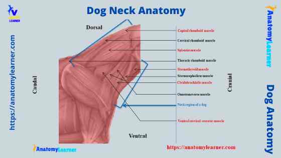
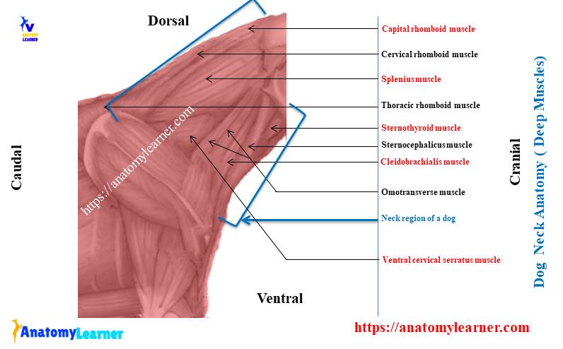
The dog neck muscle anatomy includes muscles primarily located in the neck and attached to the head or thoracic region. Many muscles are present in the dog neck region, but the most important will describe here.
You will find brachiocephalicus, sternocephalicus, omotransversarius, longus coli, longus capitis, splenius, scalenius, and serratus vetralis cervicis muscles in the neck of a dog. Here, I will describe these neck muscles from dog anatomy. But, you may learn more about the anatomy of the dog muscle from the myology section of anatomy learner.
The brachiocephalic muscle of the dog neck
The brachiocephalic is the long, flat muscle that extends between the brachium and the head and neck. You will find a clavicular intersection with this brachiocephalic muscle at the cranial end of the dog shoulder.
The brachiocephalic muscle of the dog divides into cleidobrachialis and cleidocephalicus muscles. You will find a thick cleidobrachialis muscle in a dog that arises from the distal end of the cranial surface of the humerus. This muscle has a relationship with the brachialis and biceps brachii muscles.
Again, the cleidocephalicus muscle of the dog neck extends cranially from the clavicular tendon. Further, the cleidocephalicus muscle of the dog divides into cervical and mastoid parts.
The cervical part of the cleidocephalicus muscle serves as a cranial extension of the cleidobrachialis muscle from the clavicular tendon to the dorsum of the neck. Again, the mastoid part is the deep ventrolateral part of the cleidocephalicus muscle. It extends from the clavicular intersection to the mastoid part of the temporal bone of the dog skull.
The sternocephalicus muscle of the dog neck
The sternocephalicus is another flat muscle of the dog neck that arises from the sterni manubrium. You will find two parts of this sternocephalicus muscle (occipital and mastoid parts). They were intimately joined at their origin, but they separated in the middle of the dog neck.
The mastoid part of the sternocephalius is ventral that separates as a large, elliptical muscle bundle. It unites with the mastoid part of the cleidocephalicus muscle in a large tendon that inserts on the mastoid part of the temporal bone of the dog skull.
Again, the occipital part of the dog sternocephalicus muscle is broader but thinner. It attaches to the nuchal crest as far as the midline of the neck through a thin aponeurosis.
Omotransversarius muscle of the dog
This is a flat, narrow muscle that lies at the lateral to the cervical vertebrae of the dog neck. It arises from the distal part of the scapular spine, as far as the acromion process. This muscle separates from the trapezius cervicis muscle and deep to the cervical part of the cleidocephalicus muscle.
The cranial part of the omotransversarius muscle of the dog neck becomes narrower and thicker. Again, the ventral border of the muscle limits the transverse process of the cervical vertebrae. This omotransversarius muscle of the dog neck helps to draw the limb cranially.
Longus capitis and longus colli muscles of the neck
The longus capitis muscle of the dog neck is also a flat, long muscle. It lies on the lateral and ventral side of the cervical vertebrae and lateral to the longus colli muscle.
This longus capitis muscle of the dog neck arises from the transverse process of the sixth to second cervical vertebrae and extends cranially to the axis. Again, it will cross the atlantooccipital joint and insert it on the muscular tubercles of the basioccipital bone of the dog skull.
The longus colli muscle is long in the dog neck and possesses cervical and thoracic parts. Let’s talk about the cervical part of the longus colli muscle of the dog neck. The cervical part of the dog longus colli muscle lies on the ventral surface of all the cervical vertebrae.
This muscle of the dog neck is enclosed by the right and left part of the longus capitis muscles. The cervical part of the longus colli muscle of a dog’s neck helps to flex its neck.
The splenius capitis muscle of the dog
The splenius capitis is a flat, fleshy, and triangular muscle of the dog neck. It lies on the dorsolateral part of the neck and extends from the third thoracic vertebrae to the skull.
The fibers of the splenius capitis muscle run in the cranioventral direction and cover the semispinalis capitis, longissimus capitis, terminal part of the semispinalis capitis muscles.
Do you know how this splenius capitis muscle works? This splenius muscle helps to extend and raise the head and neck of the dog. The unilateral action helps to draw the neck laterally. This muscle also functions in the fixation of the first thoracic vertebra of the dog neck.
The scalenus and serratus ventralis cervicis muscle of the dog
The scalenus muscle of the dog neck has three different parts – ventral, middle, and dorsal. This muscle bridges the space between the first three ribs and the cervical vertebrae.
The ventral scalenus muscle arises from the transverse process of the cervical vertebrae three to six. Again, the medius scalenus muscle of the dog neck arises from the transverse process of the sixth and seventh cervical vertebrae. In addition, the dorsal scalenus muscle locates between the ventral and medius scalenus and overlaps with the medius scalenus muscle.
The scalenus muscle of the dog neck also helps in flexing the neck. Fine, what about the serratus ventralis cervicis muscle of the dog neck?
The serratus ventralis cervicis muscle of the dog neck is a flat, fan-shaped muscle. It arises from the facies serrate of the scapula of a dog. And, it ends at the transverse process of the last five cervical bones of the dog neck.
This muscle covers the caudal half of the lateral surface of the dog neck. It supports the trunk and carries the trunk cranially and caudally.
Anatomy of the dog neck glands
In this part, I will try to discuss the anatomy of the dog neck glands. You will find the thyroid and the parathyroid glands in the neck of a dog. Here, you will find a little information on the anatomy of the thyroid and parathyroid glands.
The thyroid glands are paired (two lobes), an elongated, dark mass gland that attaches to the external surface of the cranial part of the trachea. This is the largest ductless endocrine gland of the dog anatomy.
The thyroid gland of the dog is positioned at the laterally and somewhat ventrally to the trachea. It extends from the five to eight tracheal rings on its respective side.
The size of a dog’s thyroid gland varies with the breed and individual. Again, the parathyroid glands of a dog are the minor endocrine gland that attaches to the cranial part of the thyroid gland. Let’s discuss the gross anatomical features of the thyroid gland of a dog.
Thyroid gland of a dog neck
You will find two lobes of the thyroid gland in the neck of the dog. These glands lie deep to the cervical fascia and are closely adherent to the trachea. You will find the sternocephalicus muscle that passes immediately lateral to the convex surfaces of each thyroid gland.
The cranial pole of the right thyroid gland lies at the level of the caudal border of the cricoid cartilage (of the larynx). It may extend caudally up to the fifth trachea ring. You will find a carotid covering that covers the gland dorsolaterally.
Here, you will also find the other vital structures of the neck region of a dog. The other vital structures of this neck region of a dog are – common carotid artery, internal jugular vein, and vagosympathetic trunk.
The left thyroid lobe of the dog varies in shape, size, and position. It may extend from the third to eight tracheal rings of the dog trachea. Again, you will find the caudal laryngeal nerve dorsal to the left thyroid lobe of a dog.
Parathyroid gland of the dog
There are four parathyroid glands in the dog that are closely related to the thyroid gland. These parathyroid glands are structurally independent, and one applies to the surface, and one embeds with the thyroid glands.
So, you will find two types of parathyroid glands in dogs – external parathyroid and internal parathyroid glands. The external parathyroid glands are located at the cranial dorsolateral edge of the thyroid gland. Again, the internal parathyroid glands are frequently found within the caudal portion of the thyroid glands.
Visceral organs of the dog neck anatomy
You will find numerous visceral structures and organs in the dog neck anatomy. The esophagus, trachea, glands, vessels, nerves, and lymph nodes. Here, I will discuss the essential visceral organs of the dog neck region.
The most clinically essential organs from the dog neck are – esophagus and trachea. Here, you will only find the anatomical features of a dog’s cervical part of the trachea and esophagus. But, you may learn the detailed anatomical features of the dog trachea and esophagus from the dog anatomy learning part of anatomy learner.
The esophagus of the dog
The esophagus is the first part of the dog alimentary canal that connects the tube between the laryngeal part of a pharynx and the stomach. The length and the diameter of the dog esophagus may vary with the breed and individual.
The esophagus of the dog passes transversely to the neck and all of the thorax. You will find three parts of the dog esophagus: cervical, thoracic, and small abdominal.
Let’s discuss on the cervical part of the dog esophagus. The cervical part of the dog esophagus mainly attache to the left longus colli and dorsal to longus capitis muscle. It will attach the trachea ventrally and to the right.
The external part of the dog esophagus is reddish due to the striated muscle coat. This striated muscle coat of the esophagus is supplied with the vagus nerve.
At the origin of the dog esophagus, you will find a left inclination. So that, at the thoracic inlet, the dog esophagus lies left lateral to the trachea. You will find different essential structures that have a close relationship with the cervical part of the dog esophagus.
The common carotid artery, vagosympathetic trunk, jugular vein (external and internal), and tracheal duct run to the lateral aspect of the esophagus.
The trachea of the dog neck
The trachea of the dog is an elastic tube that extends from the larynx to the level of the base of the heart. You will find two defined parts in the dog trachea – the cervical and thoracic parts.
The thoracic part of the dog trachea extends from the larynx to the level of the thoracic inlet. Dorsally, you will find a relationship with the esophagus of the dog. Again, some important structures like the common carotid artery, jugular vein, vagosympathetic nerves, and recurrent laryngeal nerve close to the trachea (right side).
You will find thirty-five C-shaped incompleted cartilaginous rings in the structure of the dog trachea. These incomplete tracheal rings are closed by the smooth (trachealis) muscle (transverse bundles).
Again, the annular ligament connects the tracheal rings and the membranous part. This annular ligament of the dog trachea allows considerable intrinsic movement of the trachea.
Some internal organs like the thyroid and parathyroid glands are already described. Again, the other essential structures of the dog neck will discuss in the next part of the article.
Lymph nodes of the dog neck
There is some superficial and deep lymph node in the dog neck region. Let’s discuss these superficial and deep lymph nodes of the dog neck.
The parotid lymph node locates at the rostral base of the ear. Again, the mandibular lymph center of a dog consists of mandibular and buccal lymph nodes. The mandibular lymph node of the dog lies at the ventral angle of the mandibular joint.
You will find the buccal lymph node at the rostral to the facial and superficial vein angle and dorsal to the buccinator muscle. The retropharyngeal lymph nodes of a dog consist of a dog’s medial and lateral part.
Again, the medial retropharyngeal lymph node is the largest in the dog’s neck. It is an elongated, transversely compressed lymph node with a more pointed caudal end.
The superficial cervical lymph nodes of a dog are two in numbers. One is lying dorsal to the other in the adipose tissue on the lateral surface of the serratus ventralis and scalenus muscles. Superficially, you will find the cleidocephalicus, omotransversarius, and trapezius muscle that covers the superficial cervical lymph node of the dog.
In addition, the deep cervical lymph node of the dog locates along the cervical portion of the trachea of each side. But, the presence of this cervical lymph node may vary with the dog breed and individual. Sometimes, you may find cranial deep cervical lymph nodes in some dog breeds. Again, you may find middle deep cervical lymph nodes in some dog breeds.
Dog neck blood flow anatomy
You will find the common carotid artery, internal carotid artery, and external carotid artery in the dog neck. The common carotid artery of the dog arises from the brachiocephalic trunk. You will find two branches of the common carotid artery of the dog – the right and left common carotid artery.
The left common carotid artery arises opposite to the vertebral end of the second rib and ventral to the trachea. Again, the right common carotid artery diverges from the left and obliquely crosses the ventrolateral surface of the trachea.
The internal carotid artery is the main continuation of the common carotid artery to a dog’s head. You will find different internal carotid artery branches like occipital, condyloid, cervical, cranial laryngeal, descending pharyngeal, palatine, facial, lingual, and more.
In addition, the internal carotid artery of a dog arises with the external carotid artery as the smaller of the two terminal branches of the common carotid artery. You will also find different branches in the internal carotid artery.
A vein in the dog neck
The external jugular vein of the dog originates from the brachiocephalic vein at the level of the thoracic inlet. This vein gives off cephalic, superficial cervical, and omobrachial veins on its caudal-cranial sequences.
The external jugular vein then divides into a dorsal branch (maxillary vein) and ventral vein (lingofacial vein) at the caudal border of the dog mandibular gland. Again, the cephalic vein lies at the medial part of the lateral pectoral groove. It joins with the external jugular vein of the dog just cranial to the thoracic inlet.
In addition, the omobrachial vein courses superficially upon the deltoid and cleidocervical muscles. This vein extends between the axillobrachial vein and the external jugular vein.
The vagosympathetic trunk of the dog is a large nerve that lies dorsal to the common carotid artery. This nerve conducts sympathetic fibers from the thoracolumbar sympathetic trunk to the dog’s head. Again, the parasympathetic fibers of the vagus nerve reach from the head to the body cavity of the dog.
In addition, the recurrent laryngeal nerve with its motor, sensory, and autonomic fibers turns and passes cranially to the dog neck region. You may quickly identify the recurrent laryngeal nerve on the dorsal border of the thyroid gland.
You may get more dog neck labeled diagram on social media.
Frequently asked questions on dog neck.
In this part, I will try to solve the common inquiries on the dog neck anatomy. Anatomy learners commonly ask the below-mentioned questions.
Which organs are in the dog’s neck?
Well, there are numerous organs present in the dog’s neck. You will find the most important organs like the trachea, esophagus, glands – thyroid, and parathyroid in the neck region of a dog. Again, other important structures like the common carotid artery, recurrent laryngeal nerve, external jugular vein are present in the dog neck.
You will also find some essential lymph nodes like a superficial cervical lymph node and the deep cervical lymph node in the dog neck region. All the structures and organs are described previously in this article.
Do dogs have muscles in their neck?
Yeah, there are well-developed, organized muscles present in the dog neck. You will find the brachiocephalicus, sternocephalicus, omomtransversarius, splenius, scalenus, and serratus ventralis cervicis muscles in the dog neck.
There are some other essential muscles present in the dog neck region. You may learn about these muscles from the dog muscle anatomy learning part.
Where is the neck of a dog?
The neck locates at the lateral aspect of the cervical vertebrae of a dog. It starts from the first cervical vertebra and ends at the thoracic inlet (or at the last cervical vertebra).
The dog neck’s most clinically essential structures and organs are esophagus, trachea, thyroid gland, parathyroid glands, superficial lymph nodes, carotid artery, vagosympathetic nerve, and external jugular vein.
Can dogs pull muscles in their neck?
What glands are in a dog’s neck?
You will find two major endocrine glands in the dog neck. One is the thyroid gland, and another one is the parathyroid glands. The dog’s thyroid is the largest endocrine gland that lies on the lateral aspect of the trachea (from the third tracheal ring to the seventh ring).
Again, you will find two types of parathyroid glands (total four) in the dog neck – the external and internal parathyroid glands.
Does a dog have lymph nodes in its neck?
Yeah, dogs have lymph nodes in their neck. You will find the superficial cervical lymph nodes and deep cervical lymph nodes in the dog neck. Again, the deep cervical lymph nodes are of two types – deep cranial cervical and the caudal deep cervical lymph node.
Can you feel the dog’s lymph nodes in the neck?
If the dog is healthy, it is tough to feel the lymph nodes of the dog’s neck. Again, it is also hard to feel the deep cervical lymph nodes (both the cranial and caudal) from the dog neck. Sometimes, you may feel the superficial cervical lymph nodes of the dog neck.
Conclusion
So, the dog neck anatomy contains different vital structures and organs. You might have a good piece of anatomical knowledge on these structures and organs for further clinical and surgical intervention. First, the bones and muscles of the dog neck anatomy are the basic structures that you should know very clearly.
Again, the internal organs and structures like the trachea, esophagus, course of the carotid artery, jugular vein are also crucial for a pet animal practitioner. It would be best if you also had a piece of good knowledge of the superficial lymph nodes of the dog neck.