Where is Simple Squamous Epithelium Found?
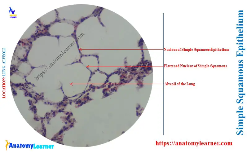
The simple squamous epithelium has a wide variety of distribution in the animal body. Here, you will know the answer to the question – ‘Where is simple squamous epithelium found’? Quick answer: the simple squamous epithelium is found in the serous membrane of the body cavities, the internal surface of the heart, and the luminal … Read more