
Looking for hollow organ histology?
Do you want to learn the histology of different organs from digestive, respiratory and urogenital system of animal? The general hollow organ histology will make your learning simple.
Hey, thank you so much for choosing anatomylearn to learn wall of a hollow organ layers histology. In this article I am going to describe wall of a hollow organ layers. After reading this article you will able to understand the general histology of hollow organs from different system.
If you want to make your histology learning easy then let’s continue this article.
Hollow organ histology
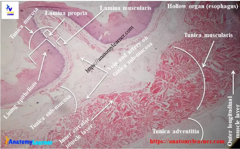
The hollow organ composed if a lumen of variable diameter. It surrounded by the following four different layers –
#1. Tunica mucosa layer
#2. Tunica submucosa or tela submucosa layer
#3. Tunica muscularis layer and
#4. Tunica adventitia or serosa
I will discuss the histology structure of these four layers in details.
This hollow organ histology is important for the digestive, respiratory and urogenital system. Let’s discuss on the wall of hollow organs layers histology.
Histology of tunica mucosa layer of hollow organ
This is the innermost layer of a tubular organ. This tunica mucosa layer consists of the following sub layers –
#1. Epithelium lining
#2. Lamina propria and
#2. Lamina muscularis
Let’s discuss on these three structures from tunica mucosa.
#1. Epithelium lining of tunica mucosa from hollow organ histology
I would like to enlist the major histological features of epithelium lining of tunica mucosa –
#1. There may present any type of surface epithelium. These surface epithelium rest on basement membrane.
#2. These surface epithelium depends on functions of different specific organs. You will find stratified squamous epithelium in oral cavity, esophagus and anal canal. Again you will find simple columnar epithelium in stomach, intestine and receteum.

Do you know about the functions of this lining epithelium?
The function of this lining epithelium in different organs varies.
In digestive system organs of animal the function of lining epithelium are –
#1. Provides a permeable barrier between the contents of tract and tissue of body
#2. It facilities transport and digestion of feed particle in animal
#3. Helps in absorption of the digest products
#4. Help to produce hormones that affect the activity of digestive organs
In respiratory organs, the function of this lining epithelium also varies –
#1. In nasal cavity you will find respiratory epithelium. This respiratory epithelium is responsible for moving mucus along epithelial surface.
#2. You will find clara cells on epithelium lining on respiratory organ. This clara cell is responsible for metabolizing airborne harmful agents
#3. Pseudostaratified columnar epithelium contains modified bipolar neuron and they are responsible for olfaction.
#4. Producing glycosaminoglycan
#2. Lamina propria of mucosa from hollow organ histology
The major characteristics of lamina propria histology from a hollow organ are –
#1. Composed of loose connective tissue – fine collagen fiber, elastic fiber and reticular fibers)
#2. Presence of blood vessels, lymphatic tissue in lamina propria
#3. Sometime you may find some mucosal gland in few organs from different system
Do you know who supply the nutrition to the epithelium lining of a tubular organ?
You already found blood vessels in lamina propria in tunica mucosa layer. These blood vessels are responsible to supply nutrition to surface epithelium.
#3. Lamina muscularis of mucosa layer
The major histological features of lamina muscularis are –
#1. Presence of thin layer of smooth muscles (one to three layers) layer in lamina muscularis
#2. These muscle layers allow independent movement of mucosa. Thus it helps to movement of luminal content.
#3. These muscle layers also assist to secret from the mucosal gland in some organs.
Sometime lamina muscularis is absent in some organs. Lamina propria is merged with the next layer tunica sub mucosa. Thus lamina propria and tunica sub mucosa form propria sub mucosa.
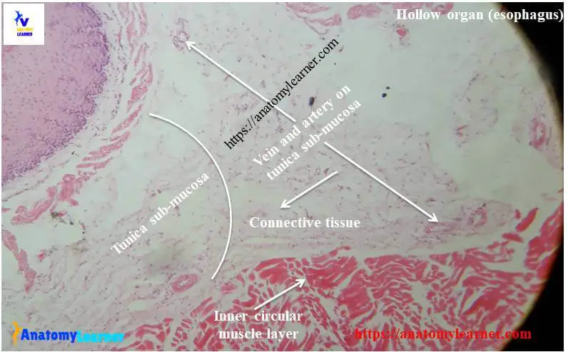
Histology of tunica sub mucosa layer of hollow organ
The main histological features of tunica sub mucosa layer of hollow organ histology are –
#1. Presence of dense connective tissue
#2. Presence of blood vessels and lymphatic vessels
#3. You will find submucosal gland in this layer
#4. Sometimes you may find some submucosal plexus at tunica sub mucosa. These plexuses are composed with ganglionic nerve plexus and autonomic nerves
Histology of tunica muscularis layer of hollow organ
You will find the following features in histology of tunica muscularis layer of hollow organ –
#1. Presence of smooth or skeletal muscle layers
#2. Muscles are arranged in inner circular and outer longitudinal direction. Sometimes you may find some oblique direction muscle layer
#3. Myenteric plexus may present
Tunica muscularis is responsible for moving ingesta and mixed with glandular secretion.
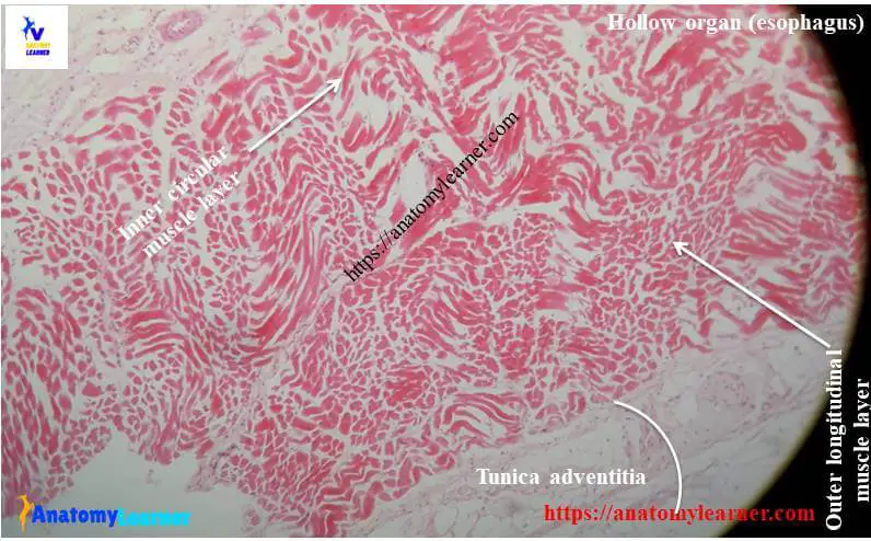
Histology of tunica adventitia layer of hollow organ
The main features of histology of tunica serosa layer of hollow organ are –
#1. There is a loose connective tissue layer
#2. Presence of simple squamous lining epithelium (mesothelium)
You will find serosa in pleura, pericardium, peritoneal cavity and other organs.
The important features of histology of tunica adventitia layer of hollow organs are –
#1. Presence of thick connective tissue layer
#2. There is no simple squamous lining epithelium
Now, you may learn the histology of different tubular organs in easy way. You should find out the exceptions pattern in wall of different hollow organs layers histology from different system.
Exception pattern in conducting portion of respiratory organs
You will find the following exception in the conducting portion of respiratory organs –
There is muscularis mucosa in conducting organs of respiratory system. Mucosa, sub mucosa layer can’t easily distinguish in respiratory organ histology.
Tunica muscularis contains cartilage in epiglottis, larynx, trachea and bronchi from respiratory organs.
These cartilages help to prevent collapse of the lumen. It ensures access of air in lung. These cartilages also provide rigidity, flexibility to organs.
You may read the following histology of organs from anatomylearn.com –
#1. Best guide to learn histology of lung from labeled lung histology slide (hollow organs)
#2. Guide to learn histology of female genital organs from labeled histological slide
Conclusion
I think you got a better idea about the hollow organ histology. Now you could able to describe any layers of hollow organ histology from any organ system.
Do you need hollow organ histology diagram pdf? You may find more update diagram related to histology of hollow organ here at social media. Please try to find out the missing label from the histology diagram pdf. You may learn more stomach histology here in this blog. Thank you so much for staying with anatomylearner.com