Epithelial tissue consists of a sheet of aggregated cells of a similar type covering the external surface of solid structure and internal surface of any hollow organs of an animal’s body. Here in this post, I will provide the basic information of epithelial tissue and show you the cells from different covering or lining epithelium with slide pictures.
Hi, do you want to learn the basic epithelial tissue histology with their identification characteristics from real slide pictures? This article will focus on identifying different types of epithelial tissues under the light compound microscope.
You will also know the basic characteristics, definition, types, location, and histology of different epithelium types. So, if you are interested to learn then, you may continue this epithelium histology article.
Epithelial tissue definition, types, and examples
I hope you already know the definition of epithelial tissue. Epithelial tissue consists of a sheet of aggregated cells of similar types that constitutes the animal’s body’s external and internal surface. These cells are firmly adherent to one another and attached to the thin layer of the basement membrane. Epithelial cells are specialized for various functions like – protection, absorption, excretion, and formation of the barrier.
Under the basement membrane, you will find the vascular connective tissue layer. According to the shape of epithelial cells and the numbers of cells layers, covering epithelial tissues are divided into four different types –
- #1. Simple epithelium tissue – have a single layer of epithelial cells resting on the basement membrane.
- #2. Stratified epithelium tissue – composed of two or more layers of epithelial cells with only the basal cell layer resting on the basement membrane.
- #3. Pseudostratified epithelium tissue – composed of one layer of cells but looks to be multilayered due to different cellular height.
- #4. Transitional epithelium is composed of multiple layers and characterized by a surface layer of dome-like cells, neither squamous nor columnar epithelial.
“Mainly epithelial tissues are divided into two groups based on their structure – the surface or covering epithelium and glandular epithelium. Here, you will find only the histology of surface or lining epithelium; if you want to know about glandular epithelium then, you might read other articles from the epithelium section.”
Epithelial tissue histology
It is an essential requirement to know epithelial tissue histology for any beginner. I will try my best to provide the best guide to identify the different types of epithelial tissues under the light compound microscope. Here, you will learn and identify the following different types of epithelium with real slide pictures –
- #1. Simple squamous epithelium histology
- #2. Simple cuboidal epithelium identification
- #3. Simple columnar epithelium histology
- #4. Non – keratinized stratified squamous epithelium characteristics
- #5. Keratinized stratified squamous epithelium histology
- #6. Stratified cuboidal epithelium
- #7. Stratified columnar epithelium
- #8. Pseudostratified ciliated columnar epithelium histology slide and
- #9. Transitional epithelium histology slide
Great, let’s learn and identify this epithelium histologyfrom real slide pictures. But, first, you might know what structures you should identify from each epithelium tissue.
#1. Layers of cells of the sample tissue
#2. The shape of cells of the provided sample tissue
#3. Basement membrane and lamina propria of the epithelium
Simple squamous epithelium identification points
If you want to identify the simple squamous epithelial tissue under light microscope, then the following identifying characteristics might help you.
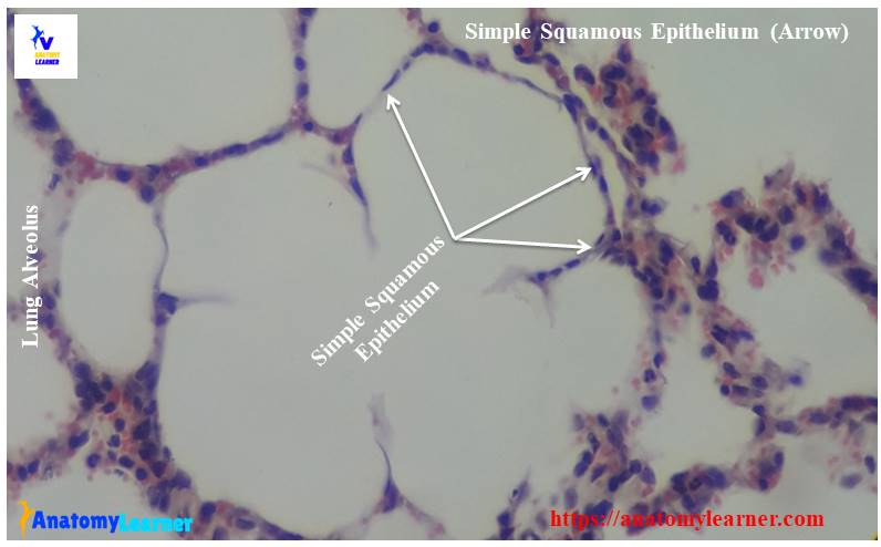
#1. The provided sample tissue shows the single layer of cells rest on the basement membrane.
#2. Cells of the sample tissue are flattened and plate-like, having considerable length and breadth but negligible height.
#3. There are centrally placed, the flattened nucleus in the cell of sample tissue
So, this is a simple squamous epithelium slide (location – the lining of lung alveoli).
Function and location of simple epithelium
The main functions of simple squamous epithelium are – protection, gaseous exchange, fluid transport, and lubrication. You will find simple squamous epithelium in the following organs or structures of an animal’s body –
#1. The lining of lung alveoli
#2. The serous lining of the body cavities – pleura, peritoneal cavity
#3. The lining of blood vessels (called endothelium)
Simple cuboidal epithelium histology
Well, let’s know the simple cuboidal epithelial tissue histology. I hope the following identification points will help you identify the simple cuboidal epithelium under the light microscope.
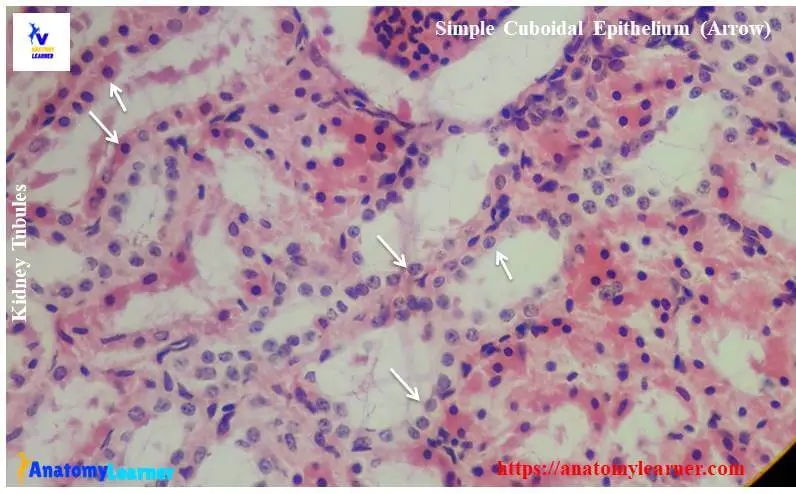
#1. The sample tissue section shows the single layer of cells located on the basement membrane.
#2. Identified cells are cuboidal in shape (have the same height and width).
#3. Presence of centrally placed round nucleus in the cuboidal cell.
So, it is a simple cuboidal epithelium slide (location – kidney tubules)
Functions and location of simple cuboidal epithelial tissue
The main functions of simple cuboidal epithelium are secretion and help in protection. You might also know the location of simple cuboidal epithelium in the organs or structure from an animal’s body. I am going to enlist the organs or structure where the simple cuboidal are found –
#1. Kidney tubules
#2. In thyroid follicles
#3. The germinal layer of ovary (lining epithelium of ovary) and
#4. A pigmented layer of retina
Identification of simple columnar epithelium
Following are the important identifying characteristics of simple columnar epithelium from histology slide under a light microscope.
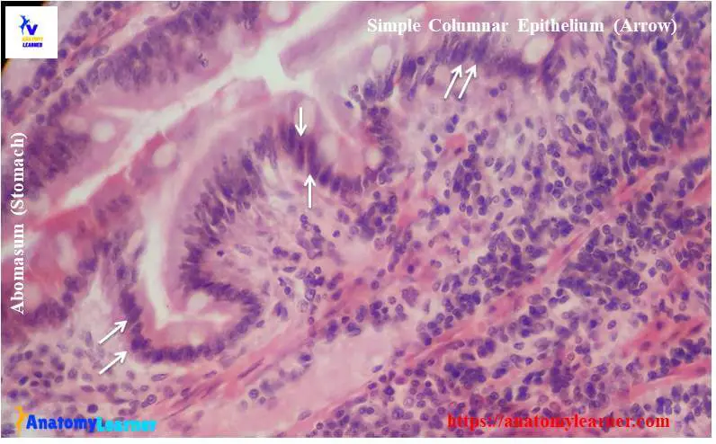
#1. The sample tissue section shows a single layer of cells rests on the basement membrane.
#2. The height of columnar cells is greater than the width (columnar-shaped cells).
#3. Presence of oval elongated nucleus located at the basal part of the cells (variable).
#4. There are some cilia with columnar cells in different organs.
So, this slide is simple columnar epithelium (non-ciliated; location – abomasum/ stomach).
Functions of simple columnar epithelium and their location
Absorption and secretion are the primary functions of simple columnar epithelium. You will find simple columnar epithelium in the following organs or structures of an animal’s body.
#1. Simple ciliated columnar epithelium in the respiratory tract, uterine tube
#2. Non-ciliated simple columnar epithelium in the stomach, gall bladder, and intestine.
Identification of stratified squamous epithelium
You will find more than one layer of cells in stratified squamous epithelium. It is again divided into two types according to the presence of keratinizing on the external surface.
Let’s identify the non-keratinized stratified squamous epithelium from the slide.
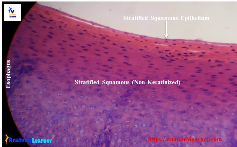
#1. The tissue section shows the several layers of cells rest on the basement membrane.
#2. Presence of cuboidal or columnar cells at the basal layer of the sample tissue
#3. The nuclei of the basal layers are oval or rounded.
#4. Presence of flattened cells with an elliptical nucleus in the superficial layer.
#5. There is no keratinize zone found in the superficial layer of cells or surface.
This is the stratified non-keratinized epithelium (location – the lining of the esophagus).
Functions and location of non-keratinized squamous epithelium
Prevention of water loss, secretion, and protection is the main function of stratified squamous epithelial tissue. You will find non-keratinized stratified epithelium in the mouth cavity, esophagus, cervix histology.
Okay, let’s identify the keratinized stratified squamous epithelium.
#1. Presence of multiple layers of cells in the sectioned tissue.
#2. The section shows dead, flat, scaly-like cells filled with keratin (known as superficial non-nucleated keratinized zone).
#3. The deep nucleated zone shows various layers of cells with different shapes.
So, this slide is keratinized stratified squamous epithelium (location – thick skin).
Location of simple keratinized stratified epithelium
You will also find the simple keratinized epithelium in the nostril, lips, hard plate.
Identification of stratified cuboidal epithelium
So, do you want to identify the stratified cuboidal epithelium under a light microscope? Well, I hope the following identifying characteristics will help you to identify the stratified cuboidal epithelium.
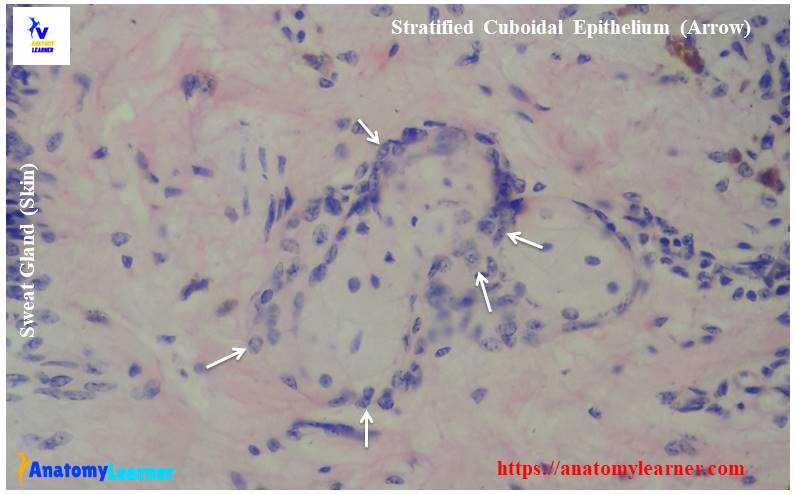
#1. Presence of double layers of cuboidal cells in the sample tissue section.
#2. Both layers of cells contain the centrally placed rounded nucleus.
This is a stratified cuboidal epithelium slide (location – sweat gland of skin).
Functions and location of stratified cuboidal epithelium
The main functions of stratified cuboidal are secretion and protection. You will find the stratified cuboidal epithelium in the following organs or structures of an animal’s body.
#1. Excretory ducts of the salivary gland, sweat glands
#2. Developing ovarian follicles
Stratified columnar epithelium histology
You may identify stratified columnar epithelium histology slide with the following identifying characteristics under light microscope.
#1. Presence of several layers of cells that rest on the basement membrane.
#2. The tissue section shows the cuboidal or low columnar-shaped cells at superficial layers.
#3. Presence of oval or elongated nuclei placed vertically (at base of cells).
This is stratified columnar epithelium (location – the mucosal lining of the male urethra).
Location of stratified columnar epithelial tissue
You will find the stratified columnar epithelium in the palpebral conjunctiva, male urethra.
Pseudostratified ciliated columnar epithelium histology
You may also ask to identify the pseudo-stratified ciliated columnar epithelium; you might find these characteristics from the sample tissue.
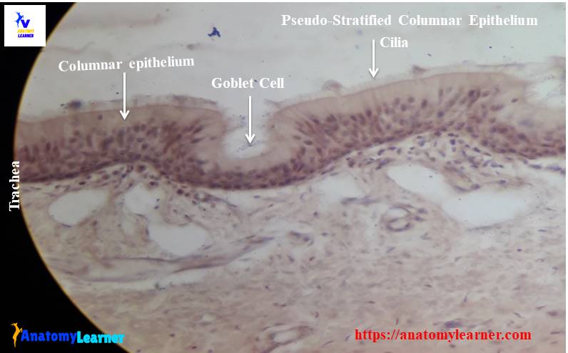
#1. Presence of a single layer of cells with different shapes and heights (many of these cells do not reach the surfaces).
#2. Presence of nuclei at different levels (gives the multilayered appearance).
#3. The tissue section shows that cells reached the surface are columnar in types and contains cilia.
This is the slide of the pseudo-stratified ciliated columnar epithelium (location – the mucosal lining of the trachea).
Location of pseudo-stratified ciliated columnar epithelial tissue
You will find the pseudo-stratified ciliated columnar epithelial tissue in the trachea, nasal cavity, and bronchi of lung histology structure.
Identification of transitional epithelium histology slide
Transitional epithelium is also a pseudostratified type cell with a wide variety of appearances. It lines the hollow organs that are capable of considerable distension. You may identify transitional epithelium histology slides under a light microscope with the following identifying characteristics.
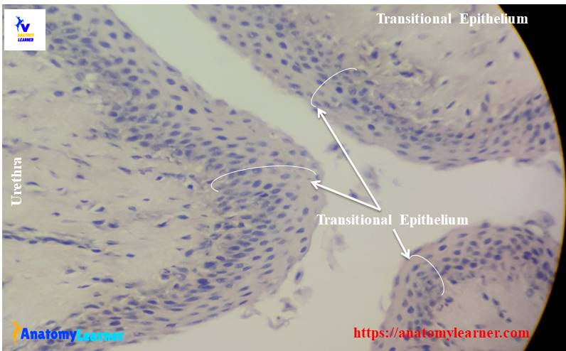
#1. Presence of various-shaped multilayered cells (generally four to six layers of cells).
#2. The superficial layer of cells shows the large rounded dome-like appearance.
#3. Presence of small-shaped cells in the deep layer of the sample tissue section.
#4. The nuclei of the cells of the deep layer are located close to each other.
So, this is a transitional epithelium (location – urinary bladder)
Functions and location of transitional epithelial tissue
Protection, preventing reabsorption of urine and allowing distension of urinary organs are the transitional epithelium’s main functions. You will also find the transitional epithelial tissue in the ureter.
Epithelial tissue drawing pictures
Now, I will share some epithelial tissue drawing pictures with you. You may try to draw the same, but please follow the instruction from the books.
If you need more epithelial tissue drawing or real slide pictures, then let me know. You may also follow anatomy learner on social media for more epithelial pictures. Don’t forget to read the other articles from anatomylearner –
#1. Identifying characteristics of loose connective tissue from slide pictures.
#2. Compact bone microscope slide and identifying features.
Conclusion
I think this short guide will help you learn and identify the epithelial tissue with real slide pictures. You might know epithelial tissue types, location, and examples carefully for further study of histology.
I will try to enrich this article regularly with more labeled diagrams and slide pictures. If you want to get updates on that, you may follow anatomy learner. Again, please don’t forget to share this article with your friends who wish to learn about epithelial tissue histology basics.