Simple squamous epithelium under a microscope consists of a single layer of thin, flat, and scale-like cells. These cells are joined together by an intercellular junction and rest on the basement membrane, whose thickness depends on the location. Here, I will show you what simple squamous looks like under a light microscope.
Again, I will also tell you almost all the locations of simple squamous animal bodies with their unique features. You will also learn the ultrastructure and function of the simple squamous epithelium with the help of an electron microscope.
At the end of this article, there is the drawing procedure and images of the simple squamous. So, don’t miss to check these drawing images and labeled diagrams of simple squamous epithelium.
Simple squamous epithelium under a microscope
The simple epithelium is the covering or surface epithelium that consists of a single layer of cells. Again, the simple squamous epithelium consists of a single layer of flattened cells with a flattened nucleus. You should know what the simple squamous epithelium looks like on the surface view and the longitudinal section view.
- Simple squamous on surface view
- The plane is perpendicular to the surface view for simple squamous
On the surface view, the simple squamous epithelium under a microscope, the cells possess an irregular shape with a slightly serrated border. These simple squamous cells fit together like pieces of a jigsaw puzzle to form a continuous sheet.
So, you will find a spherical or oval nucleus near the center of the cell that provides a slightly elevated appearance to this area. Again, the cell appears thicker in the nucleus area and possesses thin attenuated strands of cytoplasm on either side.
In addition, in-plane perpendicular to the surface, the simple squamous cell looks like spindles or flattened. The nucleus of the simple squamous become a tapper at both ends (also flattened). But, it is very hard to see the cell plasma membrane in routine stain (Hematoxylin and eosin staining).
You will find the fine details structures of a simple squamous epithelium (including its plasma membrane, nucleus, and other organelles) with the help of an electron microscope.
Identifying features of simple squamous epithelium
So, first, let’s identify the simple squamous epithelium with the help of a light microscope. You will find the below mentioned identifying features for simple squamous epithelium –
- There is a single layer of cells that rests on the basement membrane.
- The cells show a flattened appearance (having considerable length and breadth but negligible height).
- These cells appear so thin that their nuclei produce bulgings on the surface.
- A centrally placed nucleus becomes taper on both ends (flattened nucleus).
So, this is a simple squamous epithelium histology slide.
Simple squamous epithelium location
Simple squamous epithelium lines moist internal surfaces like the body cavities, the heart, and the blood and lymph vessels. Sometimes the name of the simple squamous epithelium depends on the location. The simple squamous epithelium that lines the body cavities (pleural, pericardial, and peritoneal) is known as the mesothelium.
Again, when you find the simple squamous lining in the heart, blood vessels, and lymph vessels, these epithelia are endothelium.
You will also find a special simple squamous epithelium that lines the subarachnoid and subdural spaces. These special types of simple squamous cells are mesenchymal epithelium. In addition, you will also find the mesenchymal epithelium in the anterior chamber of the eye and perilymphatic spaces of the ear.
Now, let’s enlist the location of simple squamous epithelium in the body of an animal –
- Parietal layers of the Bowman’s capsule of the kidney
- A thin loop of Henle (kidney)
- Pulmonary alveoli of the lung
- Pleura of lung
- Pericardium of hear
- Peritoneum of the abdominal cavity and others
- Internal surface of the blood vessels and lymph vessels
- The inner surface of the subarachnoid and subdural spaces
- In the anterior chamber of the eye and perilymphatic space of the middle ear
I will try to show you the simple squamous epithelium under a microscope from different organs like the lung, the parietal layer of Bowman’s capsule, the thin loop of Henle, and others.
Functions of simple squamous epithelium
The simple squamous epithelium helps in protection, lubrication, gaseous exchange, and fluid transport. Again, this type of epithelium is typical at a site that makes up blood tissue barriers. The thinness of the simple squamous epithelium also permits the diffusion and bidirectional movement of gasses, fluid, and nutrients from the free surface to the underlying tissue.
Simple squamous epithelium under microscope 40x
It is very important for the beginner how you will see the simple squamous epithelium under a microscope with 40x magnification. Here is a microscopic figure of simple squamous epithelium with 40x magnification. Let’s see this 40x magnified microscopic figure and try to understand the followings (cell, nucleus, cytoplasm, and surrounding structures).
This microscope figure shows the deep blue-colored flattened nucleus and pink color cytoplasm. But, the cell boundary (plasma membrane) is not visible under the light microscope. This is because of staining with Haematoxylin and eosin stain (H&E).
The hematoxylin stain the cell nucleus and other acidic structures that provide the blue color under a light microscope. Again, the eosin of routine staining stains the cytoplasm and collagen fibers that provide the pink color under the light microscope.
But, staining with the hematoxylin and eosin, the cell’s plasma membrane is not visible under a light microscope. So, in this microscope figure, the cell boundary is invisible under the microscope.
So, you might identify the simple squamous epithelium based on the nucleus. The microscopic image shows a single layer of the flattened nucleus (deep blue). Surrounding the flattened nucleus, you will find pink in color intracellular and extracellular matrix.
As there is a single layer of the flattened nucleus that covers the inner surface, it is the simple squamous epithelium.
Simple squamous epithelium under microscope labeled
In this portion, I will show you the simple squamous epithelium labeled diagrams from the different organs or parts, or structures of the animal’s body. The typical example of the simple squamous epithelium will be found in the lung’s alveoli, the parietal layer of the Bowman’s capsule of the kidney, and the loop of Henle of kidney tubules.
Again, the typical example of the mesothelium will find in the covering of the heart, lung, and peritoneum of the abdominal and pelvic cavity. In the blood and lymph vessels, you will find the typical example of the endothelium. In addition, the mesenchymal epithelium (simple squamous epithelium) will be found in the subarachnoid’s inner space, anterior chamber of the eye, and perilymphatic space of the middle ear.
So, let’s identify the simple squamous epithelium under a microscope from these organs or structures or parts of the body. All the labeled diagrams will represent the simple squamous epithelium.
Simple squamous in lung alveoli
You know the lung is the principal respiratory organ of an animal located on either side of the mediastinum of the thoracic cavity. The lung surface is covered by pleura, which consists of a mesothelium lining resting on the connective tissue basement membrane.
In the lung parenchyma, you will find numerous thin-walled alveoli or spaces. Again, you will also find the bronchus (intrapulmonary and extrapulmonary), bronchiole, respiratory bronchiole, alveolar ducts, and alveolar sacs in the structure of lung tissue. You may learn the details of histological features of the lung parenchyma from another article by an anatomy learner.
Histological features of lung parenchyma with microscopic slide images and labeled diagrams.
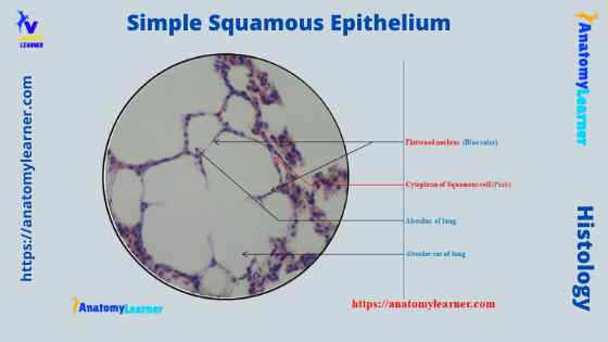
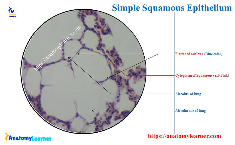
The lung’s alveoli give the honeycomb appearance in the parenchyma and lines by flattened simple squamous epithelium. These alveoli are thin-walled and fills with air.
From the lung parenchyma labeled diagram, you might identify the following structures –
- Simple squamous epithelium lining of the lung alveoli (within the parenchyma),
- A connective tissue basement membrane beneath the simple squamous epithelium lining,
- The lumen of the lung alveoli, and
- The cytoplasm of the simple squamous epithelium cells.
All these structures are identified in the labeled diagram from the lung tissue parenchyma. But, do all the components of the lung parenchyma line with the simple squamous epithelium? The answer is no; okay, let’s know the lining epithelium of other components or structures of lung parenchyma.
Lining epithelium finds in the lung parenchyma.
In the mucosa of the branches, you will find the pseudostratified epithelium with the same cellular composition as the trachea. The bronchioles represent the branches of the segmented bronchi. These bronchioles branch repeatedly and give rise to the smaller terminal bronchioles.
Again, the terminal bronchioles finally give rise to the respiratory bronchioles. You will find the simple columnar or simple cuboidal epithelium in the bronchiole. The respiratory bronchiole constitutes a transitional zone in the respiratory system that lines with the cuboidal epithelium.
Again, the initial segment of the respiratory bronchioles contains both ciliated cells and Clara cells. Here, you will find the scattered, thin-walled, out-pocketing alveoli that extend from the lumen of the respiratory bronchioles.
Another structure of the lung parenchyma (alveolar duct) is the elongated airways that line with the simple cuboidal epithelium. Again, the alveolar sacs are the spaces surrounded by the cluster of alveoli. That means the surrounding alveoli open into the alveolar sac. So, in this alveolar sac, you will also find the simple squamous epithelium.
There are three types of pneumocytes on the alveolar surface of the lung parenchyma. The type I alveolar cells (type I pneumocytes) lines the entire alveolar surface. These are the extremely thin-walled squamous cells that join with occluding junctions.
Simple squamous epithelium in kidney
The kidneys are large, reddish, and bean-shaped organ locates on either side of the spinal column in the retroperitoneal space of the caudal abdominal cavity. You will find a connective tissue capsule surrounding the surface of a kidney.
Deep into the capsule, the reddish-brown part of the kidney parenchyma is the cortex. Again, the much lighter color inner part is the medulla of the kidney.
The cortex of a kidney consists of renal corpuscles and the convoluted tubule, straight tubules, nephrons, connecting tubules, and collecting ducts. You will find the medullary ray in the medulla of the kidney that comprises straight tubules and collecting ducts.
You may learn the details of histological features of a kidney (cortex and medulla) with the microscopic slide images and the labeled diagram from another article by anatomy learner.
- Kidney histology and microscopic slide identification under the light microscope.
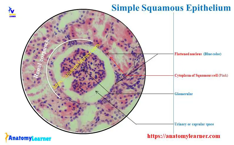
The renal corpuscle is a rounded structure that consists of a tuft of capillaries and forms a rounded glomerulus. The double-layer is covered in the glomerulus capsule – visceral and parietal layers. In the parietal layer of the glomerulus capsule (Bowman’s capsule), you will find the simple squamous epithelium under a microscope at 40x magnification.
Again, there is a simple squamous epithelium lining found in the thin loop of Henle with 40x magnification. You know the thin loop of Henle locates at the inner medulla of the kidney parenchyma.
Lining epithelium in other parts of a kidney
In the other parts of the kidney cortex, like proximal and distal tubules, you will find the lining of the simple cuboidal epithelium. But, how will you differentiate the proximal convoluted tubules from the distal convoluted tubules?
It is simple to differentiate the proximal convoluted tubules from the distal convoluted tubules under the light microscope. The proximal convoluted tubules are dark staining structures that line with the simple cuboidal epithelium that possesses the prominent brush border. Again, the lumen of the proximal convoluted tubules is indistinct under the light microscope.
In addition, the distal convoluted tubules are the lighter staining structure under the light microscope. These tubules are also lined with the simple cuboidal epithelium, but they don’t possess prominent brush borders. Again, you will find a distinct lumen in the distal convoluted tubules under the light microscope.
The kidney histology slide also shows the collecting ducts lining with the simple cuboidal epithelium. They stain lightly, and you will easily find the distinct lumen and cell boundaries under the light microscope.
Again, the thick segment of the loop of Henle lines with the simple cuboidal epithelium.
Simple squamous epithelium in the covering of heart, lung, and peritoneum
The heart is a muscular organ and pumps blood throughout the blood vessels to different body parts by repeated rhythmic contraction. You will find three layers in the heart wall where the pericardium covers the external layer.
Again, you will find a double-layer pleura that covers the lung of the animals. It is a thin and transparent serous membrane that comprises parietal and visceral parts. The visceral part invests the surface of the lung and continues with the parietal pleura at the root of the lung.
Again, the peritoneum is a serous membrane that lines the abdominal wall and is reflected over the viscera. You know the lining part is the parietal, and the reflected part is the visceral part of the membrane. Between the parietal (outer) and visceral parts of the peritoneum, you will find the peritoneal cavity that contains the fluid.
In all the membranes, as mentioned earlier, there is a simple squamous epithelium lining that may become visible with the help of a light microscope.
Covering the heart
So, you know the pericardium, fibroblasts, fluid-filled sac that holds the heart. This pericardium consists of an outer parietal layer that reflects onto the heart surface as a visceral layer. You will find the mesothelium lining (simple squamous epithelium) on these two parts of the pericardium that secrets thin clear serous fluid.
This serous and clear fluid lubricates the heart’s surface during the construction to reduce frictions. Again, the inner endocardium of the heart wall is similar to the tunica intima of the blood vessel. You will also find the simple squamous lining on the endocardium of a heart wall. This is the endothelium of the heart.
In addition, the myocardium substitutes for the tunica media of the blood vessel. Forming the bulk of the animal heart wall, it comprises mostly the cardiac muscles.
The pericardium labeled diagram shows the simple squamous lining epithelium (mesothelium).
Pleura of lung
So, the lung surface is covered by the double-layered pleura. The pleura of the lung (parietal) consists of a lining of the mesothelium (simple squamous epithelium) resting on the basement membrane (layer of connective tissue).
Here in the pleura labeled diagram (lung tissue), I tried to show you the simple squamous epithelium lining (mesothelium). I also showed you the basement membrane and cytoplasm of the mesothelium in the pleura labeled diagram.
The lining of the peritoneum
Most of the abdominal and pelvic organs are covered by the peritoneum. In the parietal part of the peritoneum, you will find the simple squamous lining under the light microscope.
Most of an animal’s digestive tract is covered by the peritoneum and shows a simple squamous epithelium lining. This is the mesothelium and locates the external surface of the thin and loose connective tissue layer (tunica serosa).
The peritoneum labeled diagram shows the simple squamous epithelium lining at the external surface of the particular organ.
You may learn the details of histological features of the three different layers of a tubular organ (where you will find the histological features of tunica serosa).
- Histological features of four different layers of a tubular organ.
Endothelium under light microscope
I hope you already know the term endothelium. When you find the simple squamous epithelium under a microscope in blood vessels or lymph vessels, it is known as the endothelium. In most cases, endothelium lines the inner surface (tunica intima) of the blood vessels, lymph vessels, and heart’s endocardium.
You may learn the details histological facts of tunica intima of a blood vessel from another article by anatomy learner –
- Basic histology of an artery (both muscular and elastic artery)
Another name for the endothelium cell is endotheliocyte. On the surface view, these endotheliocytes are polygonal and elongated along the vessel’s length. Again, these endotheliocytes possess a little cytoplasm.
Here the artery labeled diagram shows the tunica intima that consists of endothelium, basal lamina, subendothelium connective tissue, and internal elastic lamina.
You will find the endoplasmic reticulum and mitochondria in the cytoplasm of the endothelium cell under the electron microscope. Again, microfilaments and intermediate filaments also provide mechanical support to the endothelium cells.
There are tight junctions and gap junctions between the two endotheliocytes. Again, the external surface is supported by the basal lamina of the connective tissue.
Functions of endothelium
The endothelium not only lines the internal surface of the blood vessels, lymph vessels, and the inner surface of the heart but also performs several other functions. So, here I will provide some other functions of endotheliocytes –
- The endotheliocytes are sensitive to alterations in blood pressure, blood flow, and oxygen tension.
- These endotheliocytes secret various substances that can produce vasodilation by influencing the muscle tone in the wall of a blood vessel.
- They also produce the factor that controls the coagulation of the blood.
- The endotheliocytes can change the adverse condition to facilitate the passages of lymphocytes through the vessels wall.
- Again, in acute inflammation, the endotheliocytes allow neutrophils to pass from the blood into the surrounding tissue.
Simple squamous epithelium under microscope drawing
If you are a beginner in learning veterinary histology and started with the epithelium, you might have to learn how to draw this epithelium so quickly. I strongly suggest you follow my short and simple step-by-step guide to histology drawing.
Now, let’s learn how to draw the simple squamous epithelium that finds under a microscope. I will show you so simple method to draw this simple squamous epithelium.
First, you should draw the basement membrane of the simple squamous epithelium. This basement membrane is a thin, pliable, sheet-like structure of an extracellular matrix. The basement membrane provides support to the endothelium or epithelium and the tissue.
Now, you might draw the lamina propria, which consists of loose connective tissue cells, fibers, and other extracellular matrices. That’s nice, and you have drawn the basic portion of your drawing.
Now, provide the flattened cells (as shown on the diagram) on the basement membrane of the structure. So, you have drawn the flattened-shaped cell of the simple squamous epithelium.
Finally, you should provide the flattened or elongated nucleus in the central part of the cell. You may draw some of the same flattened cells and flattened nucleus in the same focus.
From your drawing sample, let’s identify the basement membrane, lamina propria, flattened cell, and flattened central nucleus.
Simple columnar epithelium under a microscope
The simple columnar epithelium under a microscope consists of a single layer of taller than wide cells. These simple columnar epithelia look closely packed and slender columns shaped. You will find the cell base on the basement membrane and the apex in contact with the lumen.
The nucleus of the simple columnar epithelium is elongated and located in the lower half of the cell. All nuclei (of these cells)are placed at the same level in neighboring cells.
In the longitudinal section of the tissue, this simple columnar epithelium is rectangular. Again, they are polygonal on the surface view (transverse section).
In addition, the simple columnar epithelium can be classified into two groups based on the surface –
- Simple columnar epithelium without cilia or microvilli, and
- Ciliated columnar epithelium
When the cell surface of a columnar epithelium does not possess any modification (cilia or microvilli), then it is the simple nonciliated columnar epithelium. Again, if you find the cilia on the cell surface, it is the ciliated simple columnar epithelium.
When you observe the simple columnar epithelium with the help of an electron microscope, you may find the microvilli–striated border or brush border. If the microvilli are regularly arranged on the cell surface, it is the striated border. Again, when the microvilli arrange irregularly on the cell surface, it is the brush border.
The lateral border of the cells has a junctional complex that includes the tight junction, intermediate junction, and desmosome. You will find the well-packed organelles in the cytoplasm of a simple columnar epithelium.
Location of simple columnar epithelium
You will find the simple columnar epithelium over the mucous membrane of the stomach and intestines. Again, you may find some striated borders in the mucosa of a small intestine. The ciliated columnar epithelium lines most of the respiratory tract, the uterus, and the uterine tube of female animals.
You may also find this simple columnar epithelium in the major ducts of glands, convoluted tubules of the kidney, small bronchi of the lung, and gallbladder. So, this epithelium is widely distributed in the animal’s body and helps to protect the wet surface. These simple columnar epithelium also possess the function of nutrient absorption and secretion.
Simple columnar epithelium labeled.
This is a labeled diagram of a simple columnar epithelium under a light microscope. I tried to show you both ciliated and nonciliated simple columnar epithelium.
These diagrams show the cilia on the cell surface, rectangular cell, and elongated nucleus. Again, another diagram shows the simple cell surface (no modification on the cell surface), elongated cells, and nucleus with the basement membrane.
On the other diagram, you will see the transverse section of the simple columnar epithelium. You may know the details of histological features of the simple columnar epithelium from another article by anatomy learner.
- Epithelial tissue histology slide with microscopic images and labeled diagrams.
Stratified squamous epithelium under a microscope
Under the light microscope, you will find two types of stratified squamous epithelium – keratinized and nonkeratinized. In the deep layer of stratified squamous epithelium, you will find the columnar epithelium that rests on the basement membrane. Again, there are polygonal cells present on the columnar layer of cells.
But, the superficial cell layer of simple squamous epithelium becomes flattened or squamous shaped. The ideal features of the nonkeratinized stratified squamous epithelium are –
- The surface of the squamous epithelium remains moist,
- The most superficial cells are living, and nuclei can see,
- No keratin substances on the cell surface.
This nonkeratinized stratified squamous epithelium lines most of the oral cavity, pharynx, epiglottis, vocal cord, esophagus, male and female urethra parts, and cornea. Secretion from the closely associated glands lubricates the surface of the nonkeratinized epithelium.
In the stratified squamous epithelium labeled diagram, I tried to show you the columnar cells, elongated nucleus, and basement membrane. You will find more epithelium-labeled diagrams on social media of anatomy learners.
Stratified squamous keratinized epithelium
When the epithelial surface becomes dry and the most superficial cell loses their nuclei, they form the keratin substances. This keratin forms the nono-living covering over the epithelium.
These areas that expose to the air and abrasion, like the skin’s epidermis, consisting of cells that lack nuclei and contain a plate of keratin protein. These cells of the epidermis are the stratified squamous epithelium.
You will find five layers of cells in the epidermis of an animal’s skin (thick and thin skin). If you want, you may learn the details of histological features of these cells from the different layers of the epidermis.
Here, in the epidermis of skin labeled diagram, I tried to show you the keratinization over the cell surface. Again, the diagram also shows the cells of the different layers of skin’s epidermis. You will also find the keratinized stratified squamous epithelium on the oral cavity of an animal.
Pseudostratified columnar epithelium under a microscope
This is not a true stratified epithelium but appears to be stratified. You know the nuclei of the columnar epithelium lie in a row toward the basal part of the cell. But, in the pseudostratified columnar epithelium, the nuclei appear to be arranged into two or more layers.
And thus, it gives the impression that the pseudostratified epithelium is more than one cell thick. The cells (simple) are attached to the basement membrane but are of different heights. Some of the cells are short and basal, while others are tall and columnar.
In some cases, you will find the hair-like projection (cilia) on the apical surface of the pseudostratified columnar epithelium. Again, mucous goblet cells may occur in the pseudostratified columnar epithelium.
In the upper respiratory tract, like nasal cavities, auditory tube, nasopharynx, larynx, trachea, and large bronchi, you will find the pseudostratified columnar epithelium. These pseudostratified columnar epithelia of the respiratory tract contain the cilia on their surface and some mucous goblet cells.
But, the pseudostratified columnar epithelium of the male reproductive tract lacks goblet cells.
Ultrastructure of simple squamous epithelium under an electron microscope
The ultrastructure of the simple squamous epithelium under the electron microscope reflects their functional diversity. You will find the riched, highly variable organelles in the cytoplasm of simple squamous cells. These organelles indicate high metabolic activities, active synthesis and secretion, and selective permeability.
A complex cytoskeleton is present in the cytoplasm of the simple squamous cells that includes the tonofilaments and microfilaments. The prominent actin containing thin microfilaments and motor protein allows changes in the cell shape and provides flexibility.
You will find the different junctions in between the simple squamous epithelium. The intermediate junction and desmosome anchor the simple squamous cells together. Again, the tight junction between the simple squamous acts as a permeability barrier to the indiscriminate passage of the materials. In addition, the gap junction between the simple squamous cells allows ionic and metabolic communication.
Frequently asked questions on simple squamous epithelium
Let’s discuss the commonly asked questions on the simple squamous epithelium. You may find your specific answers on the simple squamous epithelium in this article section.
What does simple squamous epithelium look like under a microscope?
If you want to examine the simple squamous epithelium histology slide with a normal light microscope, you may find the followings.
Suppose you are viewing the longitudinal section of the tissue, then you will find the flattened nucleus (deep blue color), a thin basement membrane, and pink colored cytoplasm. But, the lamina propira under the basement membrane is not visible under the light microscope.
Again, if you are viewing the cross-section of the sample tissue, you will see the polygonal shape with a serrated border. These polygonal-shaped cells join tightly and contain a deep blue color centrally placed rounded nucleus.
What does simple squamous epithelium look like?
In most animal body organs, you will see the longitudinal section of the simple squamous epithelium. So, you will see the simple squamous epithelium as a spindle-shaped (tapper on both ends) cell with no distinct boundary (plasma membrane).
You will find a spindle-shaped (fattened) nucleus (deep blue color) in the center of the simple squamous epithelium cell. Again, the thin basement membrane of the simple squamous epithelium may also see. The light microscope also reveals the light pink color cytoplasm of the simple squamous epithelium.
What is a simple squamous epithelium description?
If you want to describe the simple squamous epithelium, you might know the number of the cell layer, the shape of the cell, and the shape of the nucleus. You should also know the color of cytoplasm and nucleus of simple squamous epithelium with the routine stain.
I have described all the histological features of the simple squamous epithelium above. You will find a single layer of the spindle or flattened-shaped cell with a flattened nucleus.
What does stratified squamous epithelium look like under a microscope?
The cells of the stratified squamous epithelium are arranged in multiple layers. You will find the columnar basal cells and flattened superficial cells living. Again, this epithelium is present at the surface subjected to abrasion but protected from drying.
You will find the stratified squamous, nonkeratinized epithelium on the lining of the buccal cavity, oropharynx, laryngopharynx, and esophagus.
But, in the keratinized stratified squamous epithelium, you will find the tough keratin intermediate filaments that become firmly embedded in a matrix protein. This type of epithelium is present on surfaces subjected to drying or mechanical stresses.
You will find the stratified squamous keratinized epithelium on the epidermis, mucocutaneous junction of lips, nostrils, and hard palate.
Conclusion
I think you got the basic idea of the simple squamous epithelium under the light microscope. So, the simple squamous epithelium consists of a single layer of spindle-shaped (flattened) cells, a tapered or elongated nucleus that rests on a basement membrane.
All the labeled diagrams of simple squamous epithelium might help you learn them. The surface view (cross-section) of the simple squamous epithelium under the microscope is rare. But, you will find a most common longitudinal view of simple squamous epithelium in most organs.