The dog kidney anatomy consists of a capsule, hilus, and parenchyma. Here, the capsule is a thin fibrous covering at the outer surface of the canine kidney.
You will find the hilus in the medial border of each kidney of the dog. The hilus of the dog’s kidney comprises the renal vein, artery, and ureter.
Again, the kidney parenchyma or substance consists of the pale outer cortex and inner darker medulla, where you will find different essential features. In this article, you will learn these vital features from the dog kidney anatomy with the labeled diagram.
I will also show you the primary difference between the ruminant and canine kidneys at the end of this article. Again, this article has little information on the different problems (like swollen kidneys and urolithiasis) of dogs’ kidneys.
So, if you wish to learn anatomical facts about dog kidney and their common problems, let’s continue this article until the end.
Dog kidney anatomy
The dog kidneys are the paired bean-shaped organ located below the bodies of lumbar vertebrae. They are called the retroperitoneal organs of the abdominal cavity as they are located above the peritoneum.
The surface of the dog kidneys is smooth, whereas you will find the lobulation on the external surface of the ruminant kidney. From the dog kidney anatomy, you might learn the followings –
- Exact location (both surface and topographic) of both right and left kidneys,
- Anatomy of the capsule that covers both kidneys,
- The surfaces, borders, and poles of the dog’s kidneys,
- A hilus or the renal sinus that contains the renal artery, veins, and ureter,
- Kidney parenchyma (including the cortex, medulla, renal pelvis, and others),
- A structure of the nephron (unit of kidney parenchyma), and
- Blood supply and nervous innervation of the dog’s kidney,
I will discuss everything that I have enlisted above from the dog kidney. But, first, I would like to show you the essential anatomical features from both external and internal parts of the dog’s kidney with the labeled diagram you should identify at your laboratory (practical class).
Dog kidney features identification
The right and left kidneys of the dog possess 2 surfaces, 2 borders, and 2 poles or extremities. So, you might identify the followings from the external part of the dog’s kidney –
- Surfaces – both convex dorsal and ventral surfaces,
- Borders – convex lateral border, concave (slightly) medial border, and
- Poles or extremities – cranial and caudal (both convex),
You might also identify the thin fibrous capsule that covers the smooth surface of the dog’s kidney. The dorsal surface of the dog’s kidney is less convex than the ventral surface.
Here, the cranial and caudal extremities of the dog kidney join by the convex lateral and slightly concave medial border. At the medial border of the dog’s kidney, you will see the hilus (the renal sinus).
From the renal sinus or hilus (space), you might identify the following structures –
- A muscular tubular ureter,
- The thick-walled renal artery (small lumen),
- A thin-walled (larger lumen) renal vein, and
- The lymph vessels (not identified in the diagram) and nerves,
In the dog, a renal vein is paired on one or both kidneys. But, the renal artery is generally single in the structure of the dog kidney.
Here, the renal artery is most dorsally located, whereas the renal vein locates most ventrally. Again, the lymphatic vessels and nerves closely lie in the renal vein of the dog’s kidney.
You will now identify these external features from the actual dog kidney sample at your laboratory. Now, it’s time to identify the different essential features of the internal part of the dog’s kidney structure (parenchyma).
Internal features identification from dog kidney
If you want to see the internal gross anatomical features of the dog kidney, you might perform a longitudinal section, as I show in the diagram. From this longitudinal section of the canine kidney, you may identify the below-mentioned features –
- An outer pale cortex of the dog’s kidney,
- The darker inner medulla of the dog’s kidney,
- The renal pyramid in the kidney parenchyma,
- Interlobar arteries and veins in the kidney parenchyma,
- Renal pelvis and fat in this area, and
- The renal crest of the kidney (not present in the ruminant’s kidney),
Let’s continue this article to learn more about the anatomical features of the dog’s kidney parenchyma. You will know where the renal corpuscles and convoluted tubules are located in the dog’s kidney. Again, the renal pelvis and sinus formation describe the specific part of the kidney’s internal anatomy.
Unique features of the dog kidney
In comparison to the large ruminant (ox), the dog’s kidney possesses some unique features. Here, I will enlist some of these unique anatomical features of the dog kidney.
So that you will get a better idea of the dog’s kidney structure as a whole within a few minutes. Let’s see what these unique features in the dog’s kidneys are –
- The right and left kidneys of a dog are bean-shaped,
- The external surface of the dog’s kidney is smooth and covered with the fibrous capsule,
- You will see a significant variation in the location of the right and left kidneys of the dog. Here, the dog’s right kidney locates below the bodies of the first 3 lumbar vertebrae,
- And the left kidney of the dog locates below the bodies of the third to fifth lumbar vertebrae,
- The hilus locates at the middle of the medial border in both the right and left kidneys of the dogs,
- The renal pyramid of the dog’s kidney is not so prominent as the large ruminant (ox),
- You will find the renal sinus and renal crest in the internal structure of the dog kidney,
- There is a dilated renal pelvis from where the ureter arises,
So, this is the summary of the dog’s kidney anatomy. Now, you may learn the details of the anatomical features of each structure of the dog kidney.
Let’s start with the location (surface and topographic) and fixation of the dog’s kidney in the abdominal cavity.
Dog kidney anatomy location
The dog kidney locates in the sublumbar region (abdominal cavity). But, you will find a slight variation in the right and left kidney positions of the dogs.
Here, the right kidney of the dog extends from the last ribs to the third lumbar vertebra. In comparison, the left kidney locates just below the right one and extends from the third lumbar vertebra to the fifth lumbar vertebra.
Both the right and left kidneys of the dog locates below the bodies of these respective lumbar vertebrae. So, you may express the exact location of the dog kidney anatomy as follow –
- Right kidney – below the last ribs to the bodies of the first 2 or 3 lumbar vertebrae, and
- Left kidney – below the bodies of third to fifth lumbar vertebrae,
You know both the kidneys of a dog are retroperitoneal, and the dorsal surface has contact with the lumbar muscle. The dorsal surface of both the right and left kidney surround by fat.
Now, the ventral surface of both kidneys has contact with the transparent parietal peritoneum. You may also find a little covering on the cranial pole of each kidney by the peritoneum.
Both the kidneys of a dog have a cranioventral oblique direction. Again, you will see the fibrous capsule that surrounds both kidneys of the dog.
Shape and size of the canine kidney
The dog or canine kidneys are the smooth bean-shaped, reddish-brown structure in their abdomen. But, the size and weight of the dog’s kidneys may vary with the breed.
The average measurement of the kidneys of a dog is as follows –
| Parameter | Right Kidney of Dog | Left kidney of Dog |
| Length | 9 cm | 6 cm |
| Width | 5 cm | 4 cm |
| Thickness | 4 cm | 3 cm |
| Weight | 35 gm | 27 gm |
The measurement (data) that showed in table 1 are approximated. So, it may also vary in different breeds of dogs, even between male and female dogs and young and older dogs.
(Here, cm = centimeter, and gm = gram).
How is the dog’s kidney fixed in its position?
Different factors and structures keeping the dog’s kidneys in their position –
- Renal vessels and ureter that attaches to the hilus of the dog’s kidney,
- A thin fibrous capsule with the renal fat,
- Distribution of the renal fascia, and
- Pressure performed by the different surrounding organs,
Here, the renal fascia of the dog’s kidney is formed by the condensation of the extra peritoneal connective tissue. These tissues surround the dog’s kidney and continue laterally with the transversalis fascia.
As there is an excellent role of the pressure of the surrounding kidney organs to hold in the position, you may learn more about these organs from the next section of the article.
What are the organs that are related to a dog’s kidney?
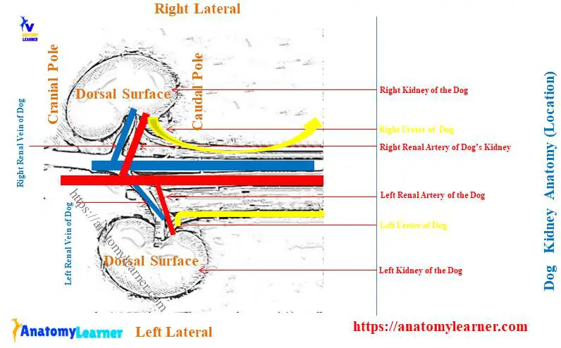
Let’s see the dog kidney labeled diagram (located in the abdominal cavity); each kidney lies lateral to the aorta and caudal vena cava. Again, the right kidney of the dog lies more cranially than the left one and has contact with the liver.
The right kidney of the dog is more firmly attached to the dorsal wall than the left. Organs related to the right and left kidneys of the dog are also somewhat different.
So, I will discuss the organs that have a great relationship with the dog’s right and left kidneys separately. But, overall, you will find the following surrounding organs that have a great relationship with the right kidney of the dogs –
- Caudate lobe of the liver,
- Aorta and caudal vena cava,
- Diaphragm and retractor costae muscle,
- Right adrenal gland of the dog,
- The right lobe of the pancreas and ascending colon,
Again, you will find the following organs that also have a great relationship with the dog’s left kidney –
- Medial surface of the spleen,
- Greater omentum and greater curvature of the stomach,
- Left lobe of the pancreas and adrenal gland,
- Quadratus lumborum, transverse abdominis, and psoas muscles,
- Descending colon and mesovarium,
- Ascending duodenum,
Okay, now, let’s discuss how these organs surround both the right and left kidneys of the dog.
Dog right kidney anatomy and relation
The cranial extremity of the dog’s right kidney embeds in the fossa of the caudate process of the caudate lobe of the liver. You know this cranial extremity of the dog’s right kidney lies below the thirteenth rib.
But, this position of the dog’s right kidney may also change depending on the gastric distension. You may also find contact with the diaphragm and costate retractor muscle with the cranial pole of the right kidney.
There is a right adrenal gland in front of the right kidney of the dog. So, it is in contact with the cranial pole of the dog’s kidney.
Dorsally, the dog’s right kidney has contact with the lumbar muscle and bodies of the lumbar vertebrae. Ventrally, this kidney has contact with the right lobe of the pancreas and descending colon.
Again, the dog’s right kidney is medially close to the caudal vena cava.
Topography of dog’s left kidney
You know the dog’s left kidney extends from the third to fifth lumbar vertebrae (below their bodies). In the anatomy of a dog left kidney, you will also find similar features – 2 poles, 2 surfaces, and 2 borders.
Here, the cranial pole of the dog’s left kidney has contact with the dorsal end of the medial surface of the spleen. You will also find the greater omentum and greater curvature of the stomach which have a close relationship with the cranial pole of the dog’s left kidney.
Now, let’s see what organs surround the dog’s left kidney dorsally, ventrally, cranially, and caudally.
Dorsally, the dog’s left kidney attaches to the quadratus lumborum, psoas, and transverse abdominal muscles. You will also find the attachment of the deep layer of the thoracolumbar fascia with the dorsal surface of the dog’s left kidney.
Cranially, the dog’s left kidney has contact with the left lobe of the pancreas. You know there is a left adrenal gland just in front of the left kidney.
Caudally, you may find a little variation in the organs that significantly relate to the male and female dog’s left kidneys. In the male dog’s left kidney, the renal peritoneum reflects onto the dorsal body wall as the parietal peritoneum.
Again, in the female dog’s left kidney, you will see the contact with the descending colon and mesovarium at its caudal aspect. Again, the peritoneum on the ventral surface of the left kidney blends with the peritoneum suspending the ovary.
The medial aspect of the dog’s left kidney also relates to the left adrenal gland, descending colon, mesocolon, and ascending duodenum. You will also find a close relationship between the descending colon with the ventral surface of the dog’s left kidney.
A capsule of dog kidney
You know the outer fibrous covering of the dog kidney is the capsule. It is the thin fibrous structure that follows the hilus to line the wall of the sinus. Thus it forms the adventitia of the dog’s renal pelvis.
This thin fibrous structure also invests the renal vessels (renal artery and vein) and nerves before they pass into the renal sinus. You may easily remove the capsule of the dog’s kidney from the parenchyma.
But, the capsule that adhered to the blood vessels and nerves of the renal pelvic or sinus is very hard to separate. You may also see the fat on the fibrous capsule that primarily attaches to the external surface.
Again, this fat may extend into the dog’s kidney renal sinus through the renal hilus. Okay, now, let’s discuss the details anatomical facts of a cavity of the dog’s kidney (renal sinus and renal pelvis).
The cavity of the canine kidney anatomy
You will see a distinct cavity on the medial aspect of both the right and left kidneys of the dogs. This is the renal hilus and sometimes considers a cavity of the kidney.
A space marked by the wall of the renal hilus of the kidney is known as the renal sinus. Now, in the renal sinus of the dog’s kidney, you will find the followings –
- A renal pelvis – funnel-shaped structure,
- A variable amount of the fat,
- Branches of the renal artery, vein, and lymphatics, and
- A ureter that is the continuation of the renal pelvis,
After passing the renal sinus of the dog kidney, the nerves, veins, and arteries enter into the parenchyma. Let’s know a little about the different structures of the renal cavity of dogs.
The renal pelvis of the dog kidney
You know the renal pelvis is the funnel-shaped structure designed for receiving urine from the papillary ducts of the kidney. Finally, the collected urine passes into the ureter through the renal pelvis of the dog.
The renal pelvis of a dog is elongated in a craniocaudal direction. It curves to conform with the lateral border of the dog’s kidney.
Now, the renal pelvis of a dog kidney extends into the renal parenchyma both dorsally and ventrally. These are the renal recesses or curved diverticula of the renal pelvis of dogs.
The number of these curved diverticula or recesses may vary in the kidney of different dog breeds. But, typically, you may find 5 – 6 curved diverticula in each renal pelvis of a dog.
Renal artery of the dog kidney
You know the kidney is the highly vascular organ whose main arterial supply comes from the renal artery. Again, you know the dog’s renal artery is the branch of the abdominal aorta.
If you see the diagram, you will find the variation in the length of the renal artery for the right and left kidneys. The length of the dog’s left renal artery is larger than the right one. But, both the right and left renal arteries of a dog separate from the lateral surface of the main abdominal aorta at the various level or asymmetrically (at the second and third lumbar vertebra).
You may also find a little variation in the diameter of the renal artery in both the right and left kidneys. Let’s see the length and diameter of the right and left renal arteries from below (Table 2) –
| Parameter | Right Renal Kidney | Left Renal Kidney |
| Length | 5 cm | 3 cm |
| Diameter | 4 cm | 3 cm |
From table 2, you may also easily understand the length and diameter of the right renal artery are more compared to the left one. But, what are the reasons behind being more lengthy for the right renal artery in dogs?
Well, you know the caudal vena cava lies to the right of the aorta, and thus, you find the greater length of the right renal artery.
Each renal artery of the dog kidney provides 2 or 3 branches to the caudal pole of the adrenal gland and a small cranial branch to the ureter. But, you may find some differences in the origin of the adrenal and ureteral arteries. They may also arise directly from the main abdominal aorta.
You may also know the different branches of the animal’s abdominal aorta (dog blood vessels) from the below-mentioned article –
Dog blood vessels anatomy with the labeled diagram,
Branches of renal artery into the dog’s kidney
Each renal artery of the dog (right and left) divides into dorsal and ventral branches. You will find a great variation in the branches of the renal artery.
The 2 primary branches of the renal artery (dorsal and ventral) may divide into the 2 – 4 interlobar arteries. Now, these interlobar arteries of the dog’s kidney branch into the arcuate arteries at the corticomedullary junction of the kidney’s parenchyma.
Again, the arcuate arteries of the dog’s kidney run towards the periphery of the renal cortex. They finally divide into numerous interlobular arteries.
You may also find the afferent and efferent arterioles in the structure of the dog’s kidney parenchyma. The afferent arteriole supply to the glomeruli of the dog’s kidney.
Now, let’s know how the blood passes from the abdominal aorta to the kidney parenchyma.
Blood enters the renal artery from the dog’s abdominal artery and goes to the different parts of the parenchyma. Here, the blood passes through the interlobar arteries, arcuate arteries, interlobular arteries, and finally goes to the glomeruli through the afferent arteries.
Now, the efferent arteriole exists from the glomeruli and courses directly into the outer layer of the medulla. It will give the long capillary network that extends to the apical pole of the renal pyramid. Again, they may also provide branches directly into the intertubular capillary networks.
Veins and lymphatics of dog kidney anatomy
The main vein that drains blood from the dog kidney anatomy is a renal vein. This vein collects blood from different parts of the kidney and finally joins with the caudal vena cava.
Here, the dog’s right renal vein is comparatively longer than that of the left one. But, the diameter of the right and left renal veins is almost similar in the dog.
The diameter and length of the dog’s right and left renal veins are shown below –
| Parameter | Right renal vein | Left renal vein |
| Length | 3 cm | 4 cm |
| Diameter | 8 mm | 8 mm |
(Here, mm = millimeter).
How do the veins collect blood from the dog’s kidney?
Well, first, the numerous stellate veins collect blood from the fibrous capsule of the dog’s kidney. These veins connect with the veins of the adipose capsule and empty into the interlobular veins.
Again, the blood from the interlobular veins drains into the arcuate and interlobar veins. Finally, the blood drains into the renal veins from the interlobar veins of the dog kidney structure.
But, you may find some other small branches of veins in the kidney parenchyma that makes the venous system so complicated.
You will also see the capsular and parenchymal lymphatics in the dog’s kidney structure. These lymphatics connect with the interlobar plexus that passes into the trunk from the kidney to the hilus.
Ureter at the dog’s kidney hilus
You know the dog’s ureter collects urine from the kidney and opens directly on the dorsolateral aspect of the urinary bladder. The length and diameter of both the right and left ureter of the dogs may vary within different dog breeds.
Here, the average diameter of the dog’s ureter varies from 0.5 – 0.7 cm in the distended condition. Again, the length of the dog’s ureter measures 10 – 14 cm (average).
But, the right ureter of the dog is slightly longer compared to the left one. This is because of the cranial location of the right kidney in the dog’s abdominal cavity.
Each ureter of the dog possesses the abdominal and pelvic parts. Here, the abdominal part of the dog’s ureter starts at the renal pelvis and receives urine from the renal crest.
This abdominal part of the dog’s ureter runs towards the urinary bladder. It lies dorsal to the testicular artery (in a male) and ovarian artery and vein (in a female dog).
Now, the pelvic part of the dog’s ureter enters between two layers of the peritoneum. It helps to form the lateral ligament of the urinary bladder and reaches the dorsolateral surface of the urinary bladder, just cranial to its neck (in the case of males).
But in female dogs, the pelvic part of the ureter reaches the lateral ligament of the urinary bladder after being associated with the broad ligament of the uterus. Now, the ureter opens into the bladder obliquely as a slit-like orifice.
Courses of dog ureter
The course of the right and left are almost similar (with a little difference) in the dog. Here, the right ureter of the dog lies close to the caudal vena cava and lateral to the abdominal aorta.
It passes through the dog’s deep circumflex iliac artery and external iliac artery. In the male dog, you will find a close relationship of the ureter with the ductus deferens (crosses each other).
The ureter of a horse does not possess any unique features compared to the dog or other animal species. Here, the renal pelvis is more dilated in horses than in dogs. This tube is narrow but more extended for the horse compared to the dogs.
Again, the ureter of a pig is slightly flexuous, and the cranial part is comparatively dilated. The rabbit ureter is simple in its structure and opens the dorsal aspect of the urinary bladder.
The ureteral artery that derives from the renal or the abdominal aorta (sometimes) is the major vessel to the dog’s ureter. You will find cranial and caudal ureteral arteries that anastomoses on the dog’s ureter.
The celiac and pelvic plexuses (autonomic nerve) innervate to the ureter of the dogs.
What is renal plexus in a dog?
It is a nerve plexus that surrounds the renal arteries, where they enter into the renal sinus. In this renal plexus of a dog, you will find the followings –
- The postganglionic sympathetic axons with their cell bodies, and
- Preganglionic parasympathetic axons from the vagus nerve,
This renal plexus in the dog’s kidney innervate the nephron, blood vessels, muscles of the renal pelvis, and other different areas of the kidney’s parenchyma.
The parenchyma of dog kidney anatomy
The parenchyma of the dog kidney consists of the pale outer cortex and internal darker medulla. In between the cortex and medulla, you will find the corticomedullary junction.
The peripheral part of the dog’s kidney’s cortex shows numerous renal corpuscles (which appear as granules) and convoluted tubules. So, these are the nephron in the parenchyma of the dog’s kidney.
You will also find the numerous arcuate arteries and veins in the parenchyma of the kidney. The thickness of the dog’s renal cortex is almost the same as the thickness of the renal medulla.
There are different longitudinal straight structures in the dog’s kidney parenchyma. The free ends of these structures face toward the renal pelvis.
All the structures project towards the pelvis and form a concave ridge on the central part of the medulla. This is the renal crest of the dog kidney.
You may also see the cone-shaped renal papillae on either side of the renal crest (in the longitudinal section) with the interlobar arteries and veins. These renal papillae are considered the apical part of the renal pyramid.
So, the base of the renal pyramid will find at the level of the renal cortex. The renal pyramid is believed to fuse from the renal crest in the dog and horse kidneys.
So, you may tell the renal pyramid extends from the cortex on the dorsal and ventral surfaces of the dog’s kidney into the center, where they fuse into the renal crest.
Again, the renal papillae open at the renal crest, which means you will find the perforation at the crest, known as the area cribrosa.
The extensions of the renal pelvis surround the renal papillae of the pyramid. These are the renal recesses in the dog kidney structure.
Dog nephron structure
The structural and functional unit of the dog’s kidney is the nephron. This structure is a continuous contorted tube that serves for urine production. It also regulates the volume and composition of the extracellular fluid.
You will quickly understand the structure of the dog’s nephron from the below-mentioned article –
- Histology of the kidney tubules (nephron and tubules) with the labeled diagram,
It is impossible to view the gross structure of the dog’s nephron from the kidney parenchyma. So, you might go through that article I mentioned earlier.
How many nephrons does a dog’s kidney have?
It varies in a different dog breeds, even in different aged dogs. But, you may find approximately 440000 – 500000 nephrons in the dog kidney.
Parts of dog nephron
The dog’s nephron posses the following segments –
- A spherical expanded double-layered Bowman’s capsule (glomerular capsule),
- Proximal convoluted tubules of the dog’s nephron,
- Loop of Henle, and
- The distal convoluted tubules of the dog’s nephron,
These tubule segments are named according to their shape, size, orientation, and functions. Let’s discuss the structures of these different segments from the dog’s nephron.
You will see the double-layered Bowman’s capsule or glomerular capsule at the beginning of each nephron. It receives the glomerular tuft of capillaries derived from the afferent artery.
Thus it forms the renal corpuscles that present only in the cortex of the dog’s kidney. Typically, you will not find any renal corpuscle in the medulla of the dog’s kidney structure.
Here, the visceral layer of the glomerular capsule is closely invested in the glomerular capillaries. In comparison, the parietal layer of the glomerular capsule consists of simple squamous epithelium that continues with the proximal convoluted tubule of the nephron.
Kidney medulla and convoluted tubules
From the glomerular capsule to the collecting tubules and papillary ducts in the kidney medulla, you will find the following structures –
- Proximal convoluted tubules,
- Proximal straight tubules,
- Thin tubule that forms a loop,
- Distal straight tubule, and
- Distal convoluted tubules,
Here, the proximal convoluted tubule of the dog’s nephron is the longest and more convoluted. You will find a single layer of low columnar or pyramidal cells in their wall. Again, the length of these proximal convoluted tubules in the dog’s kidney may vary.
A thin tubule that forms a loop is a thin and straight part of the dog’s nephron. This tubule connects the proximal and distal convoluted tubules. You will also find different segments in the thin tubule (loop of Henle) – descending part, U-turn part, and ascending part.
But, the distal convoluted tubules of the dog’s nephron are less convoluted than the proximal one. You will find a single layer of cuboidal cells in the wall of the distal convoluted tubules. Now, the distal convoluted tubule of the dog’s nephron continues with the collecting tubules.
Finally, the collecting duct joins to form the duct of Bellini or papillary duct that opens at the papilla of the renal pyramid.
Kidney of horse, ox anatomy compares to a dog
The kidneys of horses, ox, pigs, and rabbits possess somewhat little difference in their external and internal features compared to the dogs. But the basic structure and function of the kidneys of these species are almost similar.
You will find a significant difference in the large ruminant’s (ox) kidneys compare to the dog kidney anatomy. The external surface of an ox kidney shows lobulation, whereas the external surface of the dog kidney is smooth.
Again, in the internal structure of an ox kidney, you will find major and minor calyx, but no such structure is evident in the dog kidney.
The internal structures of the horse kidney are almost similar to those of dogs. But, the shape of the horse kidney is identifiable. Here, the right kidney of the horse is a heart shape, whereas the left one is an elongated shape.
Let’s see some of the important anatomical features from the horse kidney anatomy –
- The external surface of both the right and left kidneys of the horse is smooth like the dogs,
- You will find the heart-shaped appearance in the right kidney of the horse, and the left one is elongated or almost bean-shaped,
- The right kidney of the horse locates below the last 3 ribs, and the first lumbar transverse process,
- Again, the left kidney of the horse locates near the median plane below the proximal end of the last ribs and the first 2 – 3 lumbar transverse processes,
- You will not find any distinct renal pyramid in the horse kidney structure,
- The hilus of each kidney locates in middle of the medial border, which leads renal sinus,
- You will also find the distinct renal crest and renal pelvis in the structure of the horse kidney,
Now, let’s see the unique features of ox kidneys.
Ruminant (ox, cow) kidney anatomy
You will find the following unique features in the ruminant’s kidney compared to the dogs –
- Both the right and left kidneys are bean-shaped, and surfaces are smooth,
- The right kidney of the ox locates below the proximal part of the last rib and first 2 – 3 lumbar transverse process,
- Again, the location of the ox or cow’s left kidney is variable due to the condition of the rumen (part of the stomach),
- The left kidney of the ox locates below and behind the right kidney at the level of the bodies of the third to fifth lumbar vertebrae (when the rumen is empty),
- When rumen is full, the left kidney may also remain in the same location but partly at the left slide of the median plane,
- The renal pelvis of the ox kidney divides into 2 major calyces, which further divide into several minor calyces,
- You will not find any renal crest in the structure of the ox or cow kidney,
There is very little information about the ox or cow kidney. But, some small ruminants like sheep and goats possess smooth surfaces in their kidney. Again, these species possess similar features, like the internal structure of the dog’s kidney.
If you want to learn the anatomical facts of the ox kidney, you may find the information here.
Pig kidney anatomy
In the pig kidney anatomy, you will find the below-mentioned features compared to the dog’s kidney –
- Both the right and left kidneys of a pig are bean shape,
- The surfaces of both kidneys are smooth, but sometimes, you may find a little lobulation on their surface,
- Both the kidney of a pig locates below the last 4 lumbar vertebrae (transverse process),
- The internal structure (kidney parenchyma) of the pig kidney is almost similar to the ox,
Rabbit kidney anatomy
You may also compare the external and internal features of the rabbit kidney with those of dogs. Here, you will see the following important anatomical facts about the rabbit kidney anatomy compared to the dogs –
- Both the right and left kidneys of a rabbit possess the smooth and almost bean shape,
- The internal structure of the rabbit’s kidney anatomy is almost similar to the goat or dog,
- Here, the rabbit’s right kidney locates below the proximal end of the last rib and the first 2 lumbar transverse processes,
- Again, the left kidney of the rabbit locates below the bodies of the second and third lumbar vertebrae,
- You will not find any distinct renal pyramid in the structure of the rabbit kidney,
You may also learn the details anatomical facts of these kidneys from the ox, goat, horse, pig, and rabbit from the gross anatomy learning section of anatomy learner.
Dog or canine kidney anatomy diagram
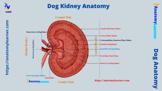
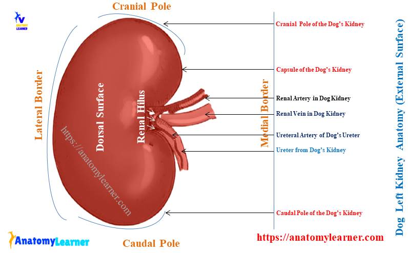
Now, in this part of the article, you will get all the labeled diagrams on the dog kidney anatomy. Let’s see both the external and internal features of the dog kidney from the below-mentioned labeled diagrams.
Here, in the first diagram, I tried to show you the shape of the dog’s kidneys (both right and left). You will also see the different borders, surfaces, and poles I identified in the labeled diagram.
Again, from the middle of the medial border of the dog’s kidney labeled diagram, I tried to show you the significant structures of the hilus. Here, I try to identify the ureter, renal artery, renal veins, and ureteral artery from the dog kidney hilus labeled diagram.
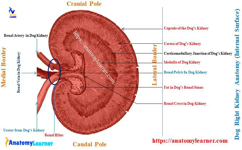
Now, let’s see the internal structure of the dog kidney with the labeled diagram. From the external surface of the dog’s kidney, I identify the fibrous capsule. I also tried to show you the peripheral pale cortex and inner darker medulla from the dog’s kidney in the labeled diagram.
Again, this labeled diagram shows the corticomedullary junction between the cortex and medulla of the dog’s kidney. Now, let’s see the internal anatomical features (features from the parenchyma) of the dog’s kidney.
Here, the diagram shows different papillae in the parenchyma of the dog’s kidney. Again, the parenchyma of the dog’s kidney shows pelvis recess, indistinct renal pyramid, interlobar artery and vein, and arcuate artery.
I also tried to identify the renal crest and renal pelvis from the dog kidney labeled diagram. You will also see some of the renal fat in the renal sinus area of the dog kidney diagram.
Finally, the formation of the renal recess and pelvis are shown in the dog kidney diagram on the social media of anatomy learner.
Dogs with kidney problems
You may commonly find 2 types of problems in the dog’s kidney – anomalies and chronic kidney problems. Chronic kidney problems in dogs are very harmful and require good veterinary care. You should call your veterinarian if your dog has chronic kidney problems.
Now, let’s see the common anomalies of the dog kidneys –
- Malformation of the kidney, which is more common in the dog,
- Congenital renal cysts in the dog’s kidney parenchyma,
- Polycystic kidney conditions in the dog are also most common,
- Hypoplasia and aplasia of the dog’s kidney, and
- Persists of fetal lobulation in the dog’s kidney,
These are the common abnormalities (specially malformation, renal cyst, polycyst, hypoplasia, and aplasia) in the dog’s kidney. But, sometimes, you may find fetal lobulation in the older dog’s kidneys.
Swollen kidneys in dogs symptoms
There is a wide range of symptoms of swollen kidneys in dogs. But, let’s try to find out the unique symptoms in the swollen kidneys of the dog –
- Increased body temperature in the dog,
- Decrease appetite and weight loss,
- Lethargy and vomition,
- Back and belly pain in the dogs (arch back condition),
- Increased thirst and urination in the affected dog,
- Blood in the dog’s urine and weakness,
- Pale gum and ulceration,
- Dog lose the balance of the body, and
- Other different common symptoms,
So, the dogs show a wide range of clinical symptoms in the swollen kidney condition. In this condition, you need to call an expert veterinarian for better care and advice for your pet dog.
Can you feel swollen kidneys in a dog?
Yes, you can feel the swollen kidneys in a dog so easily. To palpate the swollen left kidney is easier than the right kidney of the dog.
You know the left kidney of a dog lies below the body of the third to fifth lumbar vertebrae. It lies left of the median plane and peripheral part of the bodies of the lumbar vertebrae. In contrast, the right kidney lies below the last ribs and bodies of the lumbar vertebrae.
As I discussed before in this article, you also have seen the left renal artery is longer than the right one. This also confirms that the left kidney of a dog lies away from the midline of the lumbar vertebrae bodies.
On the other hand, the renal artery for the right kidney and the short and cranial part of this kidney is locked with the renal impression of the dog’s liver. This condition also confirms that the right kidney locates at the ventral to the midline of the lumbar vertebrae bodies.
So, it is convenient to palpate the left swollen kidney in the dog compared to the right one.
Common inquiries in the dog kidney anatomy
Now, I will enlist the common questions that the dog anatomy learner asks. I will try to provide concise answers to these questions on the dog kidney anatomy.
But make sure you know the basic structure of the dog’s kidney. You may read this article from beginning to end to get a basic idea of the structure of the dog’s kidney.
Let’s see the common inquiries on the dog kidney structure that the learners commonly ask.
Where are kidneys located on a dog?
The dog’s kidneys are located in an oblique direction in the abdominal cavity below the sublumbar muscle. Here, the right kidney locates more cranial to the left one and lies below the last ribs and bodies of the first three lumbar vertebrae.
This right kidney of the dog firmly attaches to the dorsal wall of the left kidney. Now, the left kidney of the dog lies just caudal and below the right one and extends below the body of the third to fifth lumbar vertebrae.
How do you differentiate the dog’s kidney from the cow’s?
You may easily differentiate the dog kidney from the cow by their both external and internal features. If you compare the external and internal features of the dog and cow’s kidneys, you will find the followings –
| Parameter | Dog kidney | Cow or ox kidney |
| Surface | Smooth | Lobulated |
| Hilus | Middle of medial border | Ventromedial for right kidney, Dorsolateral for left kidney |
| Shape | Bean shaped | Elongated bean shaped |
| Renal pyramid | Indistinct | Distinct |
| Renal crest | Present | Absent |
| Renal pelvis | Less dilated | More dilated |
| Major and minor calices | Absent | Present |
So, table 4 shows the primary difference between the dog and cow kidneys. With the help of these external and internal anatomical features, you may easily differentiate the dog kidney from the cow or ox.
The primary difference finds in the external surface and the parenchyma of the kidney (presence of the calyces in the ox kidney).
How do the dog’s kidneys keep in their position?
Different factors help keep the dog’s kidneys in their position in the abdominal cavity. Let’s see what the factors that keep the dog’s right and left kidneys in their position are –
Pedicles of the renal hilus, which contain the renal artery, vein, and ureter, help to keep the dog’s kidneys in their position. Again, the distribution of the renal fat and the deposition of the renal fascia also help them to keep their position.
The attachment of the peritoneum at the ventral surface of the dog’s kidneys and the fascia transversalis also help the dog’s kidney to remain in its position. Finally, the pressure created by the surrounding organs of the kidney plays an essential role in keeping them in their position.
Suggested reading for you from the dog anatomy organs –
- Dog heart anatomy with the labeled diagram,
- Anatomy of the dog liver – lobes, surfaces, and ligaments with the labeled diagram,
- Dog stomach anatomy with the labeled diagram,
You will also find more articles on dog organ anatomy here.
Conclusion
From this article, you have got detailed facts about the dog kidney anatomy with the labeled diagram. Both the right and left kidneys of a dog possess similar external and internal features. Only the difference is found in their location and relationship with the surrounding organs.
The external smooth surface and the location of the hilus (middle of the medial border of the kidney) are essential features to differentiate the dog kidney anatomy from an ox. Again, the presence of the renal crest and dilated renal pelvis are unique anatomical features of the dog’s right and left kidneys.