
The cow muscle anatomy includes the study of their origin, insertion, fiber direction, and action. It is essential for veterinary students for further leaning of bovine anatomy.
Here, I will help you to identify all the essential superficial muscles from the various regions of a cow with a labeled diagram.
Quick overview: there are several muscles in the body of a cow, but I will identify the most used and useful muscles from the head, neck, thorax, abdomen, and limbs. Muscles from the neck and limbs are most important for field practices.
You will find the list of muscles from different regions of the bovine body, like the head, neck, thorax, abdomen, and limbs. All these muscles from the cow will be shown in the labeled diagram and the actual samples.
So, if you want to identify the essential muscles from the cow (also from sheep and goats), let’s continue the article till the end.
Cow muscle anatomy
Muscles are the contractile organs that are responsible for the movement of the cow’s body. You will find two major types of muscles in the cow muscle anatomy – striated and nonstriated.
Here, the striated muscles of a cow include skeletal and cardiac muscle, whereas the nonstriated muscles include smooth muscle.
Before going to identify the cow muscles, you might know the below-mentioned terms –
- Origin and insertion of the cow muscles,
- The action of the muscles like extension, flexion, abduction, adduction, and rotation,
Typically, two ends (proximal and distal) of a muscle are attached to a bone (in its different areas). The attachment of the muscle, which is less movable considered the origin.
In the case of the limb muscles, the origin is usually the more proximal attachment.
Again, the attachment of the muscle that is more movable is considered the insertion. In the case of limbs, the insertion is usually the more distal attachment.
There are different types of movements of the cow muscles. But extension, flexion, abduction, adduction, and rotation are more essential movements of the bovine muscles.
An extension is a condition when increasing the angle between two adjoining bones. That means the bones go away from each other, and muscles help them with it.
When the angle between two adjoins bones decreases, the flexion movement occurs. Here, the adjoin bones come close together.
Adduction is the movement of the limbs (fore or hind) or any other structure towards the median plane of the cow’s body. Again, abduction is the movement of the limbs away from the median plane of the body.
The term rotation movement of the cow muscles means the movement of a part around its long axis.
Cattle muscles anatomy
Here, I will identify the muscles from the below-mentioned areas of a bovine species –
- Muscles of the forelimb of a cow,
- Muscles of the hindlimb of a cow,
- List of muscles from the cow’s face,
- List of muscles from the cow’s neck region,
- Muscles of the thorax, and
- Muscles of the abdomen,
For your kind information, I will only focus on the most superficial and important muscles from these regions of the cow body. But, you may learn the details of the muscles from different regions of the cow body from other articles by anatomy learners.
Again, I will not enlist the cow muscles traditionally that are followed in most notebooks of veterinary anatomy. I will share these muscles in my own style, which might help you to identify them from a real sample.
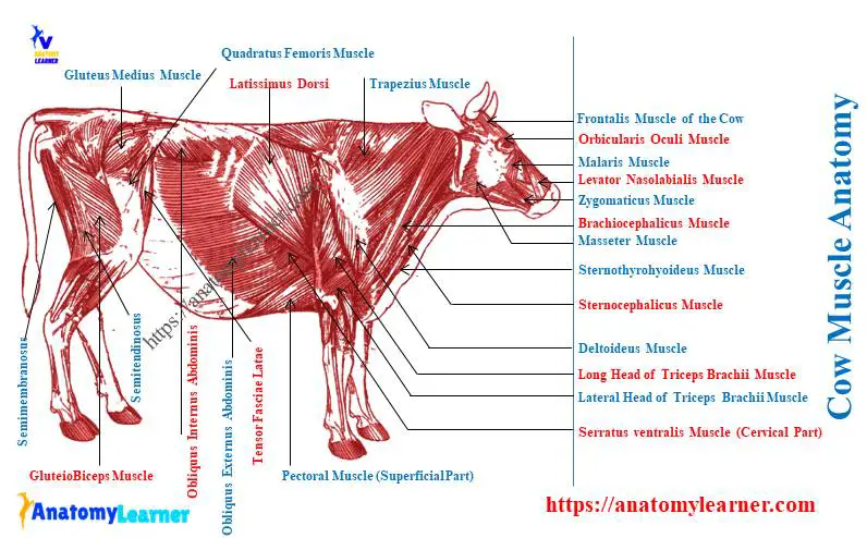
Okay, let’s see the overview of the important muscles from the various regions of the cow’s body. Here, in a diagram, I tried to show the most superficial muscles of the cow.
But, this diagram does not cover all superficial muscles from the head, neck, thorax, abdomen, and limbs. Thus, it would help if you saw them from other labeled diagrams on cow muscles that I provided in this article.
How many muscles does a cow have?
Quick answer: you will find more than 145 muscles in the cow body. But 121 muscles are major, whereas 24 muscles are minor in the cow body.
“The 145 number of the muscles are identified from the cow body by me. Some muscles may be missed, thus you may find little variation in the number of total muscles in the cow.”
But, I will not provide all these muscles from the cow’s body here. Let’s see the most superficial muscles of a cow with the diagrams.
First, start with the identification of the cow’s limb muscles.
Identification of forelimb muscles from the cow
From the forelimb of a cow, I will identify more than 25 muscles. You might go with region by region, as I show below –
Muscles from the shoulder region (lateral aspect):
- The thoracic part of the trapezius muscle,
- Deltoideus muscle of a cow,
- Teres minor muscles,
- Upper infraspinatus muscle, and
- Flatten supraspinatus muscle (extensor muscle),
Here, the teres minor is the small muscle that locates caudolateral to the shoulder and is covered by the deltoideus muscle. Now, let’s see the muscles from the medial aspect of the cow shoulder region –
- Subscapularis muscle (have three parts),
- Teres major muscle, and
- Coracobrachialis muscle,
Here, the teres major muscle is flattened and lies the caudal border of the scapula to the teres major tuberosity of the humerus bone. It also lies medial to the triceps brachii muscle of the cow.
You may learn the osteological facts of the scapula and humerus bones from the below-mentioned article –
- Scapula of an ox – the ultimate guide to learning the scapular anatomy of animals, and
- Humerus of ox – the complete guide to learning humerus bone anatomy,
All the muscles from the shoulder region (both lateral and medial) are identified in the cow muscle labeled diagram.
Muscles of the arm region of a cow
On the lateral aspect of the cow’s arm (also known as brachium), you will find the below-mentioned muscles –
- Tensor fascia antebrachii (slender and medial to the long head of triceps brachii muscle),
- The long head of the triceps brachii muscle, and
- The lateral head of the triceps brachii,
You know there are 3 heads in the structure of the triceps brachii muscle – long, lateral, and medial. Here, the long head of the triceps brachii is the most significant muscle and arises from the caudal border of the scapula bone.
You will find the lateral head of the cow’s triceps brachii muscles on the lateral aspect of the arm. Sometimes, you may find an accessory head of the triceps brachii on the lateral aspect of the arm of a goat.
After the removal of the long and lateral head of triceps brachii, you will find the below-mentioned muscles on the arm region –
- Anconeus (on olecranon fossa of ulna bone),
- Brachialis muscles,
- Biceps brachii muscle of the cow, and
- Pronator teres muscle,
Here, the brachialis muscle lies on the musculospiral groove of the humerus bone. The biceps brachii possess two heads and lie on the cranial surface of the humerus bone.
Again, the pronator teres muscle of a cow is a narrow and weak muscle. This pronator teres muscle lies medial to the elbow joint of the cows.
On the medial aspect of the arm region of a cow, you will find the medial head of the triceps. Again, the coracobrachialis is also found in the medial aspect of the arm. This is because the coracobrachialis muscle obliquely crosses the shoulder and ends on the median aspect of the humerus bone.
Muscles of the forearm of the cow
You will find 2 groups of muscles in the structure of the cow’s forearm –
- Extension group – includes the cranio-lateral muscles of the forearm of a cow, and
- Flexor group – includes the caudomedial muscles of the cow’s forearm,
There are 4 (four) extensor muscles in the forearm of a cow. Let’s see and identify the below-mentioned extensor muscles from the bovine forearm –
- Extensor carpi lateralis (largest extensor of the forearm),
- Extensor digitorum communis muscle,
- Abductor digit I longus, and
- Extensor digitorum lateralis,
Here, the extensor carpi lateralis lies on the cranial surface of the radius bone. The extensor digitorum communis muscle possesses two belies – lateral and medial.
Here, the lateral belly of the extensor carpi lateralis is slender, and the medial head is thick that lies cranio-lateral.
You will see the thin and flat abductor digit I longus between these extensor muscles. This muscle courses obliquely across the dorsal surface of the carpus bones.
The extensor digitorum lateralis is the most caudal extensor muscle in the cow forelimb. It has an extensive origin and forms a flat tendon.
Now, let’s identify the caudomedial or flexor group muscles from the antebrachium and manus region of a cow –
- Ulnaris lateralis muscle of the forearm (long and straplike; most caudally),
- Flexor carpi ulnaris muscle (wide and thin),
- Flexor digitorum superficialis (blended with the origin of flexor carpi ulnaris),
- You will find two bellies in the structure of the flexor digitorum superficial muscles – superficial and deep. Caudal to the flexor digitorum superficialis muscle, you will find –
- Flexor digitorum profundus muscle (possess three heads – radial, humeral, and ulnar), and
- Flexor carpi radialis (just caudomedial to the forearm),
The labeled diagram identifies all these extensor and flexor muscles from the cow’s forearm.
Muscles of the hindlimb of a cow
You will find some clinically important muscles in the cow hindlimb muscle anatomy. I will enlist and identify the superficial muscles from the below-mentioned regions of the cow’s hindlimb –
- Muscles of the hip and thigh regions (lateral, cranial, and medial aspect), and
- Muscles of the leg and foot (dorsolateral and planter group),
Let’s see the lateral muscles from the hip and thigh regions of a cow.
Lateral muscles of the hip and thigh of a cow
You will find the below-mentioned muscles in the structure of the cow’s hip and thigh –
- Tensor fascia latae muscle (cranial and craniolateral to pelvic limb),
- Gluteus medius muscle (fleshy and thick; between coxal tuber to greater trochanter),
- Gluteus profundus (fan-shaped; crossing caudoventrally direct over the hip joint),
- Gluteobiceps (extensive muscle and lateral to the hip region),
- Semitendinosus muscle (long fleshy, between the gluteobiceps and semimembranous muscles), and
- Semimembranosus muscle (long, thick muscle; caudomedial to the semitendinosus),
Here, the gluteus medius possesses two heads –
- Large superficial head of gluteus medius (also known as the gluteus medius), and
- Small deep head of gluteus medius (also known as the gluteus accessories),
The gluteus profudus of a cow has a wide origin in tuber coxae and insert on the neck of the femur bone. You may learn the different features of the following bones as they relate to the lateral muscles of the cow’s hip –
- Cow hip bone – osteological features of pelvic girdle bones of an ox,
- Animal femur – osteological features of the femur bone of the cow,
Cranial muscle of the cow’s thigh
You will find only the quadriceps femoris muscle in the structure of the cow thigh region. The quadriceps femoris muscle lies lateral, cranial, and medial of the cow’s thigh.
This muscle acts as a strong extensor of the stifle joint of a cow. The quadriceps femoris muscle of a cow possess the following 4 parts –
- Vastus lateralis muscle (lateral to the greater trochanter and caudolateral face of the femur bone),
- Rectus femoris muscle (cranial to the femur and just below the vastus lateralis muscle),
- Vastus intermedius (caudal to the rectus femoris muscle, you may see it from a lateral aspect after removing vastus lateralis), and
- Vastus medius muscle (caudomedial to the vastus intermedius muscle),
All these 4 parts of the quadriceps femoris muscle are identified in the cow leg muscle labeled diagram.
Medial muscles of the cow’s thigh
There are various muscles in the structure of the medial aspect of a cow thigh. But, you might identify the below-mentioned important muscles from the medial aspect of a cow thigh –
- Sartorius (cranial to gracilis muscle; strap-like and cranio-medial to stifle joint),
- Gracilis muscle (broad and flat; superficial on cranial part of the medial thigh),
- Pectineus muscle ( large fleshy and triangular shape),
- Adductor muscle (thick and fleshy; extensive),
- Quadratus femoris muscle (small and ventral to the Gemelli muscle),
- Obturator externus and internus muscles (externus is fan-shaped and ventral to ischium bone), and
- Gemelli muscle (triangular and ventrolateral to ischium to the trochanteric fossa of the femur bone),
You may also identify other different muscles from the medial aspect of the cow’s thigh. Again, to get the full idea of the cow leg anatomy (including bone, muscles, and joints), you may read the following article of anatomy learners –
Cow leg muscle anatomy
In the cow leg and foot, you will find two groups of muscles – dorsolateral and planter groups. First, let’s try to identify the dorsolateral muscles from the cow leg –
- Tibialis cranialis muscle (deepest and thin; craniolateral to the tibia bone),
- Fibularis tertius (superficial fusiform muscle),
- Extensor digitorum longus (thin and fusiform; craniolateral to the leg),
- Fibularis longus muscle (long, triangle shape, most superficial to the lateral surface of the leg),
- Extensor digitorum lateralis muscle (lateral to the leg), and
- Extensor digitorum brevis muscle (small muscle in the extensor group),
All these dorsolateral (extension) group muscles are identified from the cow leg muscles labeled diagram. Now, let’s try to identify the below-mentioned plantar group of muscles from the cow’s leg and foot –
- Gastrocnemius muscle (has two heads – lateral and medial),
- Soleus muscle (thin and ribbon-like structure),
- Flexor digitorum superficialis muscle (fleshy and fusiform muscle; lies on the deep surface of the gastrocnemius),
- Flexor digitorum profundus muscle (complex and possess three heads; lies caudolateral to the tibia bone), and
- Popliteus muscle (fleshy and triangular shape),
You will find the popliteus muscle on the distal part of the caudal aspect of the stifle joint.
Bovine head muscle anatomy
There are lots of muscles in the bovine head anatomy. But, I will show you the following muscles from the bovine face, muzzle, ear, eye, and hyoid regions –
Muscles from the face and muzzle of a cow –
- Levator nasolabialis muscle – thin and broad muscle,
- Levator labii maxillary (band type; upper),
- Depressor labii maxillary (band type, lower),
- Caninus muscle (in between the levator and depressor labii maxillary),
- Orbicularis oris (encircles on the lips),
- Malaris muscle (on the malar bone; above the zygomaticus muscle),
- Zygomaticus muscle (thin band-like muscle),
- Buccinators muscle (below the zygomaticus muscle), and
- Masseter muscle (caudal to the buccinators and more developed muscle on the masseteric region,
On the ventral aspect of the head, you will find the following muscles in a cow –
- Digastricus muscle of the cow,
- Mylo-hyoideus muscle, and
- Omohyoideus muscles of the cow,
On the ear and eye of the cow, you will find the below-mentioned muscles –
- Orbicularis oculi (encircles the eyes),
- Frontalis muscle (dorsal to the frontal bone),
- Zygomatico-auricularis (zygomatic bone to the base of the ear),
- Parotid –auricularis muscle (extends from the parotid gland to the base of the ear, a thin band like), and
- Scutulo-auricularis muscle (encircles the base of the ears),
You will also find the various muscle on the hyoid region of a cow. But, you might identify these below-mentioned hyoideus muscles from the cow –
- Stylohyoideus muscle,
- Occipitohyoideus muscle, and
- Geniohyoideus muscle,
The labeled diagram identifies all these muscles from the cow’s face, ear, eye, and hyoid region.
Neck muscles of cattle
The muscles of the cattle neck are very complex. But, I would like to enlist and identify the most superficial and clinically important cattle neck muscles.
Let’s try to identify the below-mentioned muscles from the cow’s neck –
- Brachiocephalicus muscle (possess two parts – upper cleidooccipitalis and lower cleidomastoideus),
- Omotransversarius muscles (just below the brachiocephalicus muscle),
Now, under the brachiocephalicus muscle, you will find the external jugular veins. Below the vein, you will find the below-mentioned muscle –
- Sternocephalicus muscle (possess two parts – superficial sternomandibularis and deep sternomastoideus muscles),
Below the sternocephalicus muscle, you will find the esophagus and the trachea. Let’s see the muscles below the cow trachea and esophagus –
- Sternothyrohyoideus muscle (just below trachea; possesses two parts – sternothyroideus and sternohyoideus),
- Omohyoideus muscle (just deep to the sternomastoideus muscle),
- Scaleneus dorsalis (at the level of 2nd – 4th ribs), and ventralis muscles (at the level of 1st rib that traversed by a root of brachial plexus)
- Longus coli (dorsal to the esophagus and trachea), and
- Intertransversarius muscles of the cow,
You may learn the relationship of trachea and esophagus with the longus colli muscle of the cow from the below-mentioned article –
Neck and thorax muscle of cow anatomy
The neck and thorax muscle of a cow anatomy is very complex and arranged in layers. You will find 4 or 5 layers of muscles in the caudal part of the neck of a cow.
I will help you to identify these muscles from the caudal part of the cow’s neck layer by layer. Let’s see and identify the muscles from the neck and thorax of a cow –
- The cervical and thoracic part of the trapezius muscle (first layer muscle from the surface),
- Latissimus dorsi muscle (also the superficial and first layer of muscle of the thorax),
- Rhomboideus cervicis and thoracic (2nd layer muscle; just below the trapezius),
- After removing the rhomboideus muscle, you will find the 3rd layer of muscles –
- Splenius muscle (3rd layer), and
- Semispimalis cervisis and thoracic (3rd layer),
Let’s remove the splenius muscle of the cow and find the below-mentioned muscles in the 4th layer –
- Semispinalis capitis (upper), and
- Longissimus capitis (lower or below to semispinalis capitis),
You will also find the longer muscles in the thorax of the cow –
- Longissimus cervicis and thoracic (longissimus dorsi – largest muscles in the back), and
- Longissimus lumborum (below the longissimus cervicis and thoracic),
Again, below the longissimus lumborum, you will find –
- Iliocostalis cervicis, thoracic, and lumborum muscles,
Furthermore, various spinalis muscles are just below the longissimus and semispinalis capitis (5th layer muscle in the neck). I have identified these 5th layer muscles from the cow neck anatomy.
Dorsolateral and dorsoventral muscles of cow thorax
After removing the superficial longissimus dorsi and cutaneous trunci muscles, you will see the followings –
Dorsolateral muscles of cow thorax –
- Intercostalae externi and interni muscles in the intercostal spaces, and
- Retractor costae muscles,
Ventrolateral muscles of the cow thorax –
- Serratus ventralis cervicis muscle,
- Serratus ventralis thoracic muscle,
- Deep pectoral muscle (deep to the superficial pectoral muscle), and
- Superficial pectoral muscle of the cow,
Here, the superficial pectoral of a cow has two parts – upper or cranial descending pectoral and caudal or lower ascending pectoral. Again, the thorax shows a thin and flat scalenus muscle where the nerves of the brachial plexus traversed.
Let’s learn the nerves from the brachial plexus of a cow –
Cow abdominal muscle anatomy
The cow abdominal muscles are most important compared to the thorax. You will find 4 different muscles in the anatomy of a cow’s abdomen.
To identify these 4 muscles from the cow abdomen, you might know their origin, insertion, and fiber direction. Let’s see and identify the 4 abdominal muscles from the cow abdomen –
- Obliquus externus abdominis muscle of the cow abdomen (fibers direct ventrally and caudally),
- Obliquus internus abdominis muscle of the cow abdomen (fiber directs ventrally, cranially, and medially),
- Transverse abdominis muscle (medial to the obliquus externus and internus muscles), and
- Straight abdominal muscle (form the ventrolateral wall of the cow’s abdomen),
All these 2 obliquus, 1 transverse, and 1 straight muscle from the cow abdomen are identified in the labeled diagram.
Cow muscle anatomy labeled diagram
Now, I will provide the labeled diagrams on cow muscle anatomy. In this section, you will find almost all labeled diagrams of the cattle muscle here.
I tried to show the muscles from the cow’s forelimb, hindlimb, head, neck, thorax, and abdomen with a labeled diagram. You may also find more diagrams on the cow muscle structure on social media of anatomy learners.
From the forelimb and hindlimb of the cow, I tried to identify the muscles enlisted in Table 1&2.
Table 1 shows the muscles from the cow’s forelimb with their origin and insertion. Again, Table 2 shows these muscles from the cow’s hindlimb.
The superficial muscles from the head, neck, thorax, and abdomen are also identified from the real sample.
Frequently asked questions on cow muscles
Here, you will find the frequently asked questions on the cow muscles. I will enlist only the important questions on bovine muscles that the anatomy learners ask.
Let’s see these questions on bovine muscles anatomy with the concise answer –
What is the most used muscle in a cow?
Quick answer: the most used muscles in a cow are the gluteobiceps, gluteus medius, semitendinosus, semimembranosus, and brachiocephalicus. Again, the abdominal muscles (2 obliquus, 1 transverse, and 1 straight) are also the most used muscles in the cow.
Here, the brachiocephalis is the muscles of the cow’s forelimb, and others (gluoteiobiceps to semimembranous) are found in the hindlimb.
Do cows have pectoral muscles?
Quick answer: yes, cows have well-developed pectoral muscles. The deep pectoral muscle of a cow originates from the sternum and is inserted on the lesser tubercle of the humerus bone.
Again, the superficial pectoral muscle of a cow origin from the sternum and inserts on the cranial surface of the humerus bone. Here, the deep pectoral of a cow flexes the shoulder, whereas the superficial pectoral extends the shoulder.
Conclusion
So, the cow muscle anatomy comprises more than 145 muscles from various body regions. Identifying these muscles from a cow body is required to study further the cardiovascular and nervous system (Especially for vessels and nerves).
Again, you might focus on the anatomy of the most used muscles from the bovine. Here, the muscles from the limbs, neck, and abdomen are most used and clinically important for the veterinarian.