The cardiac muscle under a microscope shows a short cylindrical fiber with a centrally placed oval nucleus. You will find some unique features in cardiac muscle that will help you to differentiate it from skeletal and smooth muscles.
Quick answer: the cardiac muscle microscope slide shows the cylindrical fiber with 1 or 2 nuclei. The unique features of the cardiac muscle are the presence of intercalated discs at irregular intervals and cross-striation.
This article might help you to get the full histological or microscopic features of an animal’s cardiac muscle with the labeled diagrams. I will also show you the real microscope slide of cardiac muscle (both longitudinal and cross-sectional views) so that you may identify it so quickly.
Again, this article might provide some differential features among 3 types of animal muscle tissue (smooth, skeletal, and cardiac). So, if you wish to know the microscopic features of cardiac muscle and want to distinguish it from the other 2 muscles, let’s continue this article till the end.
The cardiac muscle under the microscope
The cardiac muscle shows many structural and functional features, such as the skeletal and smooth muscles. This cardiac muscle shows cross striation like the skeletal muscle, but they are involuntary and contract automatically like the smooth muscle.
You will find the cardiac muscle only in the animal’s heart and some parts of the major vessels that attach to the heart. Let’s see the summary of the cardiac muscle under a microscope from Table 1 –
| Features | Cardiac muscle microscope slide |
| Muscle fibers | Short cylindrical, and branched |
| Nucleus | 1 or 2 centrally placed, oval |
| Striation | Transverse striation in cells |
| Perinuclear halo | Clear space around nucleus |
| Intercalated disc | Transverse line between 2 adjacent cells |
| Blood vessels | May see numerous capillaries around individual cell |
All the mentioned features of cardiac muscle fiber can see with the help of a light microscope (along with routine staining). Now, let’s see the real microscope slide picture of cardiac muscle and try to identify the features.
Now, let’s describe these microscopic features of cardiac muscle slides.
Unique features of cardiac muscle in a light microscope
So, from the above real cardiac muscle microscope slide, you may easily identify the shorter cylindrical muscle fibers. These muscle fibers are normally shorter than these skeletal muscle fibers.
Again, the animal’s cardiac muscle fibers show the branching pattern and anastomoses. These muscle fibers are made of cardiac myocytes (a chain of the muscle cell), each having its own nucleus.
The individual cardiac myocytes are approximately 15 – 19 micrometers in diameter and 85 – 100 micrometers in length. You may find a single or sometimes 2 nuclei in the individual cardiac myocyte.
The single or multiple nuclei of the individual cardiac myocyte locate in the center of it. Here, the cytoplasm of the cardiac myocyte shows an acidophilic appearance, whereas the nucleus shows basophilic features.
The most striking feature of cardiac muscle is the presence of a darkly stained transverse line across the fibers. These are the intercalated discs, the specialized cell junctions between the ends of adjacent muscle fibers (cells).
The cell junction (intercalated disc) provides a mechanism by which the contractile stimuli pass from one myocyte to another. In this way, the adjacent myocyte of the cardiac muscle fiber contract simultaneously. This whole process of this contraction is known as the cardiac muscle functional syncytium.
The cardiac muscle fibers also show a faint cross striation, whereas the skeletal muscle shows more visible striations. A network of fine reticular and collagen fibers surround each cardiac muscle fiber.
This fine network of cardiac muscle fiber are irregular and corresponds to the endomysium of skeletal muscle. Here, the cardiac myocytes subdivide into different groups.
A connective tissue (analogous to the perimysium of skeletal muscle) divides these groups of cardiac myocytes. Again, a well-developed capillary network surrounds the individual cardiac myocytes.
Cardiac muscle microscope slide identification
Sometimes you may be asked to identify the cardiac muscle histology slide under a light microscope. I know you already have the basics of important microscopic features of cardiac muscle.
Here, I will provide (enlist) the main identification points of the cardiac muscle microscope slide –
- The sample muscle fibers show the short cylindrical and branching pattern,
- Each of the individual muscle fibers is made of cells (cardiac myocytes),
- Individual myocytes possess a single (or two) nuclei which are located at the center of the cell,
- At the irregular interval of the muscle fibers, you will see the intercalated disc (deeply stained),
- The sample muscle fibers also show faint cross striations,
- A clear space is seen (perinuclear halo) around the single or multiple nuclei of the provided muscle fibers,
So, this is a cardiac muscle microscope slide. Now, I will describe the fine structure of cardiac muscle fibers with the labeled diagram.
The cardiac muscle under electron microscope description
Before going to the details description of cardiac muscle, you might have the basic knowledge of the skeletal muscle structure (fine or electron microscope). If you want, you may know the histological features of skeletal muscle from the below-mentioned article –
So, from the fine structure of cardiac muscle, I will show you the following features –
- Cardiac muscle cell and centered nucleus,
- Organelles and glycogen stored in myofibrils,
- Intercalated disc and different cell junctions,
- Sarcomere and sarcoplasm of cardiac muscle fibers, and
- Purkinje fibers (special cardiac conducting muscle cells),
Okay, let’s start to know the details of the fine structural features of an animal’s cardiac muscles with the help of an electron microscope.
Cardiac muscle cell and centered nucleus
The nucleus of skeletal and cardiac muscles are almost similar in their structure, but you will find the difference in their location. So, the center location of the cardiac muscle’s nucleus helps you to distinguish it from the multinucleated skeletal muscle fiber.
Under the electron microscope, you will find the myofibrils of cardiac muscle separate to pass around the nucleus. In this region of cardiac muscle, numerous mitochondria, Golgi complex, pigment granules, and glycogen may store.
In the atrial part of the animal’s heart, you will find the atrial granules. There are 2 types of polypeptide hormones in these granules – atrial natriuretic factors and brain natriuretic factors.
Both of these hormones of the heart are diuretic and affect sodium excretion in the kidney. They inhibit renin and aldosterone secretion and thus inhibit vascular smooth muscle contraction.
Mitochondria and glycogen in cardiac myofibrils
An electron microscope shows numerous mitochondria in the cardiac muscle fibers, which are densely packed. Here, the mitochondria of cardiac myocytes are larger and more considerable compared to skeletal myocytes.
These numerous mitochondria in the cardiac muscle fibers indicate the degree of aerobic metabolism in this tissue. Again, the numerous mitochondria extend the full length of the sarcomere and contain numerous closely packed cristae.
The cytoplasm of the cardiac myocytes also contains lipid droplets and glycogen. Here, the concentration of the glycogen granules is also numerous, located between the myofibrils of cardiac muscle.
Intercalated disc and different cell junctions
The intercalated disc in cardiac muscle represents the attachment between adjacent cells. Under the light microscope, these intercalated discs appear as the densely staining liner structure. Sometimes, it consists of a short segment that arranges in a step-like pattern.
But, if you see the intercalated disc under the electron microscope, you will see the transverse components that cross the muscle fibers at their right angle. These transverse components are analogous to the risers of a stairway.
Sometimes, the lateral component is perpendicular to the transverse component and lies parallel to myofibrils in the intercalated disc.
The longitudinal area of the intercalated disc contains the gap junctions that allow the transfer of chemical signals between the adjacent cells. Again, in the transverse area of the intercalated disc, you will find the desmosome and fasciae adherens.
In the desmosome of the intercalated disc, you will find the intermediate filament that extends into the cytoplasm. So, these desmosomes may result in strong attachment between the cardiac myocytes.
On the other hand, the fasciae adherens hold the cardiac muscle cells at their ends to form the functional cardiac muscle fibers (actin filaments).
An electron microscope shows an intercellular space between the adjacent cells filled with electron-dense material. Here, the fasciae adherens serve as the site at which the filament in the terminal sarcomere anchor onto the plasma membrane.
Gap junction of cardiac muscle fibers
The gap or communication junction constitutes the major structural element (SE) of the lateral component of the intercalated disc of cardiac muscle fibers. This junction provides the ionic continuity between adjacent cardiac muscle cells.
So, this junction of the cardiac muscle fiber allows informational macromolecules to pass from cell to cell. This information exchange permits the cardiac muscle fibers to show syncytium when retaining cellular integrity and individuality.
Again, the position of the gap junction on the lateral surface of the intercalated disc protects them from the forces generated during the contraction.
Sarcoplasm and sarcomere of cardiac muscle fibers
The sarcoplasm of the cardiac muscle cells (cardiac myocytes) is numerous, whereas the myofibrils are relatively few. These fewer myofibrils of the cardiac muscle merge, and thus the cross-striation of this muscle is not distinct.
The sarcoplasmic reticulum is not well-developed in the cardiac muscle compared to the skeletal muscle. Here, the T-tubule penetrates into the myofilament bundles at the level of Z line.
You will find only one T – tubule per sarcomere in the cardiac muscle structure. The small terminal cisternae of the sarcoplasmic reticulum are in close proximity to the T-tubule from the diad (at the level of Z line).
In the cardiac ventricular muscles, you will find the larger and numerous T-tubules. Again, the atrial myocytes are small and possess fewer T-tubules than the myocytes of the ventricle.
Purkinje fibers of cardiac muscle
The modified cardiac muscle cells are believed to initiate, regulate, and coordinate the conduction or heartbeat. These are the cardiac conducting cells of the cardiac muscle of an animal’s heart.
The cardiac conducting cells are organized into nodes and form highly specialized conducting fibers (Purkinje fibers). These Purkinje fibers generate and transmit the contractile impulse to a different part of the myocardium in a proper sequence.
You may easily differentiate the cardiac muscle cells from the Purkinje fiber under the microscope. The cells of the Purkinje fibers (cardiac conducting cells) are larger, and their myofibrils are mostly located at the cell’s periphery.
As the cardiac conducting cells contain a large amount of glycogen, the cytoplasm of the cell and myofibrils show a poor stain. You will not find any T-tubule in the structure of Purkinje fiber under the electron microscope. But, the heart of some animal species shows the T-tubules in their Purkinje fibers.
Cardiac muscle contraction
The cardiac muscle is stimulated to contract by a mechanism similar to the skeletal muscle. Here, the contraction is activated by the interaction of actin and myosin microfilaments.
A sequential contraction of heart chambers is stimulated by the orderly spread of the action potential through the gap junction (in the intercalated disks). Here, I will show you the short process of cardiac muscle contraction with the diagram –
- The contraction of cardiac muscle fiber initiates from the Purkinje fiber (cell membrane depolarization) and reaches to the targeted cardiac myocytes,
- When the general depolarization spreads over the plasma membrane of the cardiac myocytes, it causes the opening of voltage-gated sodium channel,
- Sodium enters to the cell, and you know the general depolarization continues through the membrane of T-tubules,
- Now, the plasma membrane of the T-tubule change its conformation to a functional calcium channel,
- Calcium is rapidly released from the sarcoplasmic reticulum, and accumulated calcium diffuses to the myofilaments,
- Now, the calcium binds to the TnC protein of the troponin complex,
- The actinomycin cross-bridges and calcium returns to the terminal cristate of the sarcoplasmic reticulum,
This is a straightforward process of an animal’s cardiac muscle contraction. But, you may learn the cardiac muscle contraction process from another article by an anatomy learner.
Similarities and differences between cardiac and skeletal muscles
First, let’s see the similarities between the cardiac and skeletal muscles under a light microscope. Like the cardiac muscle, skeletal muscle is also made of elongated fibers.
You will find numerous myofibrils in these elongated skeletal muscle fibers, like the cardiac fibers. If you see the skeletal muscle under a light microscope, you will find visible transverse striation like the cardiac muscle (less visible in cardiac muscle).
Again, the connective tissue framework and capillaries network around the individual cardiac myocytes are similar to the skeletal muscle cells. However, the electron microscope also reveals that the structure of the cardiac myofibrils is similar to these of the skeletal muscle.
Now, let’s see the differences between the cardiac and skeletal muscle fibers under the light microscope. These might help you to differentiate them (cardiac and skeletal) from the microscope slides.
The skeletal muscle fibers run parallel, whereas the cardiac muscle fiber doesn’t run stick parallel. Rather they (cardiac muscle) branch and anastomoses with other fibers to form a complex network.
Not all cardiac muscle fibers are multinucleated, whereas skeletal muscle is multinucleated. Again, you will find the nucleus in the center position in cardiac muscle fibers. But, the skeletal muscle shows the peripheral nuclei.
An electron microscope shows the less prominent sarcoplasmic reticulum in the skeletal muscle compared to the cardiac muscle. The basic difference between the skeletal and cardiac muscle fibers are shown in Table 2 –
| Features | Skeletal muscle | Cardiac muscle |
| Muscle fibers | Long cylindrical, not branched | Short cylindrical, branched |
| Nucleus | Multinucleated, peripherally | 1 or 2, centrally |
| Cross striation | Distinct | Indistinct |
| Intercalated disc | Absent | Present |
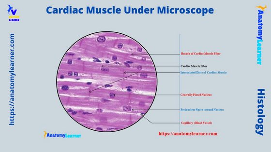
Cardiac muscle tissue under a microscope labeled
Now, I will show you the cardiac muscle tissue under a microscope-labeled diagram. Here, I will show you both cross and longitudinal sections of the cardiac muscle tissue with the labeled diagram.
I tried to show you the longitudinal section of cardiac muscle fibers in the first labeled diagram. Here, the diagram shows the short cylindrical cardiac muscle fibers with their branches.
The single centrally placed nucleus is identified from the cardiac muscle labeled diagram. Again, the binucleated fiber is also identified from the cardiac muscle labeled diagram.
The longitudinal section of cardiac muscle also shows indistinct cross-striation and intercalated discs. Here, the perinuclear sarcoplasm is also showing around the single or multiple nuclei of the cardiac muscle fibers.
The transverse or cross-section of the cardiac muscle shows the bundle of fibers with their myofibrils. Here, the individual cardiac muscle bundle encloses with the endomysium.
A thin layer of connective tissue is also seen in the cardiac muscle (transverse sectional view). The central nuclei and perinuclear sarcoplasm are in the individual cardiac muscle bundle.
But, only some of the cardiac muscle bundles (in cross or transverse section) show the nucleus. And you know this is due to the short cylindrical appearance of the individual cardiac myocyte.
When you cut this short cylindrical myocyte transversely, you will not find the nucleus in all sections. You may get more cardiac muscle fibers labeled diagrams on social media of anatomy learners. Again, you may see some of the electron microscopic figures of cardiac muscle on there.
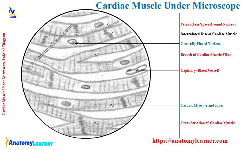
Cardiac muscle under microscope 40X, 100X, and 400X
Now, I will provide the cardiac muscle microscope slide figures with a resolution of 10X, 100X, and 400X. Here, I tried to show you all the important microscope features of the cardiac myocytes and fibers with the 40X, 100X, and 400X labeled diagrams.
First, let’s see the diagram of the cardiac muscle microscope slide with 40X. Both the longitudinal and transverse sections of the cardiac muscle microscope slide with 40X are provided here.
Now, let’s see all the microscopic features of the cardiac myocytes and fibers with 100X and 400X resolution. In the 100x and 400X cardiac muscle labeled diagram, you may easily identify the myofibrils, nucleus, and intercalated discs.
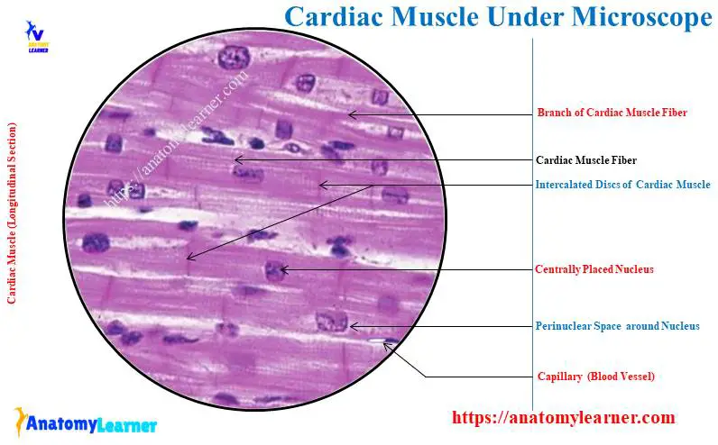
The cardiac muscle under microscope drawing
Now, I will show you the cardiac muscle microscope slide figure drawing. You may follow the full drawing tutorial for the cardiac muscle here.
Here, I will provide both longitudinal and transverse sectional cardiac muscle drawings. First, let’s see the longitudinal section of cardiac muscle fiber.
I tried to draw the short cylindrical muscle fibers along with their branches. Then you should draw the centrally placed single or multiple (2) nuclei on these individual cardiac myocytes.
Now, let’s draw the intercalated discs between the adjacent cardiac myocytes. You might also provide the perinuclear halo or space around the nucleus of the cardiac myocyte (shown in the drawing).
Again, in the transverse section of the cardiac muscle fibers drawing, you should draw the individual muscle bundle as shown in the diagram. Within the individual cardiac muscle bundle, you might draw the myofibrils.
Here, in these muscle bundles, I tried to draw the centrally placed oval nucleus. Again, I tried to show the perinuclear space surrounding the cardiac myocyte’s individual nucleus.
Frequently asked questions on a cardiac muscle microscope slide
In this section, I will provide the commonly asked questions on the cardiac muscle under a light microscope with the possible short answer. But, it is recommended to learn all the microscopic features of the cardiac muscle fibers and cells with the labeled diagram from this article (first to end).
Okay, now, let’s see the commonly asked questions on the cardiac muscle microscope slide with their concise answer –
What does cardiac muscle look like under a microscope?
The cardiac muscle looks like a short cylindrical structure under a microscope. It shows the branches network under the light microscope.
Again, you will find the single (normally) or multiple (2) nuclei on the cardiac fibers under a light microscope. In between the adjacent cardiac myocytes, you will find the intercalated discs.
But, the transverse cross striation of the individual cardiac muscle fiber is less visible with the help of a light microscope. Finally, you will see more capillaries and a perinuclear space that surround the individual nucleus of cardiac muscle.
How do you identify cardiac muscle?
You may easily identify the cardiac muscle with its unique identifying features. I have already provided the important identifying points for the cardiac muscle fibers with the help of a light microscope.
You might know the microscopic features of cardiac muscle’s fibers, cells, nuclei, and some other unique features. The unique features of the cardiac muscle include the presence of intercalated discs and cross-striation.
What are 3 characteristics of cardiac muscle?
Let’s see the 3 major characteristics of cardiac muscles –
- The muscle fibers are short cylindrical and show a branching pattern,
- Individual muscle fibers show the centrally placed single or multiple nuclei, and
- Presence of intercalated discs between the adjacent cardiac myocytes (but at irregular intervals),
Is cardiac muscle striated under a microscope?
Yes, cardiac muscle is striated under the microscope. But this striation is less visible under the microscope compared to these of the skeletal muscle.
If you see the cardiac muscle fibers with the help of an electron microscope (EM), you may easily identify the transverse cross striation and lateral striation.
What is the shape of cardiac muscle?
The shape of the individual cardiac muscle is short and cylindrical. They are known as the cardiac myocyte, which contains a single or multiple (occasionally) nuclei in the center of the cell.
How do you distinguish between a skeleton and cardiac muscle?
It is very easy to distinguish the cardiac muscle from the skeleton or bone. This is because there is a great variation in the microscopic features between the skeleton and cardiac muscle.
The microscopic features of the compact and spongy bones are also different. You may get the full guide on the microscopic features of compact and spongy bone from the anatomy learner.
The Haversian system is found in the compact bone, whereas the spongy bone contains boney trabeculae. But, you will not find such features in the cardiac muscle microscope slide.
Conclusion
So, the cardiac muscle under a microscope shows a short cylindrical fiber with a branch. The cardiac muscle fiber is made with cardiac myocytes, which contain centrally located single or multiple nuclei.
All the provided labeled diagrams on cardiac muscle under a microscope might provide a clear idea. Now, you can easily identify the cardiac muscle fibers and cells from the microscope slide and differentiate them from skeletal and smooth muscles.