The stomach of ruminants comprises four structurally distinct parts. Histologically, you will find a non-glandular mucosa having keratinized stratified squamous epithelium in the first three parts (rumen, reticulum, and omasum) of the ruminant stomach. I will show you everything about the rumen histology slide with their identification point and labeled diagram.
I will also show you the essential histological features of the reticulum and omasum parts of the stomach. So that you will make a difference among these three non-glandular parts with their histology slide labeled diagrams.
Rumen histology
Before going to the details histology of rumen, I would like to memorize the basics of a ruminant stomach. The ruminant stomach is somewhat different from that of a dog or horse and possesses four distinct parts. You will find three non-glandular parts (rumen, reticulum, and omasum) and one glandular part (abomasum) in the ruminant stomach.
The first 3 compartments of the ruminant stomach are collectively known as the forestomach or proventriculus. You will find some standard histological features and particular identifying points in these three different parts of the forestomach. Again, the fourth part of the ruminant stomach contains glandular mucosa and is histologically similar to the stomach of other species.
The rumen wall consists of four different tunics – tunica mucosa, tunica submucosa, tunica muscularis, and tunica serosa. So, make sure you know the basic structures of the different layers of a tubular organ.
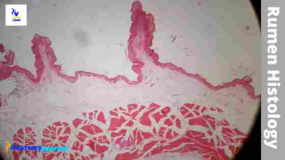
Let’s see the rumen histology labeled diagram first; this will provide you a basic idea of the different layers and other structures of a rumen.
You will find a large number of small papillae in the mucosa membrane of a rumen. They are small in young and infant but more significant in older ruminant species.
Okay, let’s continue this article to learn the histological features from a ruminant stomach.
Rumen histology slide
If you are a veterinary student, you might be asked to identify the rumen histology slide at your laboratory under the light microscope. Here, I will enlist some histological structures that you might find out from a rumen slide under a light microscope.
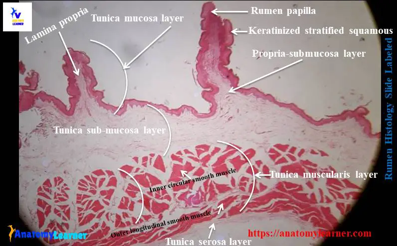
Four basic layers of rumen slide (tunica mucosa, tunica submucosa, tunica muscularis, and tunica serosa)
- Papillae of different sizes in the mucosa of a rumen slide
- Lamina propria in the core of the papillae of mucosa
- Stratified squamous epithelium (keratinized) lining in the mucosa or papillae of rumen slide.
- Stratum corneum cell layers (variable thickness) and others
I hope the rumen slide labeled diagram might help you find out the structures mentioned above easily.
Rumen histology slide identification points
Now, I will enlist the rumen histology slide identification points in this part of the article. You may write the same identifying points, or you may make them by yourself. Here, I will enlist the most important histological features from the rumen slide.
- The sample tissue section posses numerous papillae in the mucosa layer.
- There is a keratinized stratified squamous epithelium lining in the mucosal surface of the sample tissue.
- The stratum corneum varies in thickness from one to two cells to as many as ten to twenty cells.
- The dense collagen, reticular, and elastic fibers are present in the core of each papilla of the provided sample tissue.
- The lamina muscularis is absent, and there are no glands in the submucosa layer of the provided sample tissue.
- An inner thick circular and outer longitudinal smooth muscle layers are present in the tunica muscularis of the sample tissue.
So, this is a slide of the rumen of the ruminant stomach.
But, sometimes, you may find the circular, longitudinal, and oblique orientation of smooth muscle fibers in the tunica muscular layer of a rumen.
Fine, if you want to learn the details histology of the different layers of a rumen slide, continue this article till the end.
Layers of rumen slide
As I told you before, there are four different layers present in the rumen histology slide. In this part of article, you will learn everything about the histological features of different layers from a rumen. I will try to show you every structure from the different layers of the rumen with labeled diagrams.
So, what will I discuss here from the rumen slide?
- The histological features of tunica mucosa of rumen slide
- Details histological features of tunica submucosa of a rumen slide
- The histological characteristics of tunica muscularis layer of a rumen and
- The characteristics features of tunica serosa of a rumen.
Okay, let’s get started to learn the details of histological characteristics from the different layers of a rumen slide.
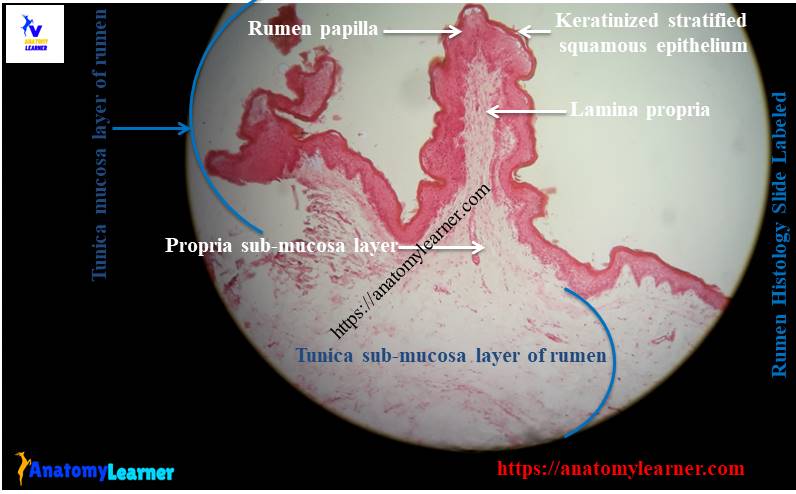
Tunica muscosa of rumen slide
You will find unique characteristics in the tunica mucosa of the rumen histology slide. There are different papillae present in the mucosal surface of the rumen. The size and shape of these papillae vary considerably from one region to another region of the rumen. It takes time to developed these papillae permanently in the rumen.
The ruminal epithelium is keratinized stratified squamous epithelium that performs some essential functions. The wall of this non-glandular compartment consists of the following cells layers and similar to that of skin.
The stratum corneum layer forms a protective shield against the rough, fibrous ingesta in the rumen. The stratum corneum layer is made up of squamous cells of variable thickness. You may find one to several layers of cells in the stratum corneum of the rumen.
The stainable nuclei may or may not be present in the stratum corneum of the rumen.
The stratum granulosum of ruminal mucosa consists of two to three cells thick. You will find distinctly flattened cells that possess keratohyalin granules in their cytoplasm. Sometimes you may find a swollen cell in the stratum granulosum with a pyknotic nucleus. The peripheral cytoplasm of these cells contains kerohyaline granules, tonofilaments, and numerous membrane-bounded, electron-dense granules.
You will find o large polyhedral cells in the stratum spinosum. These cells are slightly larger than that the basal cells. The thickness of the stratum spinosum layer varies from one to ten cells. You will find numerous mitochondria and ribosomes throughout the cytoplasm of the cells of the stratum corneum layer. Adjacent cells are connected through multiple desmosomes.
The cells of the stratum basale of rumen are columnar and extend numerous processes to the basement membrane. Its features increase the basal cell membrane surface area significantly.
Tunica submucosa of rumen slide
In the rumen histology slide, you will not find any lamina muscularis. So, the lamina propria blends with the submucosa layer forming a propria-submucosa. Each papilla of the rumen contains a core of Propria submucosa. Here in the core, you will discover a dense network of collagen, elastic, and reticular fibers.
A dense network of fenestrated capillaries lies just beneath the basement membrane of the lining epithelium of rumen. Near the tunica muscularis, you will find a more loosely arranged connective tissue in the propria-submucosa layer of the rumen.
There are also a network of blood vessels and submucosal nerve plexus present within this tunica submucosa layer. But, there are no submucosal glands present in this layer of a rumen.
Tunica muscularis and serosa of rumen slide
You will find almost similar features in the tunica muscularis and serosa layers of rumen histology slide-like tubular organ. The tunica muscularis comprises two layers of smooth muscle fibers.
You will find the inner circular and outer longitudinal smooth muscle layers in the tunica muscularis of rumen tissue. But, in a few regions of the rumen, you may find smooth muscle layers in various irregular directions. In this layer of rumen tissue, myenteric nerve plexus is present. The fibers from this tunica muscularis layer may extend into the core of the rumen papillae.
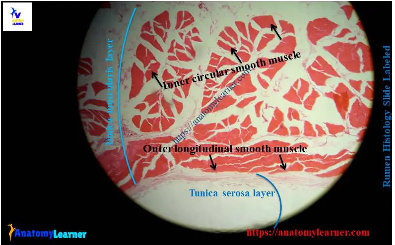
The serosa of a rumen is a loose connective tissue covered by a mesothelium (simple squamous). A varying amount of white adipose tissue and blood vessels, lymph vessels, and nerves are present in the loose connective tissue of serosa.
Rumen histology slide labeled diagram
Again, I will show you the rumen histology slide labeled diagram with the summary. Here, I tried to show you the four different layers and some of the special histological features from rumen tissue.
The rumen slide labeled diagrams represent the tunica mucosa with the lining epithelium and papillae. Again, the diagram shows the core of papillae that contain propira-submucosa. In addition, the diagram showed the other three layers of the rumen tissue. The tunica muscularis of the rumen diagram showed inner circular and outer longitudinal layers of smooth muscle.
I will provide you the updated rumen slide labeled diagram in the future. For that, you may follow anatomy learner and check these updated rumen slide diagrams on social media.
Reticulum histology slide
I will don’t describe the details histology of the reticulum slide. I will show you some of the essential features that might help you identify the reticulum slide and make a difference from the other parts.
Grossly, the interior of the reticulum is honeycomb in appearance. This is due to the anastomosing foldings of the mucous membrane of the reticulum. That means you will find the reticular folds or crests that intersect with other folds. The crests of the reticulum are in two different types – longer and shorter.
The margin of the reticular folds contains the verticle ridge. The mucosa between the crests covers with conical reticular papillae that project into the lumen.
You will find the keratinized stratified squamous epithelium lining on the mucosa of the reticulum resembles that of the rumen. The propria submucosa predominantly contains a network of collagen and elastic fibers. You will find a lamina muscularis only in the upper part of the more prominent reticular crest. The lamina muscularis is continuous with that of the esophagus.
The tunica muscularis of the reticulum consists of two layers of smooth muscle cells that follow an oblique course and cross at the right angle. In addition, the histological features of the tunica serosa of the reticulum are like that of the rumen histology slide.
Omasum histology slide
In the omasum histology slide, you will find several longitudinal folds (laminae). These laminae are arising from the internal surface of the greater curvature and side of the organ. You will discover larger laminae with thick, concave, free edges and reach the lesser curvature within a short distance.
The lining epithelium of the omasum is keratinized stratified squamous like that of the rumen histology slide. You will find a lamina propria that contains dense subepithelial capillary networks. A lamina muscularis forms a thick layer just beneath the lamina propria on both sides of the laminae of the osamum.
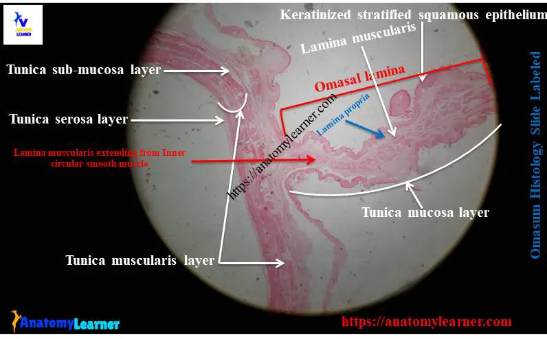
The tunica submucosa of omasum is comparatively thinner than that of rumen or reticulum. You will find thin outer longitudinal and thicker inner circular layers of smooth muscle in the tunica muscularis of the omasum. The innermost fibers of the circular layer are continued into the larger omasal laminae.
Abomasum histology slide
The lining epithelium changes from keratinized stratified squamous to simple columnar in abomasum histology slide. The lamina propria becomes less dense on the abomasal slide of the folds. In addition, the tunica mucosa of the abomasum has all the characteristics like that of a simple stomach.
How to differentiate rumen, reticulum, and omasum histology slides?
Now, I will provide a summary of the histological features of the forestomach of ruminant. These might help you to differentiate rumen, reticulum, and omasum histology slide easily.
You will find the keratinized stratified squamous epithelium lining in the tunica mucosa of the rumen, reticulum, and omasum. The lamina propria of rumen contains collagen, elastic, and reticular fiber.
It extends into the ruminal papillae. You will not find any lamina muscularis in the mucosa layer of a rumen slide.
But, in the reticulum, the lamina propria contains collagen and elastic fiber. You will find a muscularis mucosa that extends inside the more prominent fold of reticular mucosa. In addition, the lamina propria of the omasum is similar to that of the reticulum but contains a dense subepithelial capillary network.
The tunica submucosa of the rumen, reticulum, and omasum consists of loose connective tissue with blood vessels and nerves. In the tunica muscularis of the reticulum, the inner oblique layer of smooth muscle cross with the outer layer at the right angle. In addition, you may find a variation in the tunica muscular layer of an omasum histology slide.
Frequently asked questions on rumen slide.
Here, I will enlist a few frequently asked questions on the rumen histology slide that might help viva. If you have any questions on the rumen slide, let me know.
What are the special histological features of a rumen slide?
The most special histological feature of a rumen slide is ruminal papillae in the tunica mucosa layer. These ruminal papillae lines by the keratinized stratified squamous epithelium. You will find a core of lamina propria in the ruminal papillae of the rumen slide.
Is there a lamina muscularis present in the rumen histology slide?
No, there is no lamina muscularis layer present in the rumen slide. So, the lamina propria and tunica submucosa blend and forms the propria submucosa layer.
Is there any gland on the tunica submucosa of the rumen slide?
No, there is no gland in the tunica submucosa of the rumen slide. The rumen is the non-glandular compartment of the forestomach of a ruminant.
What do you mean by forestomach?
The term forestomach applies to the ruminant stomach. In ruminant, you will find four different compartments in their stomach – rumen, reticulum, omasum, and abomasum. The first three compartments (rumen, reticulum, and omasum) are collectively known as forestomach. Another name of the forestomach is proventriculus.
Conclusion
I hope this short guide on the rumen histology slide was helpful for you. Please try to draw the rumen histology slide picture and understand every single feature from each layer. You might get help from the rumen histology slide labeled diagram, and don’t forget to practice with the actual slides at a laboratory.