While studying the histological features of the seminiferous tubules and epididymis, you will see sperm cells under the microscope. They are much smaller and lie in groups along the inner margin of the Sertoli cells. In this article, you will get a details guide on the structure of sperm under a microscope with the 400x labeled diagram.
Again, you know there are different spermatogenic cells present in the seminiferous tubules of any animal. I will also help you differentiate these spermatogenic cells (especially primary spermatocyte, secondary spermatocyte, spermatid, and spermatozoa) from each other with the labeled diagram.
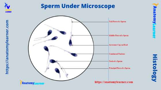
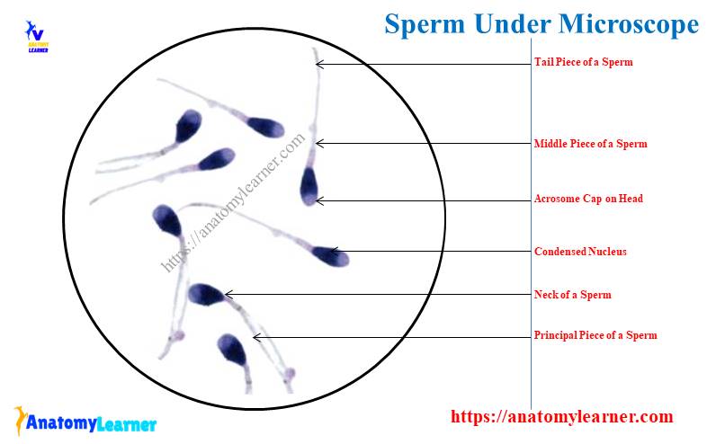
The spermatozoa or sperm has an expanded head, narrow neck, and a long principal tail. Again, you will see three pieces in the tail – middle, principal, and end under the electron microscope.
So, if you want to identify and learn the detailed histology of sperm under a microscope, let’s continue this article till the end. Don’t forget to check out the spermatozoa video from the end part of the article.
Sperm under microscope
First, I would like to show you what the sperm look like under a light microscope. I will take the example from the seminiferous tubules and epididymis of the animal. Here, the seminiferous tubules of the animal show different types of cells like primary spermatocytes, secondary spermatocytes, spermatid, and spermatozoa.
Again, the seminiferous tubules show the Sertoli cells or sustentacular cells, which are irregular outlined tall columnar cells resting on the basal lamina. These cells have oval-shaped nuclei that locate in the broad basal segment of the cells.
There are lateral tight junctions between two adjacent Sertoli cells. Let’s make it clear (structure of Sertoli cell) from the below-mentioned labeled diagram.
Here, the spermatogenic cells are adherent to the Sertoli cells. In the next part of this article, you will know and identify all of these spermatogenic cells from the seminiferous tubule along with the Sertoli cells.
Now, let’s see the body of the epididymis (cross-section) of the animal. You will see the tall columnar ciliated epithelium (stereocilia) lining the inner surface of the epididymis. Again, the duct of these epididymis shows the smooth muscle fibres arranged circularly.
The normal light microscope easily shows these stereocilia of the epididymal ducts. But, the electron microscope will show a clear view of these stereocilia. You will also see the agranular endoplasmic reticulum, lysosome, and prominent Golgi bodies in these lining epithelia of the epididymis (with an electron microscope).
- You may also learn – details of histological features of the epididymis histology slide with a labeled diagram.
Let’s see the lumen of the epididymis and there are clumps of spermatozoa in the lumen. You will not see any other spermatogenic cells in the lumen of the epididymis duct.
Sperm identifying characteristics
If you are a veterinary student or medical student, you may be asked to identify the sperm under the light microscope from the seminiferous tubules or ductus epididymis. Now, I will provide some of the important features of sperm that might help you identify it so quickly.
- The sample tissue section shows different elongated cells at the inner segment (apex) of the Sertoli cell (in the seminiferous tubules).
- These cells show an expanded head, a narrow neck, and an elongated thin (not seen clearly) tail under the microscope.
- The sample tissue also shows the other spermatogenic cells (primary spermatocytes, secondary spermatocytes, and spermatid) along with the spermatozoa.
- Again, if you see the epididymis tissue sample under the light microscope, you will find the clump of sperms at its lumen.
I hope now you can identify the spermatozoa under a light microscope with the help of the information mentioned earlier. Again, I will show you how you may also identify the other spermatogenic cells from the seminiferous tubules of an animal.
How to differentiate sperm from other spermatogenic cells under a light microscope
If you observe the sperm from the seminiferous tubules, you will see some other spermatogenic cells. This is very easy to differentiate the sperm from other spermatogenic cells from the seminiferous tubules.
In this part of the article, I will show you the characteristics and histological features of all spermatogenic cells, along with the sperm. So that you may differentiate the sperm under a light microscope from primary spermatocytes, secondary spermatocytes, and spermatids.
Before that, you may also read the below-mentioned article to get a full idea of the structure of seminiferous tubules –
- Histological features of the seminiferous tubules with the labeled diagram
Okay, first, let’s see the different histological features of the seminiferous tubules of an animal. The seminiferous tubules are the long, convoluted tubules that line with the germinal epithelium (stratified).
In the germinal epithelium of the seminiferous tubule, you will see two main types of cells –
- Spermatogenic cells that produce the sperm, and
- The supporting cells (Sertoli or sustentacular cells) nourish the developing sperm cells.
- Again, the spermatogenic cells of the seminiferous tubules divide into four main groups –
- Spermatogonia (A and B types),
- Primary spermatocytes – largest cells,
- Secondary spermatocytes – an intermediate shaped cells, and
- Spermatid – much smaller type cells,
Finally, the spermatid cells convert into spermatozoa by the process of spermiogenesis. Again, you will also see some of the other structures in the seminiferous tubules or between two seminiferous tubules.
The other tubule structures include Sertoli cells, interstitial or Leydig cells, septa or interstitial connective tissue. All these structures are identified in the seminiferous tubules 400x labeled diagram.
Okay, let’s see the main difference between the different types of spermatogenic cells and sperm.
Spermatogonia under the light microscope
In the germinal epithelium of a seminiferous tubule, you will find spermatogonia (stem cells) at its base. Again, the other spermatogenic cells are arranged in the order of the development process.
The spermatogonia of the seminiferous tubules are immature cells that undergo several mitotic divisions. This is why you will see a different stage of development of the spermatogenic cells under the light microscope.
The spermatogonia differentiate into Type – A and Type – B cells. You know the Type – A is the dark Type of spermatogonia, whereas the Type – B is the pale Type.
The dark Type: A spermatogonia serve as the stem cells of the germinal epithelium of the seminiferous tubule. Again, the pale Type – B spermatogonia goes for maturation to form the primary spermatocyte.
But, how you will confirm the Type – A and Type – B spermatogonium under the light microscope? The Type – A (dark) spermatogonium possesses an oval nucleus with an eccentric nucleolus. Again, the Type – A spermatogonium’s nucleoli may sometimes attach to the nuclear membrane.
These cells have a great intensity of staining their nuclei with the routine stain (Hematoxylin and Eosin). So, under the microscope, you will identify the Type – A spermatogonium as follow –
- The spermatogonium is located at the base of the seminiferous tubules,
- Larger cells with an oval nucleus (cells are larger compared to other cells of the seminiferous tubule),
- Nucleus takes deep stain (posses deeply stained nucleoli),
Again, the Type – B (pale) spermatogonium is the larger cell that possesses the spherical nucleus. You will also find the eccentrically placed spherical nucleolus.
Again, you may see the light Type – A spermatogonium that divides to form more light Type A spermatogonium. In contrast, Type B divides several times to form primary spermatocytes.
Primary spermatocytes under a microscope
So, you know the primary spermatocytes of the seminiferous tubules have resulted from the mitosis division of the Type B spermatogonium. They are the largest cells with a larger spherical nucleus than the spermatogonium.
These primary spermatocytes lie in the cell layer luminal to the spermatogonia (the middle region of the seminiferous tubules). The nucleus of the primary spermatocyte shows a coarse chromatin clump. Again, they undergo the first meiotic division and form two secondary spermatocytes.
Here in the first meiotic division, the chromosome reduces to half, meaning each secondary spermatocyte possesses a haploid number of chromosomes.
The prophase of the first meiotic division within the primary spermatocyte is prolonged. They show a considerable alteration of the nucleus in the different stages of the prophase (leptotene, zygotene, pachytene, and diplotene).
This is why you will see the primary spermatocytes at various stages of development in the seminiferous tubule under a light microscope. So, the main identifying features of the primary spermatocytes from the sperm under the light microscope are –
- Larger cells than the spermatogonia that locate in the middle of the seminiferous tubule,
- May see the chromatic clump in the nucleus of the primary spermatocytes,
- Observe different stages of prophase (development) under the light microscope,
I hope you will perfectly identify the primary spermatocytes from the seminiferous tubules of any animal.
Secondary spermatocytes in the seminiferous tubule
The secondary spermatocytes are smaller or intermediate between the primary spermatocytes and spermatids. So, you will also see a smaller nucleus than the primary spermatocyte. Again, the nucleus of the secondary spermatocyte shows the less dense chromatin in their nuclei.
You will find these secondary spermatocytes at the luminal surface of the seminiferous tubules. The secondary spermatocytes undergo the second meiotic division and immediately form two spermatids.
Here, the number of chromosomes remains the same, which means each secondary spermatocyte possesses the haploid number of chromosomes. But in the light microscope, they have rarely seen in the seminiferous section as they undergo the second meiotic division as soon as they are formed.
Spermatid under the light microscope
The spermatid is the small cells compare to the spermatocytes that lie in the luminal part of the seminiferous tubules. They are rounded cells that possess an initially eccentric, very light nucleus.
But, the chromatin of the nucleus may condense during the maturation of the spermatid into spermatozoa. At this time, the nucleus becomes smaller than the nucleus of spermatocytes and spermatogonia, which show a dark stain.
Now, the spermatid changes shape and forms a spermatozoon. This process of the formation of the spermatozoa from the spermatid is known as spermiogenesis (the last stage of spermatogenesis).
Spermatogenesis is the whole process of formation of spermatozoa (from spermatogonia to the spermatozoa), known as spermatogenesis. This is a continuous process that occurs along the length of the seminiferous tubules. So, in spermatogenesis, you will find the following steps –
- Spermatogonia or stem cell near the basal lamina,
- Formation of the primary and secondary spermatocytes,
- Formation of the spermatids and
- Spermiogenesis – terminal phage of spermatogenesis,
I hope you will identify the spermatid cell under the light microscope easily. So, the main identifying points of the spermatid cell from the seminiferous tubules are –
- Smaller rounded cells with small spherical or oval nuclei compared to other cells of the seminiferous tubules,
- They lie in a group in association with the Sertoli cells (at the luminal part),
But, how will you differentiate the nucleus of Sertoli cells from different types of spermatogenic cells? I will show how you differentiate the Sertoli cells from spermatogenic cells.
Sertoli or sustentacular cells of seminiferous tubule
The Sertoli or sustentacular cells of the seminiferous tubules are the irregular outline of tall columnar cells that rest on the basal lamina. You will see a clear, large oval nucleus that locates the centre of these Sertoli cells.
In the cytoplasm of the Sertoli cell, inclusion products are present (known as the crystalloid of Charcot Bottcher). The basal part of the Sertoli cell is broad, and the apical part of the cell is narrow.
The lateral cell membrane of the Sertoli cell possesses complex infolding that is impossible to view under the light microscope. Again, this lateral infolding involves a group of spermatogenic cells that can easily identify.
The apical cell membrane of the Sertoli cell also possesses the infolding that project into the luminal surface of the seminiferous tubules. You will see the sperm locked in the Sertoli cell’s apical folding under the light microscope.
Again, the electron microscope shows a more smooth endoplasmic reticulum and a less rough endoplasmic reticulum in the cytoplasm of the Sertoli cell. The cytoplasm of the Sertoli cell also shows the numerous mitochondria, well-developed Golgi complex, and vesicles.
You will also see the numerous cytoskeleton in the cytoplasm of a Sertoli cell under the electron microscope. These cytoskeletons of the Sertoli cell provide structural support for the developing spermatozoa.
The lateral membrane of the two adjacent Sertoli cells forms the tight junction and subdivides the lumen of the seminiferous tubule into two compartments –
- Basal compartment – narrow and locates basal part of the seminiferous tubules, and
- Adluminal compartment – wider compartment,
Here, the junctions of the adjacent lateral membrane of the Sertoli cells form the blood-testis barrier. A cross-section of a normal seminiferous tubule may show more than twenty Sertoli cells.
Basement membrane and lamina propria
You will also see the basement membrane beneath the germinal epithelium of a seminiferous tubule. In this basement membrane, you will find the club-shaped projection that extends into the basal infolding of the Sertoli or sustentacular cells.
There is a lamina propria below the basement membrane of the seminiferous tubule. This lamina propria comprises collagen, elastic fibres, fibroblasts, lymphocytes, and monocytes. You will see these lymphocytes and monocytes in the germinal epithelium of a seminiferous tubule.
The lamina propria of the seminiferous tubule also shows the Myoid cells, interstitial cells, and different capillaries. You will also find the one to five layers of peritubular cells beneath the basement membrane.
These peritubular cells of the seminiferous tubules contain actin filament bundles responsible for the contraction.
Functions of the Sertoli cells of the seminiferous tubule
The Sertoli cells of the seminiferous tubule perform the below-mentioned functions –
- Provide the physical and nutritional support to the developing spermatozoa,
- Formation of the barrier (blood-testis) in between the adjacent Sertoli cells,
- Synthesis and release of the androgen binding protein that facilitates an increase in the concentration of the testosterone,
- Synthesis and release of the antimularian hormone and inhibin,
- Phagocytosis of cytoplasm eliminated during spermiogenesis,
Okay, now, see the main identifying features of the Sertoli cells under the light microscope –
The Sertoli cells possess a large, oval nucleus at the basal part of the seminiferous tubule (vertical position). You will not see any other nucleus along the line of the Sertoli cell’s nucleus.
Again, the Sertoli cell’s nucleus is exceptional as it contains a prominent nucleus at its centre. In the Sertoli cell labelled diagram, you will see the nucleus of the Sertoli cell that differs from the different spermatogenic cells.
How a sperm is formed – a process of spermatogenesis
You already know how the sperm is formed if you read the previous information in this article. There are various types of spermatogenic cells – spermatogonia, primary, secondary, and spermatid.
These various cell types result from the process of cell maturation, and this is called spermatogenesis. That means the spermatogonia (stem cell of the seminiferous tubule) converts into the spermatozoa or sperm cells through the different maturation processes. The formation of the spermatozoa from spermatogonia is known as spermatogenesis.
The full spermatogenesis process may divide into three main phages –
- Spermatocytogenesis – this is the process where the spermatogonia differentiate into the primary spermatocytes,
- Meiosis phage – this is the phage where the reduction division of the chromosome occurs. The diploid primary spermatocyte reduces their chromosome and forms the haploid spermatids.
- Spermiogenesis phage – in this phage, the spermatid transforms into the mature spermatozoa.
I have already described all of the spermatogenic cells previously in this article. So, I will not repeat these microscopic features of these spermatogenic cells (spermatogonia, primary spermatocyte, secondary spermatocytes, and spermatids).
Again, I provide a short guide on the meiosis phage in the primary and secondary spermatocytes. So, I would like to describe only the third phage of spermatogenesis (spermiogenesis phage).
Okay, let’s know the details of the spermiogenesis phage of the spermatogenesis.
Spermiogenesis process
The spermatid of the seminiferous tubules is a more or less circular cell containing a nucleus, Golgi complex, centriole, and mitochondria. These small spermatids from the cluster occupy a position near the lumen of the seminiferous tubule.
Transforming spermatids into spermatozoa accumulate different enzymes, reduces the number of organelles, and forms the flagella and other structures. You will see the main four phages in the process of spermiogenesis –
- Golgi phage – acromial granules and vesicles appear, the flagellum begins to form,
- Cap phage – formation of the acrosomal cap and the flagellum develop,
- Acrosomal phage – nucleus becomes condensed, enlarge or elongation of the cells, and mitochondria appear,
- Maturation phage – the transformation of fully developed spermatozoa,
So, the most important morphological changes during spermiogenesis are the formation of the acrosome, condensation of the nuclear chromatin, growth of a motile sperm tail, and loss of excessive spermatid materials.
Now, let’s see the changes in the different phages of spermiogenesis.
Golgi phage of spermiogenesis
This is the first phage where the spermatid begins to form a spermatozoon. In this phage of spermiogenesis, the proacrosomal granules appear in the Golgi vesicles. Again, these proacromosal granules fused from a single acrosomal granule within a single acrosomal vesicle.
The acromial granular and the vesicle help to form the anterior pole of the future sperm head. After the formation of the acrosomal vesicle, the centriole leaves the vicinity of the nucleus. You will find the flagellum axoneme in this phage of spermiogenesis.
After the generation of the microtubules, the centriole return to the vicinity of the nucleus and from the connecting piece.
Cap phage of the spermiogenesis
In the cap phage of spermiogenesis, the full growth of the acrosomal vesicles occurred. You will also see the head cap develop that covers the anterior two-thirds of the nucleus.
But, in the late cap phage of spermiogenesis, you may find the spherical polarized spermatid. The polarised spermatid’s nucleus and head may shift to the eccentric position.
You may also see the two centrioles at the posterior pole of the nucleus. The distal centriole of the polarized spermatid gives rise to the flagellum.
Acrosomal phage of the spermiogenesis
In the acrosomal phage of spermiogenesis, you will see several alterations in the morphology of the spermatid. The nucleus of the spermatid becomes condensed, the cell goes elongated, and mitochondria may shift their location.
Again, the chromosome of the spermatid become tightly condensed and packed. The nucleus volume decreases as the total volume of the chromosome also decreases. In addition, the nucleus in this phage becomes flattened and directed towards the periphery of the tubules.
In this phage, you will see the spermatozoa’s developing tail towards the seminiferous tubule’s lumen. Now, the spermatid is separated from the lateral Sertoli cells and embedded into the apical part of these cells.
The microtubule appears as a cylindrical structure that helps elongate the spermatid or newly formed primary spermatozoa. In this stage, you may see the electron-dense, ring-like annulus under the microscope between the middle and principal pieces of the sperm.
Again, a mitochondrial sheath forms around the axoneme of the middle piece of the tail of the spermatozoon. You may see nine columns of dense outer fibres around the axoneme. A dense fibrous sheath surrounds the dense outer fibre of the axoneme.
Maturation phage of the spermiogenesis
This is the final stage of spermatozoa development, where the shedding of the spermatid cytoplasm occurs. Again, the nuclear condensation becomes completed in this maturation phage.
Most of the mitochondria gather around the axoneme (a middle piece of the spermatozoon) in a helical manner. Again, the outer fibres and fibrous sheath of the principal piece become more developed in the maturation phage.
Because of the autolysis, the number of the spermatid in the late maturation phage is less than the cap-phage spermatid. The cytoplasm of the newly formed spermatozoon is less than the cytoplasm of the spermatid.
Structure of sperm under a microscope
Under the light microscope, the sperm consists of two main portions – the head and the tail. But, the electron microscope shows four different parts in the tail of spermatozoa. The four different parts of the tail of sperm are – the neck, middle piece, principal piece, and end piece.
So, I will describe the following different parts of the spermatozoa that you will find under the light and electron microscopes –
- The head of the spermatozoa or sperm,
- A neck of the sperm,
- The middle piece of the sperm,
- A principal piece of the sperm, and
- The end piece of the sperm or spermatozoa,
Now, let’s see the main histological features of the different segments of mature spermatozoa.
The head of a sperm
The head of sperm is covered by a cap known as the acrosomic cap, anterior nuclear cap, or galea capitis. If you see the acromosal cap from the front, you will see it as an oval structure. Again, if you view it from the side, you will see the acrosomal cap as the pointed structure.
The shape of the head of sperm may vary in different species. Do you know who determines the shape of the head of a sperm? Well, the shape of the nucleus and acromose determine the shape of the head of a sperm.
The head of the sperm consists of chromatin that is extremely condensed. Thus it appears to have a homogenous structure that is highly resistant to various physical stress.
Again, the acromasal cap of the sperm consists of several hydrolytic and proteolytic enzymes. These enzymes are necessary during the acrosomal reaction in the capacitation process.
These acrosomal enzymes are needed to penetrate the zona pellucida during fertilization. The post acrosomal sheath at the base of the sperm head consists of sulfur proteins.
The acrosomal sheath of the inactive sperm stains intensely with the eosin or bromophenol dye. Again, the post acrosomal head possesses some receptors in its plasma membrane. In addition, the posterior surface of the head possesses some grooves for implantation of the tail of the sperm.
The neck of the spermatozoa
The neck of the spermatozoa is a relatively short and narrow structure between the head and middle piece. It consists of a centrally located centriole and a funnel-shaped basal body.
In the basal body of the sperm’s neck, you will see the principal structure. This basal body of the sperm is also known as the connecting piece as it helps to union the head with the other pieces of the spermatozoa.
The basal body of the neck consists of nine peripheral, longitudinally oriented coarse fibres that continue with the coarse outer fibres of the middle piece. You will also see the articular surface on the proximal side of the basal body that fits into the depression of the head.
Again, on the outer surface of the neck, you will see a plasma membrane that continues up to the end part of the sperm.
The middle piece of the spermatozoa
The middle piece of the sperm also shows the typical structure in their flagellum. You will see two central microtubules and nine peripheral doublets microtubules in the middle piece of a sperm. Thus, a complex axial filament is formed in the middle piece of a sperm.
Again, the axial filament of the middle piece is surrounded by the nine longitudinally oriented, tapered outer fibres. These fibres are connected to the fibres of a connecting piece of the sperm’s neck.
So, you will find the mitochondria in a helical arrangement in the structure of the middle piece of a sperm. You will see a ring-shaped thicken of the plasma membrane in the middle piece of the sperm. This thick plasma membrane marks the limit between the middle piece and the principal piece of the spermatozoa.
The principal piece of the sperm’s tail
This is the longest part of the sperm’s tail and possesses an axial filament. The structure of the axial filament is identical to that of the middle piece and surrounds the continuing outer fibres of the middle piece.
You will see a variation in the shape and size of the fibres in the principal piece of the sperm. They become tapper gradually towards the end of the principal piece.
The structural protein of the principal piece fuses to outer fibres to form the peripheral fibrous sheath of the principal piece of the sperm.
The end piece of the sperm’s tail
The end piece of the sperm tail is composed of a central axoneme surrounded by the plasma membrane. Proximally, you will see this axial filament complex that possesses nine peripheral doublets.
Again, the fibres become reduced gradually to singlets distally. They may terminate at the various levels of the end piece.
But, how will you identify the starting part of the end piece from the principal piece? You know the principal piece contains the fibrous sheath. This fibrous sheath terminates at the beginning of the end piece. So, where you will see the termination of the fibrous sheath, you may consider it as the starting portion of the end piece.
Sperm in the epididymis
You may also see the spermatozoa under the light microscope while studying the histological features of the epididymis. You know, the epididymis section shows multiple tubules that surround the connective tissue. There is a thin, smooth muscle layer that also surrounds the connective tissue layer of the epididymis.
If you notice the epithelium lining of the epididymis of any animal, you will find the pseudostratified columnar epithelium. These epithelia of the epididymis show the tall columnar principal cells with the stereocilia and the small basal cells.
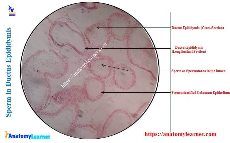
The lumen of these epididymis shows the cluster of sperm under the light microscope. But, there are no spermatogenic cells in the lumen of the epididymis. The structural features of these sperm in the lumen of the epididymis are identical to that of the seminiferous tubules.
Cyclic events in the seminiferous tubules
You know that spermatogenesis is a continuous process within the seminiferous tubules of animals. So, before completing one series of spermatogenesis, another series of spermatogenesis may start.
You will see different spermatogenic stages or events in the seminiferous tubules of the bulls, rams, boar, and other different animals. These stages or events depend on the changes in the shape and staining of the nuclei during the cell division and the release of the sperm into the lumen of the seminiferous tubules.
Okay, let’s see what the cyclic events that are found in the seminiferous tubules of the animals are –
You will see two larger primary spermatocytes basally after the spermiation. Again, the spherical spermatid lies nearest to the lumen of the seminiferous tubules.
The spermatid, along with its dark-stained nuclei, is elongated. These elongated spermatids are arranged in bundles and lie in deep apical recesses of the Sertoli cells. Again, you will see the second generation of primary spermatocytes in the basal region of the seminiferous tubule.
The first and second mitotic division occurs, and two-generation spermatids may present in the seminiferous tubules. You will see the elongated older and newly formed spherical spermatids in the luminal part.
Now, the bundle of the older spermatid have moved away from the vicinity of the Sertoli cell nuclei. The spermatozoa leave the tubular epithelium after separation from their residual bodies.
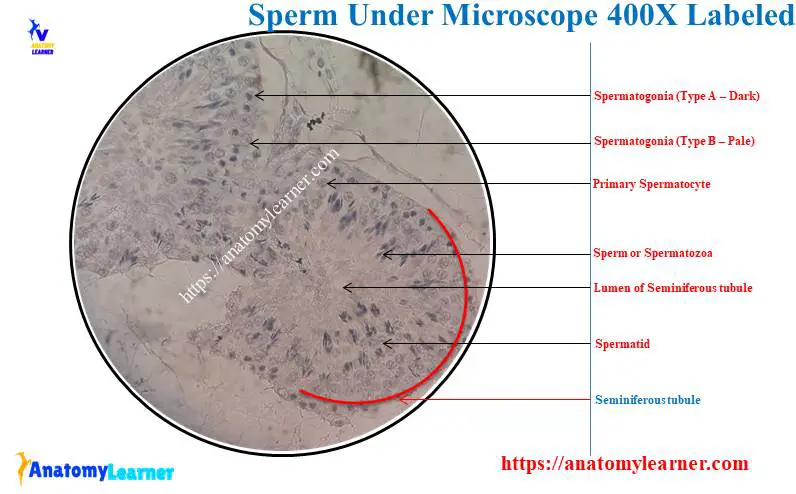
Sperm under microscope 400x labeled
I will show you the sperm under a microscope 400x with the labeled diagram. Here in the diagram, you will see some seminiferous tubules lined by the thick germinal epithelium.
The picture shows the dark Type A and pale Type B spermatogonia located at the seminiferous tubules’ basal part. Again, the primary spermatocytes are also identified from the labeled diagram.
The spermatid in a different stage of development is embedded in the germinal epithelium closer to the lumen. Again, the higher magnification (400x) shows the developing spermatid’s tail protruding into the seminiferous tubules lumen.
Again, you will see the prominent supportive Sertoli cells (nuclei) located throughout the seminiferous tubules’ germinal epithelium.
There is a fibromuscular interstitial connective tissue that surrounds the seminiferous tubule. In the labeled diagram, I tried to show you the testosterone secreting interstitial cells in the fibromuscular interstitial connective tissue layer.
Again, the higher magnification of the seminiferous tubule (400x) will also show different fibrocytes and an inner basement membrane surrounding each seminiferous tubule. In addition, the fibrocytes, blood vessels, nerves, and interstitial Leydig cells are present between the seminiferous tubules.
You will find more sperm-labelled diagrams here on social media for anatomy learners.
Sperm with 40x and 100x labeled diagram
I will also show you the sperm with the 40x and 100x magnification. I will provide the image of the sperm with 40x and 100x magnification both from the seminiferous tubule and epididymis.
The 40x magnification of the seminiferous tubule shows the germinal epithelium and the smaller sperm cells. These sperm cells of the seminiferous tubules are located at the luminal surface, and their tails project towards the lumen.
But, the tail of these sperm cells is not visible with the help of light microscopy (40x magnification). You will get almost all the structures of the seminiferous tubule with 40x magnification.
Again, the seminiferous tubules with 100x magnification clearly show head and tail portions of the sperm. You will see the deep–stained head of the spermatozoa under the microscope with 100x magnification (10×10; objective lens 10x and ocular lens 10x).
But in the epididymis slide with 40x and 100x magnification, you will only understand the cluster of the spermatozoa in their lumen. It is difficult to identify the head and tail parts of the spermatozoa from the epididymis with the help of the light microscope.
Sperm under the electron microscope
If you observe the sperm under an electron microscope, you will easily identify every single part. So, the spermatozoa that appear like the ordinary light microscope will show many details differences under an electron microscope.
But, which electron microscope you should use to observe the details features of the spermatozoa? You may use the scanning electron microscope to view the details features of the spermatozoa.
You know there are the head, neck, middle piece, principal piece, and tailpiece in spermatozoa. The electron microscope will clearly show the spermatozoa’s expanded head and constricted neck region. Again, with the help of the scanning electron microscope, you will see the middle, principal, and tail pieces so clearly.
Here, the spermatozoa labeled diagram shows the head, neck, middle piece, principal piece, and tailpiece with the help of an electron microscope.
Dog sperm under a microscope
You may use the routine or spermac stain to observe the dog sperm under the light microscope. If you use the spermac stain to observe the dog sperm, you will see a red nucleus, whereas the other parts (like the acrosome, middle piece, and tailpiece) show a green colour.
So, you will easily identify the different parts of the dog spermatozoa under the light microscope (as they show different colours with the spermac stain). The middle piece of the dog sperm will show a paler stain as there are no mitochondria.
The acrosomal cap of the dog sperm is a cap-like structure covering most of the head. You may also see an apical thickening on the acrosomal cap of the dog’s sperm.
Again, the post-acrosomal sheet covers the remaining part of the sperm head. The neck of the dog sperm possesses a connecting piece, the complex cross-striated column.
In addition, the mitochondria of the dog sperm are also arranged helically distal end to the annulus. The structure of the flagellum of the dog sperm is identical to these of the ruminant sperm.
Abnormalities of the dog sperm
You may see different types of abnormalities in the dog spermatozoa. Mainly, the abnormalities may be seen in the head, acrosome, middle piece, and tailpiece. Again, the sperm agglutination may find in the dog’s spermatozoa.
The major head abnormalities include the macrocephalic, microcephalic, pyriform, ridged sperm, and double form. Again, the minor head abnormalities of the dog sperm include a narrow head, head-based defect, and detached head.
The acrosomal abnormalities may occur in the form of lipped and crysts. Again, you may see the abnormal distribution of the acrosome in the head of the dog sperm. In addition, you may see the swelling in the acrosomal part of dog sperm. Sometimes the dog sperm shows losses of the acrosome.
The middle piece of a dog sperm may show the retained cytoplasmic droplet, rupture middle piece, and pseudodroplet defect. Again, you may find a minor defect (distal droplet) in the sperm of a dog.
The tail of a dog sperm also shows different abnormalities – like dag defect. Again, the more common abnormality of the dog sperm is the presence of a double tail.
You may also find the simple bend, coiled tail, and terminal coil tail in the dog spermatozoa. Again, the dog sperm may show different agglutination like head to head, tail to tail, head to tail, and other different attachments with different parts of the sperm.
You may find more information on the abnormalities of the dog sperm here with labeled diagrams.
Unhealthy sperm under a microscope
While studying the histology slide of the sperm, seminiferous tubule, and epididymis, you may find some abnormal sperms. This is very difficult to show you all types of abnormal sperm under a light microscope. Here, I will try to show you some of the common abnormal conditions of the animal sperm.
- Defect in the head of the sperm,
- Unhealthy sperm with acrosomal abnormalities,
- Unhealthy sperms with middle and tailpiece abnormalities, and
- Sperm agglutination conditions,
You may see the enlarged head in some sperm (known as the macrocephalic sperm). These macrocephalic sperm may possess double tails. Again, some sperm may possess a small head known as the microcephalic sperms. Some of the sperm possess a pyriform-shaped head.
In addition, you may find the narrow and small heads in some sperm. But, you can not view these unhealthy sperms with the help of a normal light microscope. It would be best if you used the electron microscope to view these spermatozoa abnormalities.
It is common to observe the detached head in most unhealthy sperm.
You may see the irregular distribution of the acrosomal material in the sperms under a light microscope. Again, if you find the decreased acrosomal staining during the viewing of the sperm, it results from the damaged acrosome.
If the sperm can not mature fully, then the retention of the cytoplasmic droplets may occur. Generally, you may find some membranous and granular materials in the cytoplasmic droplets.
The mitochondrial damage may result in the ruptured middle piece of the spermatozoa. Coiled tails and double tails are more common abnormalities of the unhealthy sperm of the animal.
The sperm agglutination may occur as head to head, tail to head, tail to tail, and you may observe it clearly under the light microscopy.
Sperm histology slide
If you are asked to write a short note on the sperm histology slide, you should write the microscopic view of it. I have already described all the histological features of the sperm with the labeled diagram.
First, you should write the identifying points of the sperm histology slide. Then, it would help if you went through the spermatogenesis process (optional). Finally, you should provide the details structure of the spermatozoa.
That means you must describe the histological features of the different parts of spermatozoa – head, neck, middle piece, principal piece, and tailpiece.
You may also write the different abnormalities of the spermatozoa if possible.
Frequently asked questions on sperm under a microscope
So, in this part of the article, I will try to provide specific answers to the frequently asked questions on spermatozoa. You may find more information on the sperm and their histological features in the description part of this article (above).
Okay, let’s see what the common questions on the animal sperm that the histology learners ask are –
Can you see sperm under a regular microscope?
You may see the sperm under a regular microscope easily with the routine stain. All the spermatogenic cells and the sperm may be visible under light microscopy.
The late spermatid may be locked into the apical surface of the Sertoli cells. Again, the spermatozoa or sperm may see on the apical surface that the tail protrudes into the lumen of the seminiferous tubule.
Again, if you see the sperm histology slide, you may easily identify the head, neck, and different pieces under the light microscopy.
Can you see sperm with your eye?
Yes, you can see sperm with your eye through the light microscope. With the help of a light microscope, you will see the head, neck, and different pieces like the middle, principal, and tail of a sperm.
Again, the microscopic slide of seminiferous tubules and epididymis will also show sperm towards their lumens.
What does inactive sperm look like under a microscope?
The inactive sperm shows the head, neck, and tailpiece under a microscope which is similar to the structure of normal healthy sperm. The labelled diagram has already described all the structures of sperm in this article.
Conclusion
So, this article provides the details structural features of sperm under the light microscope. All the labeled diagrams might help you identify the sperms from seminiferous tubules and epididymis of an animal. The sperm’s head, neck, and different parts of the tail will be easily identifiable under light microscopy.
I hope you got the idea of the details of every single structure of the spermatozoa. The sperm under a microscope with 40x, 100x, and 400x labeled diagrams might help you clear the basic concept.