The cow’s knee is a composite joint in its forelimb. Here, I will discuss the cow knee joint anatomy with diagrams showing the bones and ligaments involvement with it.
Quick answer: the cow knee joint is formed by the union of the distal end of the radius and ulna, carpals, and the proximal end of the metacarpals.
I will help you to differentiate the common and special ligaments of the cow’s knee from the horse’s knee joint.
Cow knee joint
The cow knee constitute all joints between the carpal bones. It is almost similar to the structure of a synovial joint except for its special ligaments and bone involvement.
Another name for the cow’s knee joint is the carpal joint.
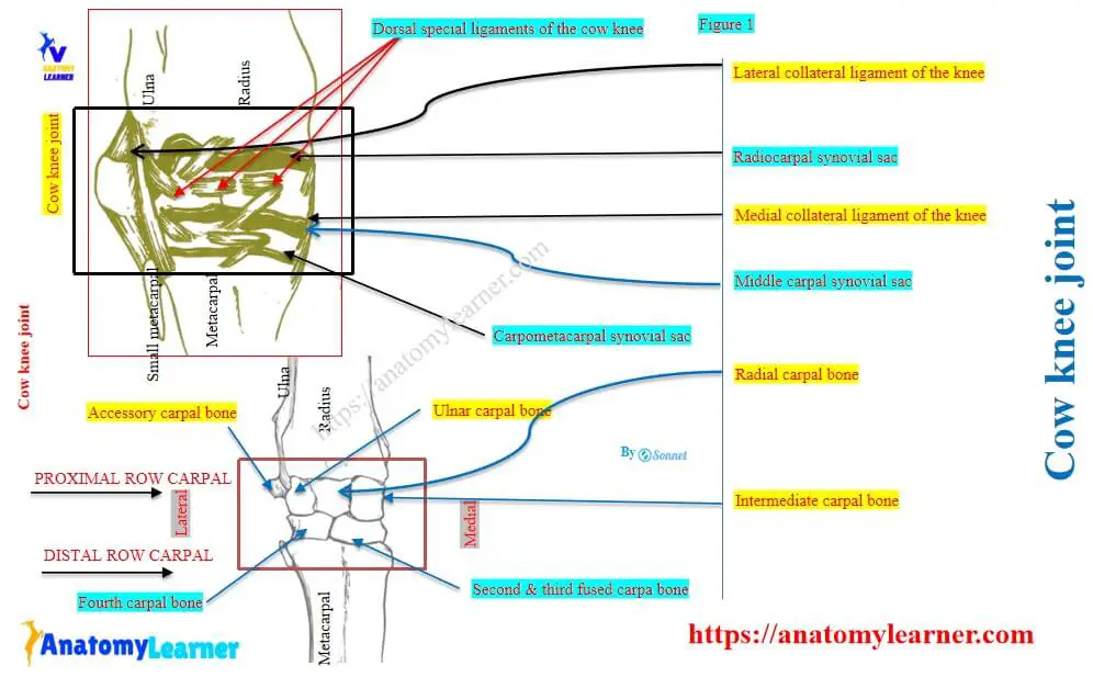
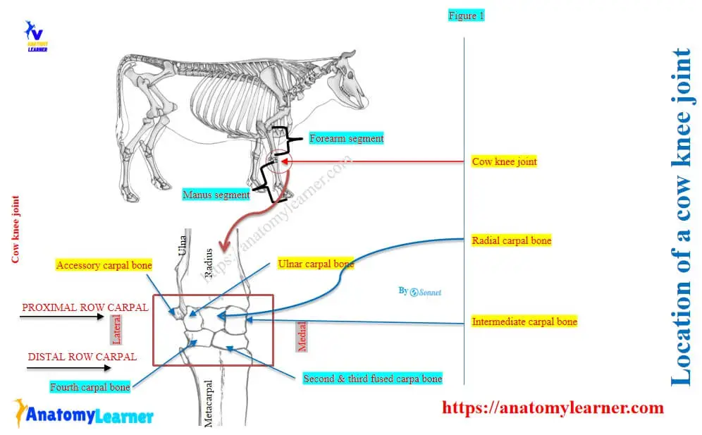
Location and formation: the cow’s knee joint is located between the forearm and manus segments of the forelimb. This joint is formed by the union of the distal articular end of the radius and ulna, two rows of carpals, and the proximal articular end of the metacarpal.
Here, Figure 1 shows the location and formation of the cow’s knee joint between the forearm and manus segments of the forelimb. It also shows the arrangement of the five carpal bones in the carpus sub-segment.
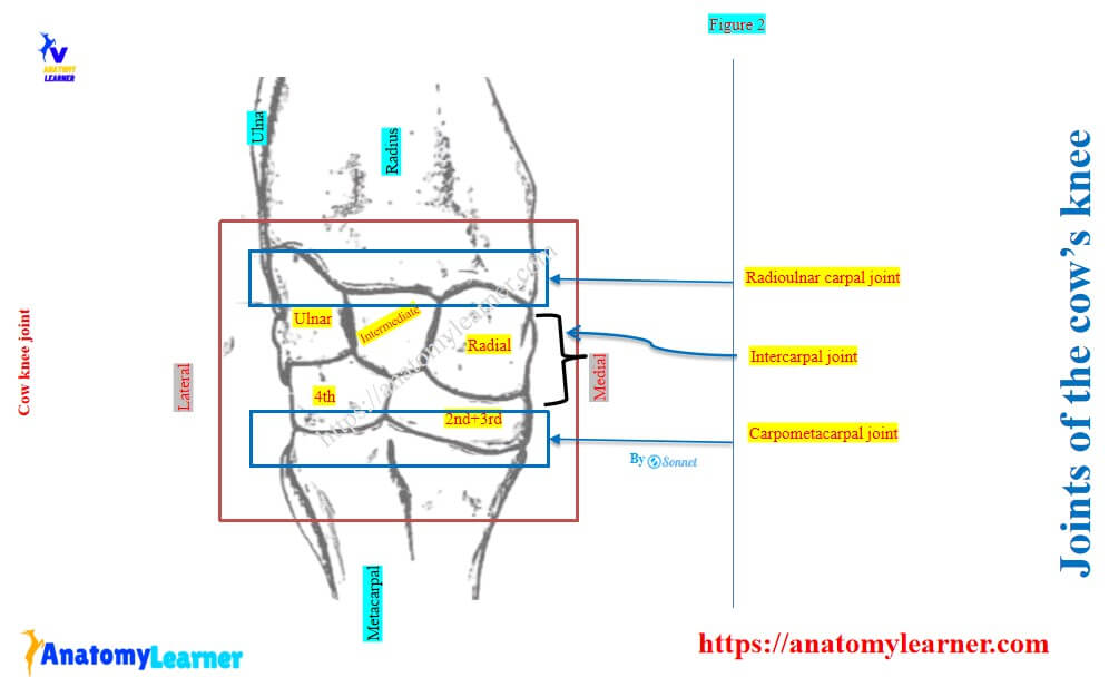
As a composite joint, the cow’s knee subdivides into followings –
- Radioulnar carpal joint / antebrachio-carpal joint: between the distal end of the radius and ulna and proximal carpals,
- Intercarpal joints: among the proximal and distal row carpals, and
- Carpometacarpal joint: between the distal row of carpals and the proximal end of the metacarpal,
Here, the Figure 2 presents the three joints of the cow’s knee among the upper radius and ulna, five carpals, and lower metacarpals.
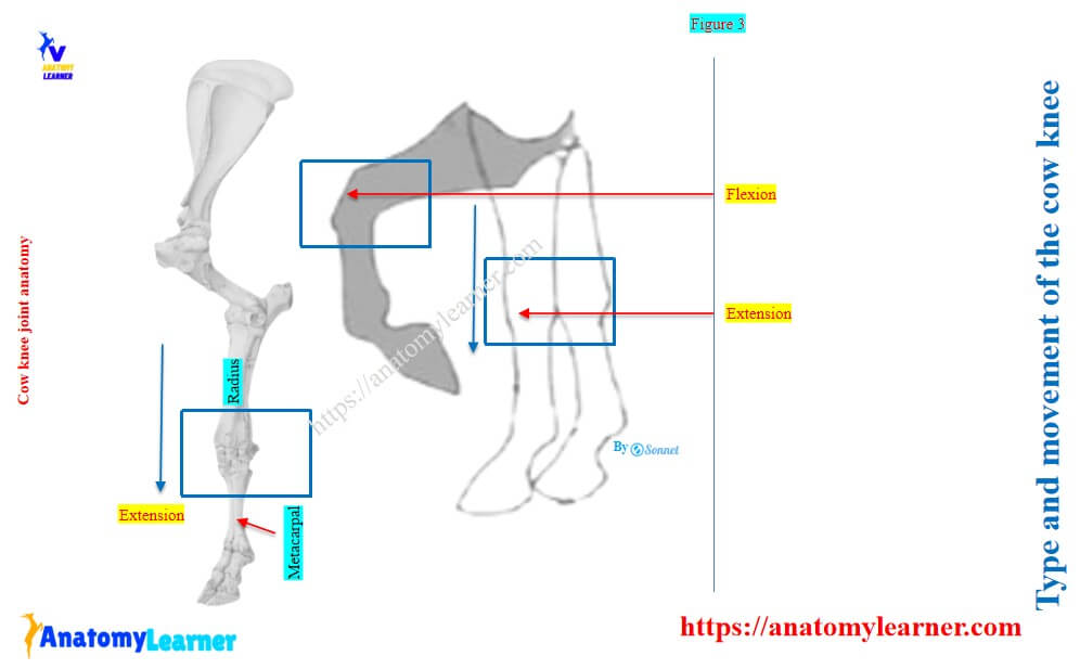
Type and movement of the cow’s knee
Type of joint: the cow’s knee is a hinge/ginglymus type of joint. It is grouped under the uniaxial synovial joint of animals.
This type of joint permits the movement of the bone in one plane of the body. The main movements of the animal’s hinge joints are especially extension and flexion.
Movement of the cow’s knee: the carpometacarpal joint of the cow’s knee shows a slightly gliding movement. Again, the other two joints of the knee show the extension and flexion movements.
The gliding/ translation movement of the animal’s joint occurs when one surface of the bone slides over another surface of another bone.
Here, Figure 3 shows the type and movements of the cow’s knee joint.
Bones’ involvement in the cow knee joint
Bones from the forearm and manus segments are involved in the formation of the cow’s knee joint. Here, I will discuss the bones involved in the cow’s knee with their articular surfaces.
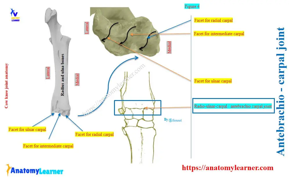
Radio-ulnar-carpal joint of the knee
It is also known as the antebrachio-carpal joint of the cow’s knee. The following bones are involved in the formation of this radio-ulnar carpal joint in the cow’s knee –
- Distal articular ends of the radius and ulna,
- Proximal row of the carpal bones (radial, intermediate, and ulnar carpals),
There are three facets on the distal end of the cow’s radius and ulnar bones. They are arranged from medial to lateral as facet for radial carpal, intermediate carpal, and ulnar carpal.
Here, Figure 4 shows the facets from the distal end of the cow’s radius and ulna bones. It also shows the radio-ulnar-carpal (antebrachicarpal) joint of the cow’s knee structure.
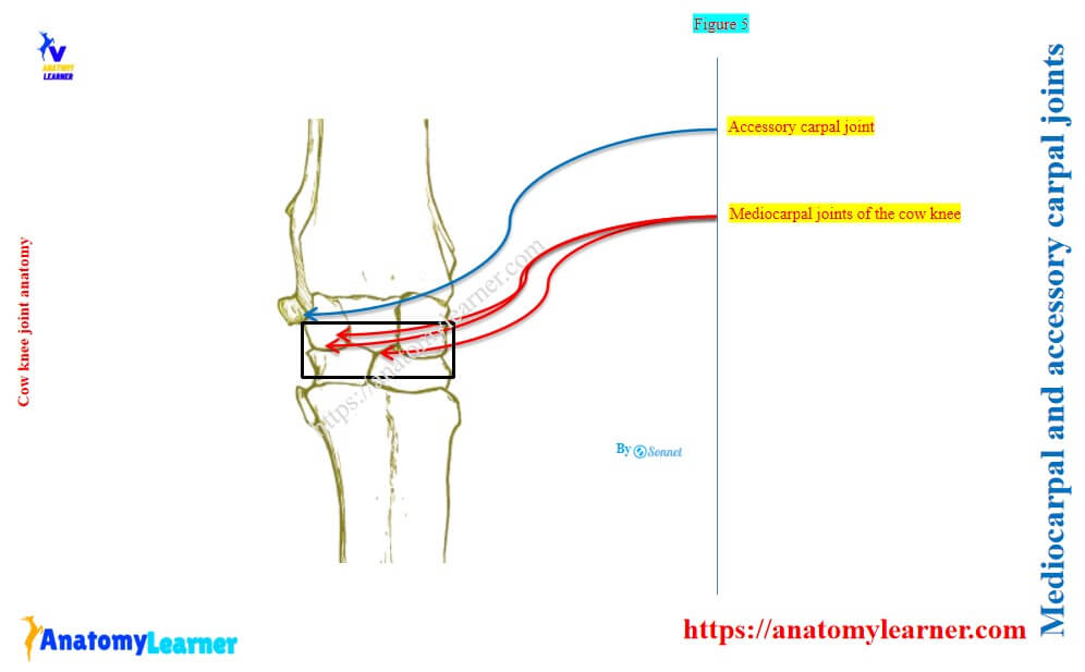
Intercarpal joint of the cow knee
The intercarpal joint of the cow’s knee is formed between the two rows of carpal bones (proximal and distal row carpals). This intercarpal of the cow’s knee again consists of the following joints –
Mediocarpal joint: this is a complex synovial joint between two rows of carpals (except the accessory carpal), and
Accessory carpal joint: the lateral accessory carpal bone forms a joint with the caudolateral aspect of the ulnar carpal bone.
Here, Figure 5 shows the mediocarpal and accessory carpal joints of the intercarpal joint of the cow’s knee.
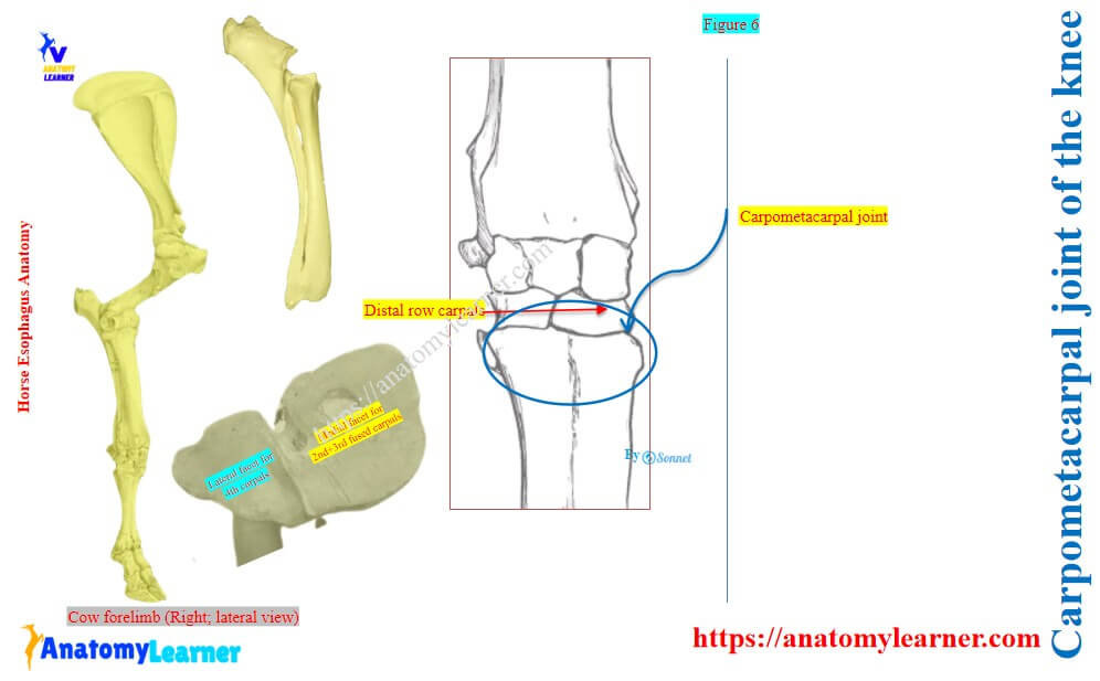
Carpometacarpal joint of the cow’s knee
The carpometacarpal joint is formed between the distal row of the carpals and the proximal articular end of the metacarpal bone. Here, the distal row of the cow’s carpus presents the second and third fused, and fourth carpal (medial to lateral).
Thus, the medial aspect of the distal part of the cow’s metatarsal presents the larger facet.
Figure 6 shows the distal row carpal bone of the cow’s carpus and the proximal end of the metacarpal with its facets. It also presents the carpometacarpal joint of the cow’s knee.
Here, the antebrachiocarpal and intercarpal joints of the cow’s knee are considered as the hinge joint. But, they are not the typical example of the hinge/ginglymus in the animal’s joint.
However, the carpometacarpal joint of the cow’s knee is plane / arthrodial. You will also find the arthrodial joints in the cow’s knee. They are also formed between the adjacent bones of similar rows.
Cow knee joint ligaments
The cow’s knee joint presents two types of ligaments: common and special ligaments. Here, some of the ligaments involved in the whole knee joint structure are known as the common ligaments.
Again, some ligaments are attached to individual bones of the cow’s knee. These smaller ligaments of the cow’s knee are the special ligaments.
Now, I will discuss these common and special ligaments from the cow’s knee joint with diagrams.
Common ligaments of the cow’s knee
The common ligaments of the cow’s knee are –
- Joint capsule / capsular ligament,
- Lateral collateral ligament, and
- Medial collateral ligament,
Joint capsule of the cow’s knee
The joint capsule is the fibrous part of the outer ligament and is common to all three joints of the cow’s knee. This ligament attaches close to the margin of the articular surface of the radius proximally and the metacarpal distally.
The deep part of the joint capsule attaches to the carpal bone and also to the small ligaments of the cow’s knee. Again, this joint capsule divides into two distinct parts –
- Anterior part: it is known as the dorsal carpal ligament. It is loose and forms the fibrous canals for the extensor tendons of the knee.
- Posterior part: it is known as the volar carpal ligament. It is very thick, dense, and closely attached to the carpal bones.
The posterior volar carpal ligament leaves the irregularities of the bone and forms a smooth anterior wall of the carpal canal. This ligament continues downward and forms the inferior cheek ligament in the knee. The cheek ligament bends with the tendon of the deep flexor of the digits.
Synovial membrane of the cow’s knee
The synovial membrane forms three distinct sacs for three joints –
- Radiocarpal synovial sac,
- Intercarpal synovial sac, and
- Carpometacarpal synovial sac,
Here, the radio-ulnar-carpal synovial sac is larger than these other sacs. It encloses the joints formed by the proximal row of carpals and the accessory carpal bones. This sac also includes the special interosseous ligament of the knee.
The intercarpal synovial sac extends from the proximal to the distal/lower row of the carpal bones. It also includes the special interosseous ligament of the cow’s knee. Finally, this sac communicates between the third and fourth carpal bones with the carpometacarpal synovial sac.
Here, the carpometacarpal synovial sac is very limited in the cow knee joint. This sac extends from the distal row of the carpals to the proximal end of the cow’s metacarpals.
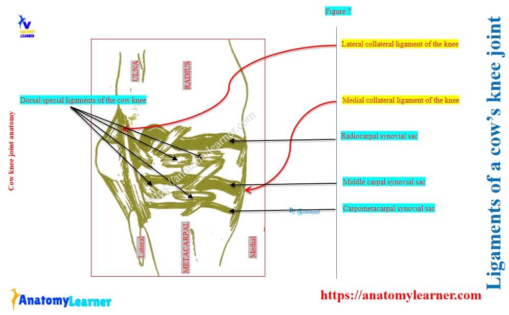
Lateral collateral carpal ligament of the cow knee
The lateral carpal ligament of the cow’s knee attaches to the lateral tuberosity of the distal end of the radius. Again, the long superficial part attaches below to the proximal end of the small metacarpal bone.
But, you may also find some of the fibers that extend to the proximal end of the large metacarpal bone.
There are two deep parts of the lateral carpal ligament in the cow’s knee. One deep part attaches to the ulnar carpal bone on its lateral aspect. Another deep part of the lateral carpal ligament attaches to the fourth carpal with the metacarpal bone.
Medial collateral carpal ligament of the cow’s knee
The medial carpal ligament of the cow’s knee is stronger and wider than the lateral carpal ligament. This ligament is proximally attached to the medial tuberosity of the distal end of the cow’s radius bone. Again, it ends below on the proximal end of the large metacarpal bone.
There are two small parts of the medial carpal ligament. These two small part attaches to the radial and second and third fused carpal bones of the cow’s carpus.
The posterior part of this ligament attaches to the transverse ligament of the cow’s knee. It helps to form the canal for the flexor carpi tendon.
Here, Figure 7 shows the lateral and medial collateral ligaments of the cow’s knee joint.
Special ligaments of the cow’s knee joint
You will find different short ligaments that attach two individual bones of the cow’s carpus. Let’s see the short/special ligaments of the cow’s knee –
- Radio-ulnar-carpal joint: it presents two types of special ligaments – two anterior and three posterior.
- Special ligaments in proximal carpals: there are two anterior, three posterior, and one interosseous ligament in the proximal rows of carpals.
- Intercarpal joint: it presents two anterior and three posterior special ligaments.
- Special ligaments in distal carpal bones: there is one anterior and one interosseous ligament in the distal/lower row of the carpal bones of the cow’s knee.
- Carpometacarpal joint: it presents two posterior and two interosseous special ligaments.
But the number and size of these special ligaments in the cow’s knee joint vary.
Comparative anatomy of the knee joint between the cow and the horse
The general structure of the cow and horse knee are almost similar. But there are great variations in the structure and number of the bones involved in the horse’s knee joint.
Thus, you will find a variation in the appearance and attachment of the different ligaments compared to the cow’s knee. Some of the key variations in the ligaments of the knee between cow and horse are shown in the table –
| Features | Horse knee | Cow knee |
| Lateral collateral ligament | Stronger and wider | Small and weak |
| Medial collateral ligament | Ends at small and large metacarpal bones | Ends at the large metacarpal |
| Joint capsule | Strong and thick | Strong and thin |
| Synovial membrane | Voluminous | Less voluminous |
FAQ’s on cow knee
Yes, the cows have the knee joints in between the forearm and manus segments of its forelimb. It is a composite synovial joint that is mainly responsible for the extension and flexion movements.
Radio-ulnar-carpal, intercarpal, and carpometacarpal are the 3 joints of the cow’s knee. Here, the carpometacarpal joint is arthrodial, whereas the radio-ulnar-carpal and intercarpal joints are hinge-type joints.
The knee joint is called the carpus joint/articulatio carpi in animals. Most of the animals like horses, sheep, goats, and cow shows almost a similar structure in their knee joints, with a few exceptions in their ligaments.
Conclusion
So, the cow knee joint comprises the radio-ulnar-carpal, intercarpal, and carpometacarpal joints. Thus, it is considered a composite synovial joint and is formed by the fusion of the distal end of the radius and ulna, carpals, and the proximal end of the metacarpals.
There are common and special types of ligaments in this cow’s knee joint. Joint capsule, lateral, and medial carpal ligaments are the common ligaments in the cow’s knee. Whereas, there are different short special ligaments present in the anterior and posterior aspects of the carpal bones of the cow’s knee.
References
- Choudhary OP, Saini S: Anatomy of the knee joint in cattle (Bos taurus): Gross, radiographic and computed tomographic insights. Kafkas Univ Vet Fak Derg, 31 (2): 215-221, 2025.
- Supriya B, Rao TSC, Ramayya PJ: Anatomy of the carpal articulation of buffalo calves (Bubalus bubalis). Buffalo Bull, 35 (4): 653-660, 2016.
- Chauveau A., Comparative Anatomy of the Domestic Animals. 2nd ed., 194-225, Appleton Company New York, 1891.
- Sisson S: Ruminant Syndesmology: The Anatomy of the Domestic Animals. 5th ed., 787- 790, W.B. Saunders Company, Philadelphia USA, 1975.
- König HE, Hans-Georg LH: Veterinary Anatomy of Domestic Animals: Textbook and Colour Atlas. 7th ed., 171-217, Stuttgart: Georg Thieme Verlag, Germany, 2020.
- Ghosh RK: Primary Veterinary Anatomy. 9th ed., 85-103, Current Book International Kolkata, India, 2024.
- Karimi H, Ardalani GH, Moghaddamm G: Anatomical structure of buffalo’s carpus. J Fac Vet Med Univ Tehran, 57 (4): 17-22, 2002.