Fetal circulation is somewhat different than the general blood circulation in the animal’s body. I will provide the fetal circulation flow chart diagram in this short article.
Here, you will also find a concise explanation of this short fetal circulation flow from the foetus. Let’s see how fetal blood circulation differs from the normal blood circulation of the body.
Fetal circulation flow chart
In fetal circulation, most of the blood from the right atrium goes to the left atrium instead of going to the right ventricle through the foramen ovale. Now, the blood from the left ventricle goes to the different parts of the body, limbs, organs, and head through the aorta.
A little amount of blood that goes to the right ventricle from the right atrium passes through the pulmonary artery. Now, from the pulmonary artery, most of the blood goes to the descending aorta through the ductus arteriosus.
From the head, neck, and forelimb of the fetus, blood returns to the right atrium through the cranial vena cava. Again, blood that goes to the viscera and hind limb passes through the umbilical arteries to the placenta.
From the placenta, blood reaches the caudal vena cava (explain in the next part). Finally, the fetal blood goes to the right atrium of the heart through the caudal vena cava.
I hope the below-mentioned fetal circulation flow chart might help you to understand this circulation perfectly –
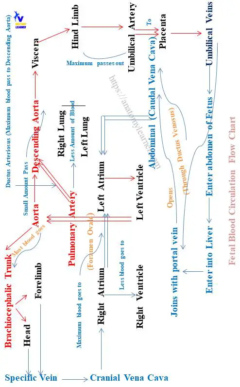
You may easily identify the structures or organs that involve this system directly from this circulation flow chart. Let’s see the organs or structures that send blood from the different parts of the fetal body –
- Structure sends blood to the body – left ventricle, aorta, abdominal aorta, brachiocephalic trunk, and also the pulmonary trunk,
Again, the structure that returns the blood to the fetus’s heart –
- Fetal caudal and cranial vena cava,
Unique features of fetal circulation
You will find 7 unique features in the fetal blood circulation compared to the general circulation of the animal’s body –
- Most of the blood passes to the left atrium directly through the foramen ovale,
- A little amount of blood passes to the right ventricle,
- Blood that passes from the right ventricle to the pulmonary trunk finally reaches the descending aorta (most of the blood),
- A small amount of the blood goes to the inactive fetal lungs,
- From the viscera, maximum blood passes to the placenta through the umbilical arteries,
- The umbilical veins enter the abdomen and liver and join with the portal vein,
- Finally, blood reaches the caudal vena cava through the ductus venosus,
I hope you got the basic idea of fetal circulation from these features. Now, you may easily compare the fetal and general blood circulation of the animals.
Now, let’s explain the flow chart of this foetal circulation.
Fetal circulation flow chart explanation
Now, let’s explain the fetal blood circulation flow chart with the diagram. Let’s start with the right atrium of the fetus. In the right atrium of the fetus, blood returns from the head, forelimb, viscera, and placenta.
You will find the foramen ovale in the interatrial septa of the fetus’s heart. Through this foramen ovale, Maximus blood goes to the left atrium of the fetal heart. A little amount of blood goes to the right ventricle of the fetus’s heart.
You know the foramen ovale of the fetus closes at birth.
Now, blood from the left atrium directly goes to the left ventricle of the fetus’s heart. And you know, from the left ventricle, blood goes to the different parts of the body through the aorta.
So, from the aorta of the fetus, blood sends to the descending aorta and the brachiocephalic trunk. Now, from the abdominal aorta of the fetus, blood passes to the viscera and hind limb.
Again, through the brachiocephalic trunk, blood sends to the head and forelimb region of the fetus. In addition, a little amount of blood comes to the pulmonary trunk from the right ventricle of the fetus.
Maximus blood from the fetal pulmonary trunk passes to the descending aorta through the ductus arteriosus. You know this ductus arteriosus of the fetus becomes ligamentum arteriosus (band-like structure) after birth.
Blood passes to the viscera and hind limb that finally comes to the placenta (maximum amount) through the umbilical arteries.
Blood returns to the fetal heart
From the placenta, oxygenated blood will carry through the umbilical veins into the fetus. You know there are 2 umbilical veins in the fetus where only the left one persists.
Now, the umbilical veins enter into the abdomen of the fetus and finally enter into the liver. These umbilical veins join with the portal vein and hepatic veins.
Finally, they open into the abdominal vena cava (caudal vena cava) through the ductus venosus. And you know, blood will then passes into the right atrium of the fetus’s heart from the caudal vena cava.
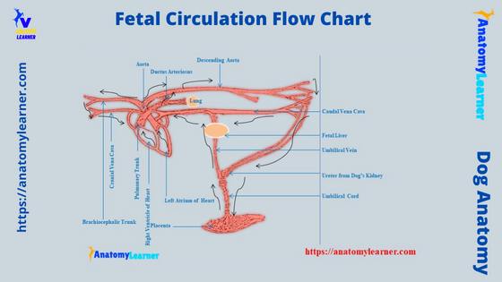
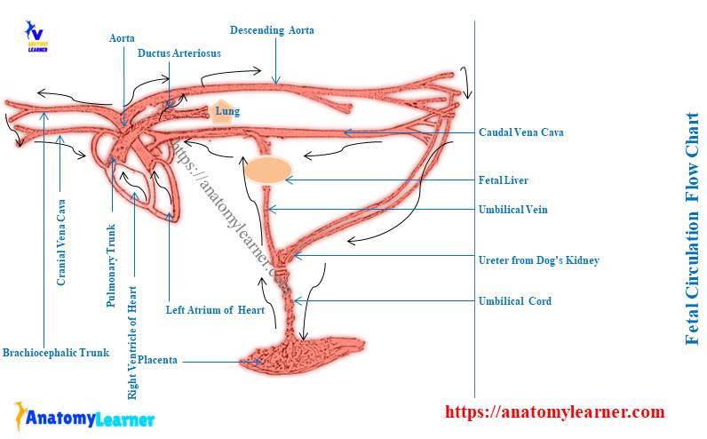
Fetal blood circulation flow chart diagram
Now, I will also provide a fetal blood circulation flow chart diagram that might help you better understand this circulation. Here, in the fetal circulation diagram, I tried to show the blood pathways from the fetus’s heart to the different viscera of the body, limbs, and neck.
Again, the fetal circulation flow diagram shows how the blood goes to the placenta from the viscera and hindlimb. The umbilical veins that collect blood from the placenta and enter the fetus’s abdomen are also shown in the diagram.
In addition, the umbilical veins enter the liver and join with the portal veins are also shown in the diagram. Finally, the diagram expresses the flow of the blood that passes from the caudal vena cava to the heart and also from the cranial vena cava to the right atrium of the fetal heart.
You will find more diagrams on pulmonary and systemic blood circulation here.
What is the pathway of fetal circulation?
Here, you will find 3 major pathways in fetal blood circulation –
- Blood passes from the left atrium to the head and body,
- Blood passes to the placenta, and
- The blood returns back to the left atrium – through caudal and cranial vena cava,
A short overview of the fetal blood circulation pathways has already been shown and described. Please follow that short guide and chart to get a full concept of fetal circulation.
What are the five structures of fetal circulation?
If you see the fetal circulation flow diagram, you will find the below-mentioned important and exceptional structures involve in this circulation –
- Foramen ovale (not persists after birth),
- Ductus arteriosus (become ligament arteriosus after birth),
- Ductus venosus (communicate with the abdominal vena cava),
- Placenta – a combination of fetal and maternal circulations, and
- Umbilical veins and arteries – umbilical veins collect blood from the placenta, and arteries help to pass blood to the placenta from the viscera and hindlimb,
Hopefully, you will find all these five structures of fetal circulation in the labeled diagram I provided.
What are the three shunts in fetal circulation?
In the fetal blood circulation, you will find the below –mentioned 3 shunts –
- Foramen ovale shunt,
- Ductus arteriosus shunt, and
- Ductus venosus shunt,
Here, the foramen ovale acts as a shunt that passes oxygenated blood from the right atrium to the left atrium of the fetal heart. Thus, it prevents excess blood from passing into the pulmonary trunk. And it indirectly presents the overload in the inactive lungs of the fetus.
The ductus arteriosus acts as a shunt that passes the maximum blood to the descending aorta and prevents excess load for the inactive lungs. Finally, the ductus venosus acts as a shunt that helps to communicate the umbilical vein (joins with hepatic) with the caudal vena cava.
What does fetal circulation mean?
Fetal circulation means the circulation in the fetus or unborn baby. You will find a great variation in the blood circulation between the fetus and newborn.
I have already discussed the basics of fetal blood circulation with their exceptions. Here, the lung of a fetus receives a little amount of blood that prevents it becomes overloaded. Again, you will see the active participation of the placenta in the fetus blood circulation.
In addition, the umbilical veins play an important role in conveying blood from the placenta to the caudal vena cava. But, there is no such event found in the normal or general blood circulation in the animal.
Suggested reading for you from anatomy learner –
- What is the difference between pulmonary and systemic circulation
Conclusion
So, this short guide might help you get fetal circulation basics. Here, the fetal blood circulation flow chart differs from the general systemic and pulmonary circulation.
Unique blood flow in the fetal circulation finds through the foramen ovale, ductus arteriosus, and ductus venosus. But, you will not find such features in the general blood circulation after birth.