The dog pancreas anatomy consists of 2 lobes – right and left which are united by a body. It is a solid gland located mainly at the right side of the median plane and possesses both exocrine and endocrine functions.
In this article, I will describe the anatomical facts of the dog pancreas with a labeled diagram. Again, you will find the comparison of pancreases of other different animals with the dogs.
Quick summary of a dog’s pancreas anatomy: pancreas is a pinkish-gray large lobulated and elongated solid gland in a dog. Anatomically, it possesses the right and left lobes and locates at the right side of the median plan along with the stomach and duodenum.
If you want to learn the details and anatomical facts of the dog pancreas, let’s continue this article until the end.
Dog pancreas anatomy
The dog pancreas anatomy is somewhat externally different from the ruminant and horse. You will see a V-shaped pancreas in the dorsal part of the cranial and right lateral abdominal region of a dog.
First, let’s see some of the important anatomical facts about the dog pancreas from the labeled diagram –
- The flat and rounded right lobe of the dog pancreas,
- Left lobes of the dog pancreas, which possess – dorsal and ventral surfaces,
- The body of the dog pancreas,
- 2 excretory ducts of the pancreas – pancreatic and accessory ducts,
- Cranial and caudal pancreaticoduodenal arteries of the dog pancreas,
- Pancreaticoduodenal veins, and
- Nerves related to the dog pancreas,
All these external features from the dog pancreas are identified in the labeled diagram. Now, let’s see the unique gross features of the dog pancreas –
- There are 2 branches or parts in the dog pancreas – right and left lobes,
- These 2 lobes of the dog pancreas are united by the body at an angle of approximately 45 degrees,
- Again, both these right and left lobes of the dog pancreas meet together behind the pylorus and form a V-shaped structure,
- There are 2 pancreatic ducts found in the dog pancreas structure,
- The main pancreatic ducts of the dog pancreas open into the duodenum (just 4 centimeters behind the opening of the bile duct),
- Again, the accessory duct of the dog pancreas joins with the common bile duct,
But, sometimes, the accessory pancreatic ducts of the dog pancreas open into the duodenum separately. When the right and left lobes of the pancreas meet together to form the V-shaped structure, they may extend up to the corresponding kidneys.
Now, let’s get into the details of external anatomical facts of the dog pancreas with labeled diagrams.
Dog anatomy pancreas location
In short, the dog pancreas is located in the dorsal part of the cranial and right lateral abdomen. It forms a V-shaped structure in a dog, lies on the right aspect of the stomach, and attaches to the duodenum.
Anatomically, this solid pancreas gland of the dog divides into –
- Thin and slender right lobe – lies on the duodenum, and
- The short, thick, and wider left lobe – lies on the greater omentum,
These two lobes of the dog pancreas join at the body, which lies just caudomedial to the pylorus. Thus, you will find a Y-shaped structure of the pancreas in the pylorus of a dog.
Sometimes this joining structure (2 lobes with the body of the pancreas) is known as the pancreatic angle.
Where is a dog’s pancreas diagram?
Now, the dog’s pancreas diagram might help you understand its exact location. Here, the dog’s organ anatomy diagram shows the exact location of the dog’s pancreas in its abdomen.
You may also know the details of other different abdominal organs of a dog from the below-mentioned article –
- Dog organ anatomy with the labeled diagram,
Here, the diagram shows the location of the dog’s pancreas in the abdomen. You will find the right lobe of the dog pancreas just caudal to the liver and stomach.
Again, just caudal to the pancreas, you will see the kidney of the dog.
External features of dog pancreas (color, shape, size, weight)
From table 1, you will find the specific color, shape, size, and weight of the dog pancreas –
| Dog pancreas anatomy | Mesurement/ appearance |
| Color | Yellowish / pinkish gray |
| Shape | V-shaped |
| Appearance | Solid, lobulated, elongated gland |
| Length | 25- 30 centimeters |
| Right lobe length | 9 – 15 centimeters |
| Left lobe length | 8 – 10 centimeters |
| Width (right lobe) | 1 – 2 centimeters |
| Width (left lobe) | 3 – 4 centrimeters |
| Weight | 15 – 25 grams |
| Lobes | Right and left lobes |
| Accessory pancreas | Rarely occur in dogs |
| Ducts | Pancreatic and accessory ducts |
So, the color of the dog pancreas may vary in different conditions. You may find the yellowish-gray color in the dog pancreas when they preserve for a long time.
Again, you may find the pinkish-gray color in the freshly obtained pancreas of the dog.
You will also see the larger lobulation and elongated appearance in the dog pancreas. These lobules are pointed and form a nodular surface with irregular margins.
Again, the shape of the dog pancreas is almost V-shaped because of the position of the right and left lobes.
The average weight of the dog’s pancreas is about 15 – 25 grams. But, the weight of the dog’s pancreas may vary in size and breed of the dogs.
The total length of the dog’s pancreas (including the right and left lobes) is about 25 – 30 centimeters. But, the average length of the specific lobes (right and left) of the dog pancreas may vary.
In table 1, I have already shown the average length and width of the right and left lobes of the dog pancreas. But you will learn the other gross anatomical facts from the dog pancreas’ right and left lobes in the next section of this article.
The right lobe of canine pancreas anatomy
The right lobe of the dog pancreas anatomy is comparatively larger than these of the left lobe. This lobe of the canine pancreas lies in the mesoduodenum.
You will find the contact of the right lobe of the canine pancreas with the dorsal part of the right abdominal wall. Topographically, this part of the pancreas extends from the ninth intercostal space (9th) to the fourth lumbar vertebra.
The rounded and flattened caudal extremity of the right lobe of a dog pancreas lies in the concavity of the duodenal loop. You may easily separate this pancreas from the duodenal loop of the mesoduodenum.
Towards the pylorus, the dog pancreas becomes more flattened and narrow dorsoventrally. Thus, you will find both the dorsal and ventral surface in this right lobe of the dog pancreas.
The caudal part of the right lobe (large) is related to the sublumbar region of the dog. Here, you will find a great relationship between the right lobe of the pancreas with some of the important organs of dogs.
This right lobe has contact with the uterus, the ventral surface of the right kidney, and also with the caudate lobe of the dog’s liver. Again, you will find the following relationship between the right lobe of the dog pancreas with other different organs –
- Ventrally – it relates to the parts of the dog’s ileum,
- Caudally – relates to the part of the cecum, and
- Cranially – relates with the ascending color of the dog,
Again, the ventral surface of the right lobe of a dog’s pancreas relates to the jejunum loop. Although, most of the ventral surface of the right lobe has great contact with some of the other visceral organs.
Size of the right lobe of a dog pancreas
You know the size of the right lobe of the dogs pancreas may vary with the age and breed. So, I will provide the average size (length, width, and thickness) of the dog’s pancreas.
A medium-sized dog has a right lobe in the pancreas about 9 – 15 centimeters long. Again, the thickness and width of the right lobe of a dog’s pancreas vary as the shape changes from cranial to caudal extremities.
Averagely, you may find a variable thickness (1 – 3 centimeters) in the dogs pancreas. But, the width of the right lobe is extremely variable as this part of the pancreas sometimes becomes round and again converts into a flattened structure.
Again, the average weight of the right lobe of the dog pancreas varies from 10 – 15 grams.
Left lobe of the dog’s pancreas
The left lobe of the dog pancreas is somewhat structurally different than these of the right lobe. Again, this left lobe of the canine pancreas is comparatively larger than the right lobe.
Now, let’s see the location of the left lobe of the dog’s pancreas exactly. You will find the left lobe of the pancreas in the deep wall of the greater omentum of the dog.
This lobe arises from the body segment of the pancreas and runs caudosinistrally. This lobe is a flattened structure with distinct dorsal and ventral surfaces.
Summary of the surfaces of the left lobe of the dog pancreas –
- The dorsal surface (small) of the left lobe – has contact with the liver, aorta, portal vein, caudal vena cava, and
- A ventral surface of the left lobe of the pancreas – has contact with the colon and stomach of the dog,
The dorsal surface of the left lobe of a dog’s pancreas lies slightly right of the median plane of the body. You will find the contact of this left lobe of the pancreas with other different organs of the dog abdomen.
Followings are the organs that have contact with the left lobe of the dog pancreas –
- Caudate process of the liver (on the right side), and
- Portal vein, caudal vena cava, and aorta (on the left side),
The left part or lobe of the dog pancreas ends in the left aspect of the sublumbar region. Here, you will see the close relationship of the left lobe with the cranial pole of the left kidney.
Again, you will find the close relationship of this lobe of the dog pancreas with the middle part of the spleen.
Ventral surface and size of the left lobe of dog pancreas
The ventral surface of the left lobe of a dog pancreas has contact with only 2 organs. You will see the contact of the left lobe with the transverse color ventrocaudally.
Again, the dorsal wall of the dog stomach contacts with the left lobe of the pancreas ventrocranially.
The length of the left lobe is about two-thirds of the total length of the dog pancreas. Averagely, you may find the length of the left lobe of a dog pancreas as 8 – 10 centimeters.
The thickness of the left lobe of the dog pancreas also varies from 2 – 4 centimeters. Again, the weight of the left lobe may vary from 8 – 18 grams.
Body of the dog pancreas anatomy
The body of the dog pancreas is a smaller portion that unites the right and left lobes. It forms a Y-shaped structure and binds these lobes at an angle of approximately 45 degrees.
The body of the dog pancreas lies close to the caudosinistral part of the pyloric region. Here. You will find a larger concave impression on the cranial part of the pancreatic body.
The body of the dog pancreas becomes 1 centimeter thick and 3 – 4 centimeters wide, just caudal to the concave impression. You will see the portal vein that crosses the dorsal part of the body of the pancreas.
There are pancreaticoduodenal and gastroduodenal veins that cross on the right side of the body. Again, they disappear into the dog pancreas at the caudal to the concave depression of the body part.
Do dogs have accessory pancreas?
Yes, the dogs occasionally have an accessory pancreas. So, the accessory pancreas is not normal in dogs.
This accessory pancreas of the dog (if they possess it) locates in the wall (lateral) of the gallbladder and in the caudal part (1) of the mesentery.
Ducts of the dog or canine pancreas
You will find 2 excretory ducts in the dog or canine pancreas. Sometimes, you may find 3 (2 pancreatic and 1 accessory) ducts in the dog or canine pancreas.
Within the pancreas of the dog, these 2 excretory ducts are intercommunicated. Sometimes, they (2 ducts) may cross each other within the parenchyma of the dog pancreas.
Let’s see what the excretory ducts in the dog or canine pancreas are –
- Pancreatic duct (normally 1 in number, sometimes 2 in some of dog) – drain the right lobe of the pancreas, and
- Accessory pancreatic ducts (1 in number) – drain the left lobe of the dog’s pancreas,
In the embryonic condition, the pancreatic duct drains the embryonic ventral pancreas. Whereas the accessory pancreatic ducts drain the embryonic dorsal pancreas.
Let’s know a little about the pancreatic and accessory ducts of the canine or dog pancreas.
A pancreatic duct of the dog’s pancreas
The pancreatic duct (large) of the dog pancreas is smaller than the accessory pancreatic duct. You may find only 1 larger pancreatic duct in the dog pancreas.
But, occasionally, you may also find the smaller pancreatic duct in the dog pancreas.
- Small pancreatic duct (if persist in the dog pancreas) – aires from the right lobar duct, and
- Larger pancreatic duct – arises from the left lobar ducts,
The opening of the dog pancreatic duct is always closely associated with the bile duct. This larger pancreatic duct of the dog opens in the opening of the bile duct and enters into the duodenum on the major duodenal papilla.
After the larger pancreatic duct forms from the left lobar ducts of the dog pancreas, it crosses with the accessory pancreatic duct.
Externally these 2 ducts of the dog pancreas form a Y-shaped junction and enter the duodenum at the major duodenal papilla.
The accessory pancreatic duct (main) of the dog pancreas
The accessory pancreatic duct is the larger duct in the dog pancreas. This main duct of the dog pancreas is formed by the union of two lobar ducts from the right lobe.
This accessory pancreatic duct may occur at the level up to the intestinal wall. It (accessory pancreatic duct) opens into the duodenum on the minor duodenal papilla.
They actually open through the mesenteric wall of the proximal part of the descending duodenum. And you know this is located to the left of the cranial pancreaticoduodenal vessels.
So, the opening of the pancreatic and accessory pancreatic ducts of the dog pancreas are separate. But, the opening of these ducts in other different species may vary.
Vessels in a dog pancreas anatomy
The right and left lobes of the dog pancreas anatomy receive different vessels. Here, the right lobe (identified) of the dog pancreas receives the pancreatic branches of cranial and caudal pancreaticoduodenal arteries.
These 2 arteries form anastomoses in the pancreas gland.
So, the main arteries of the right lobe of dog pancreas – pancreatic branch of cranial and caudal pancreaticoduodenal arteries,
The left segment of the left lobe of a dog pancreas receives the pancreatic branch of the splenic artery. So, this branch of the splenic artery is the main arterial supply to the left lobe of the dog pancreas.
Again, the left lobe of the dog pancreas also receives arterial supply from the hepatic artery. This pancreatic branch of the hepatic artery runs the dorsal surface of the left lobe of the dog pancreas.
The body of the dog pancreas receives one or more branches from the gastroduodenal artery. Again, the free end of the dog pancreas also receives pancreatic branches from the celiac artery.
The right pancreatic lobe of the dog pancreas drains blood through the caudal pancreaticoduodenal vein. This pancreaticoduodenal vein joins with the cranial mesenteric vein of the dog.
Again, the left lobe of the dog pancreas drains blood through the two branches of the splenic vein. The small branches of the cranial pancreaticoduodenal vein also drain blood from the body of the dog pancreas.
Now, the lymphatics from the dog pancreas drain into the duodenal lymph nodes. Again, the lymphatics also may drain into the hepatic, splenic, and jejunal lymph nodes of the dog.
Nerve innervation to dog pancreas
The sympathetic axons from the celiac plexus innervate most parts of the dog pancreas. These sympathetic axons travel with the pancreatic branches of the cranial pancreaticoduodenal and celiac arteries.
Again, the caudal part of the dog pancreas innervates with the sympathetic axons of the cranial mesenteric artery. This axon runs along with the caudal pancreaticoduodenal artery.
You may also find the parasympathetic axon from the vagus nerve that innervate the different areas of the dog pancreas.
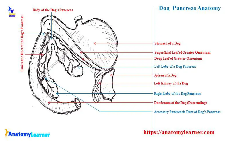
Dog pancreas anatomy labeled diagram
Now, let’s see the labeled diagram of the canine pancreas anatomy. Here, the diagram shows the different parts of the stomach, spleen, pancreas, and different parts of duodenum of the dog.
The left lobe of the dog pancreas is identified in the labeled diagram. Here, the left lobe of the pancreas is attached to the deep leaf of the greater omentum of the dog.
The labeled diagram also shows the body of the dog pancreas that join the right and left lobe and forms an angle. Again, in the diagram, the right lobe is identified from the wall of the descending duodenum of the dog.
The pancreatic duct, accessory pancreatic duct, and major and minor duodenal papillae are also identified from the dog pancreas labeled diagram.
You may also find more diagrams on the dog pancreas and some related organs here on the social media of anatomy learners. Again, you may learn about the histological features of the exocrine and endocrine parts of the dog pancreas from the below-mentioned article of anatomy learner –
- Pancreas histology slide – histological features pancreatic Islet with the labeled diagram,
Histologically, you will find the compound glands (both serous and tubular) in the parenchyma of the dog pancreas. Externally, you will find a thin layer of connective tissue capsule in the dog pancreas histology slide.
This thin layer of connective tissue enters into the parenchyma of the dog pancreas and divides into different lobules. Each of these lobules consists of numerous acini and numerous pancreatic lobar ducts.
You will find several pyramidal-shaped cells in each acini of the dog pancreas. Again, there are some centoacinar cells found in the dog pancreas. The Islet of Langerhans is the endocrine part of the dog pancreas, which histologically possesses different types of cells.
Pancreas in animals (Cow, horse, and pig compare to dogs)
You will find a significant difference in the anatomy of cows, horses, and pigs’ pancreas compared to these of dogs. The cow or ox pancreas possesses a great variable feature to compare to the dogs.
First, let’s see the anatomical features of the cow or ox pancreas compared to the dogs. Again, you will find little information on the anatomical facts of the pancreas of the following species –
- Horse pancreas anatomy compare to the dog,
- Anatomical facts of the sheep, goat, cow, and
- Pig pancreas anatomy compare to the dog,
Cow pancreas anatomy
The cow pancreas is a soft gland located chiefly on the right side of the median plane of the body. You will see the below-mentioned anatomical facts in the cow or ox pancreas compare to these of the dogs –
- The cow pancreas attaches to the ventral surface of the liver by the layer of the mesentery,
- You will find the reddish-yellow pancreas in the cow,
- The cow or ox pancreas is a flat and irregular quadrilateral organ,
- You will see 2 surfaces and 4 borders in the pancreas of the cow,
- On the dorsal surface of the cow pancreas, you will find the attachment with the liver, diaphragm, kidney, abdominal vena cava, and mesenteric arteries,
- Again, the ventral surface of the cow pancreas relates to the dorsal sac of the rumen, color, and hepatic artery,
- The cow pancreas possesses cranial, caudal, lateral, and medial borders,
You may easily identify these cranial, caudal, lateral, and medial borders of the cow pancreas with the help of their unique features. Here, the cranial border of the cow pancreas is more or less straight.
You will see the notch in the caudal border of the cow pancreas to accommodate the portal vein.
The medial border of the cow pancreas is long compared to the lateral and runs parallel to the duodenum. Again, the lateral border of the cow pancreas is convex and irregular in shape.
The main arterial supply for the cow, sheep, or goat pancreas are branches of the celiac artery, hepatic artery, and ruminal artery. Again, the sympathetic axons from the celiac-mesenteric plexus innervate the cow pancreas.
Horse pancreas anatomy – unique features
The horse pancreas is somewhat triangular in shape. You will find the pancreas in a horse transversely at the dorsal part of the abdominal cavity at the right side of the median plane of the body.
In the anatomy of the horse pancreas, you will also find the dorsal and ventral surfaces. Here, the ventral surface of the horse pancreas relates to the colon and cecum.
You will find 2 pancreatic ducts in the horse pancreas – large and small pancreatic ducts. Here, the large pancreatic duct opens into the duodenum along the side of the hepatic duct.
The site where the pancreatic and hepatic ducts open in a horse is known as the hepato-pancreatic ampulla. Again, the smaller pancreatic duct of the horse pancreas opens into the duodenum on a papilla just opposite the larger pancreatic duct.
Pancreas of pig and rabbit
You will also find the roughly triangular-shaped pancreas in the pig as you found in the horse. The pancreas of the pig also possesses right and left lobes like the dogs.
The right lobe of the pig pancreas relates to the duodenum. In contrast, the left lobe of the pig pancreas attaches to the stomach. You will also find the other different organs of the pig that are related to the pancreas.
The parenchyma of the pig pancreas has exocrine and endocrine parts like the dogs. Again, you will find more amount of fat in the parenchyma of the pig pancreas compared to the dogs.
The pancreatic ducts of the pig pancreas open into the duodenum just a few centimeters away from the pylorus. Other anatomical facts of the gross pancreas of a pig are almost similar to these of dogs.
The rabbit has a diffuse and delicate pancreas compared to the dogs. You will find this gland in the fold of the mesentery and descending part of the duodenum of the rabbit.
There is only one pancreatic duct found in the rabbit pancreas. Again, this single pancreatic duct of the rabbit pancreas opens into the duodenum.
Frequently asked questions on dog pancreas anatomy
Now, let’s see the frequently asked questions on the dog pancreas anatomy with their short answer. Here, I will try to enlist the commonly asked questions on the dog or canine pancreas by the learners.
But, you might read the full guide on the dog or canine pancreas that is already provided in this article. Okay, let’s see the common questions on the anatomical facts of the canine pancreas –
What are the functions of the pancreas in a dog?
Like the liver of the dog, the pancreas also possesses both exocrine and endocrine functions. As an exocrine gland of a dog, the pancreas secrets pancreatic juice, which is considered the most important secretion of the digestive system.
This pancreatic juice is a clear alkaline secretion that contains protein, fat, and carbohydrate. Again, the pancreatic juice produced by the pancreas is conveyed to the descending part of the duodenum through ducts.
The islet of the Langerhans of the dog pancreas produces insulin (a protein) as an endocrine secretion. This endocrine section of the dog pancreas keeps the sugar content of the blood at a constant level.
Can a dog live without their pancreas?
Yes, a dog can live without their pancreas for a long time or throughout its life. You already got the idea of the functions of the dog pancreas as an endo and exocrine gland.
But, these exocrine and endocrine functions performed by the dog pancreas can also be produced by other tissues or cells. So, if you remove it or if your dog has no pancreas, it may lead the life without any harm.
What is the anatomy of the pancreas dog?
In short, the dog or canine pancreas anatomy means its location, size, shape, parts, and composition. Here, the composition of the dog pancreas possesses the endocrine and exocrine parts, different pancreatic ducts, pancreaticoduodenal arteries, and veins.
All the external anatomical facts of a dog’s pancreas are already described in this article. It will help if you read the article to get a basic idea of the canine pancreas anatomy.
Where is the location of a dog’s pancreas?
The location of the dog’s pancreas extends from the deep leaf (medial) of the greater omentum to the wall of the mesoduodenum. Here, the left lobe of the dog’s pancreas lies on the greater omentum of the dog.
Again, the right lobe of the canine pancreas lies on the external dorsal surface of the duodenum. The body of the canine pancreas locates at the level of the pylorus and forms 45-degree angle between the right and left lobes.
Conclusion
So, I think you got the basic idea of the dog pancreas anatomy with the labeled diagram. Here, the location of the right and left lobes, along with the body of the dog pancreas, are essential to know their anatomical facts.
All the pancreatic ducts (small and larger pancreatic and accessory pancreatic ducts) should accurately identify from the live sample of the dog pancreas. The labeled diagrams on the dog pancreas help you understand all the anatomical facts easily.