The dog spleen anatomy consists of 2 extremities, surfaces, and borders. These extremities, surfaces, and borders of the dog spleen possess different structures that make it unique.
You will see a significant variation in the shape and location of the dog spleen compared to the ruminant. In this article, I will show (with a picture) the exact location of the dog’s spleen and its essential anatomical facts.
Quick summary of a dog’s spleen: spleen is the reddish-brown, roughly tongue-shaped largest lymphatic organ situated in the left hypogastric region of the dog. Anatomically, the dog spleen possesses 2 extremities (dorsal, ventral), 2 surfaces (parietal, visceral), and 2 borders (cranial and caudal).
If you want to know the normal size, location, and details of other anatomical facts about dog spleen, let’s continue this article till the end.
You might check the comparison table for a dog, ruminant, and other animals’ spleen from the last section of this article. Okay, let’s learn the anatomical facts of canine spleen anatomy.
Dog spleen anatomy
The dog spleen is the hemolymph organ that filtrates blood and lymph. You know this organ forms erythrocytes in fetal life and lymphocytes after birth.
First, let’s see some of the essential features of dog spleen anatomy –
- The spleen of a dog is a reddish-brown, tongue-shaped (some authors defined it as human footprint-shaped) organ,
- You will see the wider ventral extremity compare to the dorsal extremity,
- The parietal surface of the dog spleen is convex, whereas the visceral surface is concave,
- The visceral surface of the dog spleen possesses a ridge, and within this ridge, you will find the hilus,
- The position of the dog spleen is variable (will describe in the next section),
It will be better if you identify the different anatomical structures of the dog spleen with the labeled diagram. Let’s try to identify the below-mentioned structures from the dog spleen –
- The rounded and wedge-shaped dorsal extremity of the dog spleen,
- The wider ventral extremity of the spleen,
- A concave and lateral parietal surface of the dog spleen,
- Concave and the medial visceral surface of the spleen,
- Long hilus on the visceral surface of the spleen,
- Thin, irregular cranial and caudal borders of spleen,
- Phrenicosplenic and gastrosplenic ligaments,
- Outer covering or capsule of the dog spleen,
- Splenic artery and vein, and
- Left gastroepiploic artery and vein,
All these structures from the dog spleen are identified in the labeled diagram. Some other structures and organs related to the dog spleen will be described in a specific section of this article (details anatomical facts of dog spleen).
Dog anatomy spleen location
The dog’s spleen is typically located in the left hypogastric region of the dog’s abdomen. That means the dog’s spleen is located approximately parallel to the greater curvature (larger curve) of the stomach.
But, the location of the dog spleen varies in dogs. Normally, the location depends on the size of the dog’s spleen and the position of the other abdominal organs.
The below-mentioned article might provide an idea of the abdominal organs and structures of the dog –
- Dog abdomen anatomy – abdominal muscles and organs, and
- Dog organs anatomy with labeled diagram,
The dorsal extremity of the dog spleen remains fixed below the proximal end of the last rib. But, you may find variation in the location of the dog’s spleen depending on the condition of the stomach.
In the empty and extended stomach, you will see the following location of the dog’s spleen –
- When the dog’s stomach is empty – the spleen is entirely cranial for the left costal arch, and
- When the dog’s stomach is full or extended – the spleen locates completely into the flank region and may reach the pelvic inlet,
I hope you can understand the location of the dog’s spleen in empty and distended stomach conditions. Now, let’s see dogs’ normal spleen size and shape with the labeled diagram from the next part of this article.
Normal spleen size in dogs
Typically, the size of the spleen varies in different breeds of dogs. The average size of the dog spleen is about 6 centimetres long, 2 centimetres wide (but varies in dorsal and ventral extremities), and 1 centimetre thick.
Here, the dorsal extremity of the dog spleen is about 1 centimetre wide, whereas the ventral extremity shows average 2 centimetres wide. But, the ventral extremity’s middle area shows more wideness than others.
You will also see a great variation in the size of other animals’ spleens compared to the dogs. Let’s see the average size of the spleen from a dog, horse, pig, and cow in table 1 –
| Spleen | Dog | Horse | Cow | Pig |
| Length | 6 cm | 7 cm | 40 cm | 30 cm |
| Wide | 2 cm | 3 cm | 10 cm | 6 cm |
| Thickness | 1 cm | 1 cm | 3 cm | 2 cm |
These measurements are calculated here from averaged size animals (dogs, horses, pigs, and cows) and expressed in centimetres (cm).
What does a spleen look like in a dog?
The spleen of a dog looks like a tongue (tongue-shaped structure). It is considerably longer than it’s wide and slightly constricted in the middle.
Dog anatomists also described the dog spleen as the human foot-print shaped structure. In the cross-section of the dog spleen, it looks like a triangular shape.
The consistency of the dog’s spleen is comparatively more rigid than that of the cow’s or horse’s spleen. You will find a trabecular framework in the structure of the dog spleen.
Dog spleen anatomy description
Now, I will describe the details anatomical facts of the dog spleen with the labeled diagram. First, let’s try to identify the lateral (parietal) and medial (visceral) surfaces of the dog spleen.
Here, the comparatively longer surface of the dog spleen anatomy is the lateral or parietal surface. You will find the details anatomical facts of the following different surfaces, extremities, and borders of the dog spleen –
- Anatomy of the extremities of the canine spleen,
- Anatomical facts of the surfaces of dog spleen, and
- Anatomy of the border of the canine spleen,
You know, the spleen of the dog possesses 2 surfaces (parietal and visceral), 2 extremities (dorsal and ventral), and 2 borders (cranial and caudal). Let’s see the anatomical facts from these surfaces, extremities, and borders of the dog spleen with the diagram.
The dorsal extremity of canine spleen anatomy
If you see the diagram of the dog spleen, you will find the narrow dorsal extremity. This end or extremity of the dog (canine) spleen is almost rounded and wedge-shaped.
Let’s see the location of the dorsal extremity of the canine spleen. This dorsal extremity of the dog spleen lies the ventral crus of the diaphragm.
Thus, you will find the dorsal extremity of the spleen in between the fundus of the stomach and the cranial pole of the left kidney. But you may also find a little variation in the location of the dorsal extremity of the dog or canine spleen.
The variation in the location for the dorsal extremity of the dog spleen is less than the ventral extremity. This is because of the relatively fixed position of the left kidney in the dog’s abdomen.
You will know the actual location of the right and left kidneys of the dog from the below-mentioned article –
- Dog kidney anatomy – right and left canine kidneys location with diagram,
Sometimes, you may find the dog spleen in the dorsal half of the abdominal cavity between the last thoracic and first two lumbar vertebrae. If the dog’s stomach is moderately full, the last two or three ribs may cover the dorsal extremity of the spleen structure.
Now, let’s see the location and other anatomical facts of the ventral extremity from the dog spleen with a diagram.
The ventral extremity of the dog spleen
The shape and location of the ventral extremity of the dog spleen vary greatly. Here, the ventral extremity of the dog spleen shows uniform round-shape features.
It is more widespread than any part of the spleen (dorsal and middle). But, the dog spleen’s middle half shows twice as wide as the dorsal extremity.
You will find the most pointed part in the most cranial segment of the ventral extremity of the dog spleen. The direction of this pointed part varies from cranioventral to craniodorsal.
You will find it in a different location in the contracted and distended conditions of the dog spleen. You will see the dog’s spleen completely hidden deep in the middle of the caudal border of the rib cage.
But, in the distended condition, the location of the dog spleen is somewhat different. You will find the dog’s spleen just beyond the thoracic cage in the distended condition.
The ventral extremity of the extended spleen moves beyond the midventral line to the right side of the floor of the abdomen. Again, you may also find the variation in the location of the ventral extremity of the dog’s spleen depending on the stomach conditions.
Here, the ventral extremity of the dog spleen may extend from any level of the caudal sternum to the caudal of the umbilicus. The ventral extremity of the dog spleen may project beyond the costal arch when the stomach remains moderately full.
It may reach the ventral end of the seventh to tenth ribs and caudally to the transverse plane of the body. I hope you can understand the location of the dorsal and ventral extremities of the dog’s spleen clearly.
The parietal surface of the canine spleen
The parietal surface of the canine spleen is longer and more convex. This is the lateral surface and faces the diaphragm. It also faces the lateral abdominal wall on the left side.
First, this surface of the dog spleen is in contact with the diaphragm. And then with the transverse abdominis muscles. The parietal surface of the dog spleen extends from the vertebral end of the last 2 ribs just opposite to the left eleventh intercostal space.
The parietal surface of the spleen crosses the medial surface of the costal arch. Again, it passes obliquely ventral along the medial surface of the cranial part of the abdominal wall.
You will not find any remarkable anatomical facts on the parietal surface of the canine spleen. This surface of the dog spleen covers with the thick connective tissue capsule.
The visceral surface of the dog spleen anatomy
The visceral surface of the dog spleen anatomy is somewhat smaller compared to the parietal surface. This surface of the canine spleen is concave and faces medially.
The visceral surface of the dog spleen possesses a longer hilus (where the artery, vein, and lymphatics enter or exist). This long hilus of the spleen divides its medial or visceral surface into two equal longitudinal segments.
Here, the cranial part of the spleen’s hilus has great contact with the greater curvature of the stomach. Again, the caudal part of the hilus is related to the left kidney proximally.
In addition, the caudal part of the hilus also has contact with the middle segment of the dog’s colon. Finally, this part of the spleen’s hilus is related to the mass of the small intestine ventrally.
There is an impression of an organ on the visceral surface of the dog’s spleen. As there is a huge omentum on the medial or visceral surface of the dog spleen, they present direct contact with this surface with various visceral organs.
Cranial and caudal borders of the dog spleen
The dog spleen’s cranial and caudal borders are thin and irregular. You may find shallow or deep fissures on both cranial and caudal borders of the dog spleen.
You will find the concave area on the proximal part of the cranial border of the dog spleen. Sometimes this concave area may extend the whole border of the cranial aspect.
Again, you may find angular depression on the cranial border of the dog’s spleen. The middle half of the cranial border becomes convex and forms a sigmoid-like structure.
There are 2 important ligaments that attach to the visceral surface of the dog’s spleen – phrenicosplenic and gastrosplenic ligaments. Here, the phrenicosplenic ligament of the spleen leaves the crus of the diaphragm between the oesophagal hiatus and celiac artery.
The caudal part of the phrenicosplenic ligament becomes wide and passes to the hilus of the spleen. Then this ligament passes to the greater curvature of the dog’s stomach and forms an extensive ligament.
This extensive ligament of the spleen is known as the gastrosplenic ligament. You will also see different veins and arteries in the dog spleen’s visceral surface (hilus).
The more remarkable arteries and veins from the visceral surface of the dog spleen are – gastroepiploic and splenic arteries and veins.
Summary of dog spleen anatomy
Here, I will summarize all the anatomical facts of the dog spleen in table 2. Let’s see the summary (important anatomical points) of the canine or dog spleen –
| Dog spleen anatomy | Description |
| Location | Variable, left hypogastric area (below last 2 ribs) |
| Size | 6 cm length, 2 cm width, and 1 cm thickness |
| Shape | Tongue-shaped / human foot-print |
| Surfaces of dog spleen | 2; parietal and visceral |
| Extremities of spleen | Two; dorsal and ventral |
| Borders | 2; cranial and caudal |
| Larger surface | Parietal or lateral surface |
| Concave surface | Visceral surface of dog spleen |
| Location of hilus | Visceral surface of the spleen |
| Main ligaments | Phrenicosplenic and gastrosplenic ligaments |
| Arterial supply | Splenic artery |
| Nerve supply | Form splenic plexus |
| Histology features | Have red and white pulps |
| Main functions | Phagocytosis, haemopoises, immune response, store RBC |
All the information mentioned above is enough to get a basic idea of the dog or canine spleen. This information will also help you to identify the dog spleen structure from the live sample.
Blood supply and nerve innervation to dog spleen
The splenic artery is the main arterial supply to the dog spleen that come from the celiac artery. Again, the blood into the gastrosplenic vein from the dog spleen through the splenic vein.
Numerous branches of the splenic arteries enter the spleen through the longer hilus on the medial aspect. These branches of the splenic arteries reach the trabeculae and become surrounded by lymphoid tissues.
And you know these lymphoid tissues are the white pulp of the dog spleen. You will find smaller, straight penicilli arteries in the red pulp of the dog spleen.
So, you may say the main arterial supply of the dog spleen structure – is the splenic artery (branch of the celiac artery).
Now, let’s see the main nerve innervation in the dog’s spleen.
Followings nerve innervate to the dog spleen –
- Celiac nerve plexus, and
- Branch of the vagus nerve,
Here, in the celiac plexus of the dog, you will find the nonmyelinated postganglionic sympathetic axons. Again, you will also find some myelinated axons in the dog’s celiac plexus structure.
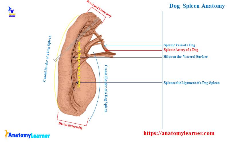
Dog spleen anatomy labeled diagram
I will show you the different structures (gross) from the other parts of the dog spleen with the labeled diagram. First, I tried to show you the exact location of the dog’s spleen in the abdominal cavity.
Here, the labeled diagram also shows the relative position of the other organs that have a great relationship with the spleen. The surfaces (parietal and visceral) are identified from the dog spleen in the labeled diagram.
The narrow dorsal extremity and the wider ventral extremity from the dog spleen are also identified in the labeled diagram. Here, the dog spleen labeled diagram also shows the irregular cranial and caudal borders.
The concavity or sigmoid-shaped structure on the cranial border of the dog spleen is also identified in the labeled diagram. Again, the longer hilus of the dog spleen is shown on the visceral surface.
Various arteries and veins are shown from the visceral surface of the dog spleen on this labeled diagram. In addition, 2 important ligaments – phrenicosplenic and gastrosplenic are also identified from the visceral surface on this labeled diagram.
If you need more labeled diagrams of the dog or canine spleen structure, you may find them on the social media of anatomy learners.
Other suggested articles on dog anatomy from anatomy learners –
- Dog thymus anatomy with labeled diagram,
- Dog lung anatomy – canine right and left lungs lobes with labeled diagram,
Dog spleen histology structure
Externally, the dog’s spleen is surrounded by the fibrous capsule, which contains smooth muscle fibres. The histological section of the dog spleen shows thick fibromuscular trabeculae that arise from the outer fibrous capsule.
These thick fibromuscular trabeculae extend into the parenchyma of the dog’s spleen. External to the capsule of the dog spleen, you will also find the peritoneal covering.
Here, I will briefly describe the histology of the dog’s spleen. But, the below-mentioned article might help you to know details of the dog spleen with the microscopic pictures –
- Spleen histology slide – white pulp and red pulp histology with a labeled diagram,
So, you will mainly find the following essential features in the dog spleen histology (microscope slide) under a light microscope –
- A connective tissue capsule (that is rich in elastic and smooth muscle fibres),
- Larger and fibromuscular types trabeculae (thick), and
- A parenchyma that consists of red and white pulps,
If you notice the histology slide of a dog spleen, you will find the arteries leave the trabeculae and become surrounded by the reticular tissue. Again, you will also see the lymphocytes surrounding the trabeculae and forming the white pulp.
The lymphocytic infiltration is not similar throughout the white pulp of the dog spleen. Sometimes these infiltrations may be more significant and form the lymphatic nodules within the white pulp of the dog spleen.
On the other hand, in the red pulp of the dog spleen microscope slide, you will find the splenic sinusoids and cords.
Dog spleen functions
The spleen of a dog performs a wide variety of functions. Let’s see some of the essential functions that are performed by the dog spleen –
- Storage of erythrocytes – the spleen of the dog store and concentrate erythrocytes and release these cells when needed,
- Filtration and phagocytosis – it helps to filtrate blood and remove unwanted particles from erythrocytes,
- Production of blood cells – dog spleen extracts iron from hemoglobulin used by the red bone marrow and produces new erythrocytes. Again, the dog’s spleen helps to produce many lymphocytes and monocytes.
- Immune response – the fewer lymphocytes and monocytes produced by the dog spleen are responsible for antibodies production,
The canine spleen also contains the megakaryocytes that are responsible for the production of platelets. But, the spleen is not a mandatory organ in dog life.
How to differentiate a cow’s spleen from a dog’s spleen?
If you are asking to differentiate the cow’s spleen from the dog’s, you may perform it easily based on their external features. The cow spleen is elliptical in outline, whereas the dog spleen is tongue or human foot-printed shape.
The surfaces, borders, and extremities are similar in both 2 spleens, but the size is somewhat different. Again, the location and attachment with other important organs greatly vary in cow spleen compared to dogs.
The visceral surface of the cow spleen locates on the dorsal surface of the rumen (part of the ruminant stomach). On this dorsal surface of the rumen, this spleen passes obliquely (Cranio-ventrally).
On the other hand, the parietal surface of the cow spleen is convex and related to the diaphragm. In its cranial border, you will not find any peritoneal covering in a small part of the cow spleen.
The dorsal extremity of the cow spleen locates below the proximal ends of the last two ribs. In comparison, the ventral extremity of the spleen locates below the lower third of the eighth and ninth ribs.
The location of the hilus on the cow’s spleen is also different from the dog’s. You will see a smaller hilus (compared to the dogs) at the upper segment of the visceral surface (just close to the cranial border).
Now, let’s see the main distinguishable features between a dog’s and cow’s spleen from table 3 –
| Features | Cow spleen anatomy | Dog spleen anatomy |
| Shape | Elliptical | Tongue shaped |
| Relation to stomach | Lies dorsal surface of rumen | Greater curvature of stomach |
| Cranial border | Little concave | Concave |
| Hilus | Small | Longer |
Horse spleen anatomy
The anatomical facts (especially the size and shape) of the horse spleen are somewhat different than these of the dogs. In the horse, you will find a roughly triangular spleen that locates at the cranial and left part of the abdomen between the fundus of the stomach and the diaphragm.
- Essential and unique external features of horse spleen – rough triangular shaped,
- You will also find some other exceptional anatomical features in the horse spleen compared to the dog –
- The proximal end of the horse spleen is wide and considered the base,
- The ventral extremity or end is pointed and considered the apex,
- You will find a hilus in the groove that locates at the base of the horse’s spleen,
- The parietal surface of the horse spleen is also convex,
Again, the visceral surface of the horse spleen possesses gastric impression cranially and intestinal impression caudally,
I hope you got the basic idea of the anatomical facts of the horse spleen, which might help you to differentiate it from others.
Pig spleen anatomy
If you want to compare the pig spleen with dog spleen anatomy, you will find the below-mentioned gross features –
- The shape of the pig spleen is somewhat elongated and narrower compared to the cow,
- This organ extends dorsoventrally along the left segment of the greater curvature of the pig’s stomach,
- You will also find a remarkable variation in the size (length, wide, and thickness) of the pig spleen compared to the dogs,
- The visceral surface of the pig spleen is also concave and possesses the smaller hilus on its upper part,
- Again, the parietal surface of the pig spleen is smooth and convex like other species,
So, the major difference between dogs and pig is their shape, size, and location. Again, the location of the hilus on the visceral surface of the spleens is different in dogs and pigs.
Spleen of rabbit
The shape of the rabbit’s spleen is elongated and spatula-like. You will find the dark brown color in the spleen of the rabbit.
The length of the rabbit spleen is about 3 centimetres long. This spleen of the rabbit extends dorsoventrally along the greater curvature of the stomach.
The proximal extremity of the rabbit spleen is pointed, whereas the distal extremity is blunt. Again, the cranial border of the rabbit spleen is convex, whereas the caudal border is irregular.
Frequently asked questions on dog spleen anatomy
Now, let’s see some of the commonly asked questions on dog spleen anatomy by anatomy learners. Here, I will enlist the frequently asked questions on the dog spleen with their concise answer.
But, I recommend you to read the full guide on the anatomical facts of the dog spleen from the start to the end of this article. Okay, let’s see these questions on dog spleen with their short answer –
What does the spleen do in dogs?
The spleen does a lot of functions in the dog’s body. It helps store and concentrate erythrocytes, produce new blood cells, and possess a phagocytic activity.
The spleen of the dog also filtrates blood and removes worn-out erythrocytes from circulation. They help to produce antibodies and also platelets in a dog’s body. You will find more functions of the dog spleen in this article.
What is the anatomy of the dog spleen?
Anatomically, the dog spleen shows 2 surfaces, 2 extremities, and 2 borders. Again, these surfaces, extremities, and borders possess some important structures and features.
You might first identify the thin and rounded dorsal extremity and wider ventral border to understand this organ’s orientation. The attachment of the spleen with other organs and long hilus are essential features for you to understand its relative position.
What function does the spleen have in a dog?
The spleen has several functions in a dog – phagocytosis, haemopoiesis, immune response, storage, and blood filtration. A dog’s spleen also takes iron from haemoglobin and helps produce new erythrocytes.
How long do dogs live after spleen removal?
The dogs may live for a long time after removing the spleen. They may survive throughout their life without their spleen.
But how or why can they live after the removal of the spleen? Well, the functions performed by the dog spleen may also be performed by the other tissues.
So, after removing the dog’s spleen, its normal functions may continue through the body’s other tissues.
Conclusion
So, you got the basic idea of the dog spleen anatomy with the labeled diagram. Here, the different surfaces, extremities, and borders, along with their essential features, are important to know about the dog’s spleen.
The information provided in this article might help you to differentiate the dog spleen from other species like dogs, cows, rabbits, and pigs. Now, please try to practically identify all of the dog’s spleen anatomy essential features.