The horse radius and ulna are the long bones of the forelimb. Here, I will describe the osteological features of the radius and ulna bones from the horse with the diagram.
Quick overview: The radius is a typical long bone and much larger than the ulna of the forearm in a horse. Again, the ulna is the reduced long bone situated behind the radius and partially fused with it.
First, I will describe the features of the horse radius bone and then the ulna bone. Thus, you will easily identify them from the forelimb of a real horse skeleton.
What is the radius bone of a horse?
The radius is a typical long bone that forms the forearm of a horse. However, the forearm also contains the ulna bone, which remains fused with the radius on its caudolateral aspect.
Here, the radius is a much larger bone than the ulna bone. The ulna bone is an ill-developed and reduced long bone that doesn’t reach the distal end of the radius bone.
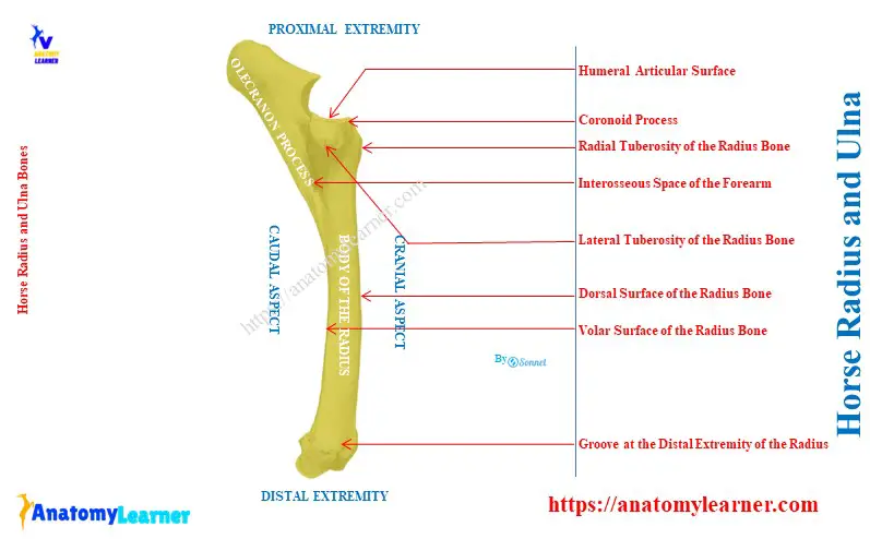
The radius of a horse extends vertically from the elbow to the carpal joint. Thus, this bone proximally articulates with the humerus and distally with the carpus.
The radius of a horse is gently curved. Its cranial or dorsal surface is slightly convex, which provides this curved appearance.
Horse radius bone anatomy
As a typical long bone, the horse’s radius consists of a curved and flattened body and two extremities. Before describing the bone, first, let’s identify the below-mentioned important osteological features –
- Curve and flattened body of the radius,
- Surfaces of the radius: convex dorsal and concave volar or palmar surface,
- Borders of the radius: slightly concave medial and strongly curved lateral border,
- Proximal interosseous space of the forearm,
- Humeral articular surface with the coronoid process at the proximal extremity,
- Tuberosities of the radius at its proximal extremity: radial or bicipital, medial, and lateral tuberosity,
- Three carpal articular surfaces at the distal extremity and
- Grooves and ridges at the dorsal and volar aspect of the distal extremity,
All of the above-mentioned osteological features are identified from the horse radius bone. Now, I will describe these features in detail from the body and extremities of the radius.
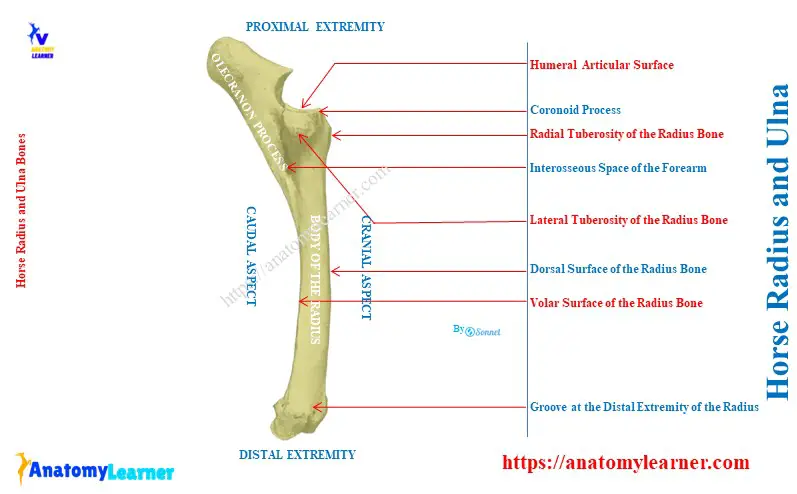
Body of the horse radius bone
The body of the horse radius is slightly curved in its length. It is also somewhat flattened from before backward. Again, the body body of the radius bone becomes wider at the extremities.
The body the equine radius consists of two surfaces and two borders. Here, the surfaces are the dorsal and palmar (volar), and the borders are lateral and medial.
The dorsal surface of the body is slightly concave and smooth in its length. It is also rounded side by side.
The volar surface is correspondingly concave and flattened in its transverse direction. At the proximal part of this volar surface, there is a smooth and shallow groove.
This groove concurs with the ulna bone and forms the interosseous of the forearm. Below this smooth and shallow groove of the radius bone, a nutrient foramen is present.
Below the interosseous space, the volar surface has a narrow, rough, and triangular area. Within this surface, the cranial surface of the ulna bone attaches to the interosseous ligament.
But in the older horse, the ulna bone is completely fused with the radius bone in this area.
Borders of the radius bone
The medial border of the body is slightly concave in its length. At the proximal end of this medial border, there is a smooth area. The tendon of the horse’s brachialis muscle is inserted within this smooth area.
Again, there is a small rough area just below this smooth area at the medial border. The muscles and long medial ligament of the elbow joint attach to this small, rough area.
The lateral border of the body of the radius bone is strongly curved, but there are no important osteological features at this border.
The proximal extremity of the horse radius bone
The proximal extremity of the horse radius is also known as the head. It is flattened in its dorso palmar aspect and widens transversely.
This extremity has a large humeral articular surface that articulates with the distal end of the humerus. A sagittal ridge crosses this humeral articular surface.
You will find a synovial fossa at the posterior part of this cross-sagittal ridge. This synovial fossa ends at the prominent part of the cranial aspect. This prominent part is known as the coronoid process of the radius bone.
Two facets at the caudal aspect of the proximal end articulates with the ulna. Between these facets and interosseous space, there is a quadrilateral rough area. In this area, the radius and ulna are united by the interosseous ligament.
There are three tuberosities present at the proximal end of the horse radius bone –
- Radial or bicipital tuberosity: It is located on the medial side of the dorsal surface. The tendon of the biceps muscle inserts into this radial tuberosity.
- Medial tuberosity: it is continuous with the radial tuberosity. It furnishes attachment to the short part of the medial ligament.
- Lateral tuberosity: it is more salient in the radius bone. It provides attachment ot the lateral ligament and to the common and lateral extensor muscles of the horse’s digits.
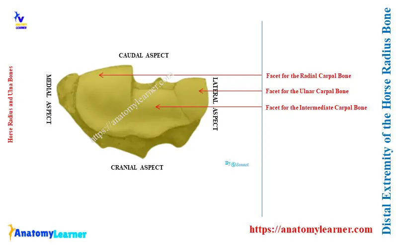
The distal extremity of the radius bone
The distal extremity of the horse radius bone is also compressed from before backward. This extremity presents the articular surfaces for the carpal bones.
This articular surface is divided into three parts –
- The medial one is the largest and quadrilateral. It is also convexoconcave from and articulates with the radial carpal bone.
- The middle one is also similar to the previous one but smaller in size. It articulates with the intermediate carpal bones of the horse.
- Finally, the lateral one is small and convex. It articulates with the ulnar carpal below and with the accessory carpal behind.
Features from the dorsal and volar surface of the distal extremity
The dorsal surface of the distal extremity presents three grooves that are separated by the ridges. Here, the medial one is small and oblique and lodges with the tendon of the extensor carpi oblique muscle.
The middle one is vertical and gives passes to the tendon of the horse’s extensor carpi radialis muscle. Finally, the lateral one is similar to the middle one and contains the tendon of the common digital extensor muscle.
The volar surface of the distal extremity of the radius bone is crossed by the rough ridges. You will find the tuberosities at the either side at the distal extremity.
The collateral ligament of the carpus joint attaches to these tuberosities correspondingly.
What are the features of the ulna bone in a horse?
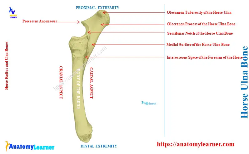
As a reduced long bone, the horse ulna differs slightly from the ox ulna bone. First, let’s identify the osteological features of the horse ulna bone –
- Three-sided body of the ulna bone,
- Enlarge proximal and small distal extremities,
- Surfaces of the shaft of the ulna bone: dorsal, medial, and lateral,
- Borders of the body of the ulna bone: lateral, medial, and volar borders,
- Lateral and medial surface of the proximal extremity of the ulna bone,
- Anconeus process at the dorsal border of the ulna bone,
- Semilunar or trochlear notch of the horse ulna bone,
- Enlarge olecranon process of the ulna bone and
- Olecranon tuberosites,
The labeled diagram identifies all the above-mentioned osteological features of the horse ulna bone. Now, I will describe all these features of the shaft (body) and extremities of the horse ulna bone.
Body or shaft of the horse ulna bone
The body of the horse ulna bone is three-sided and tapered distally. It presents three surfaces and three borders.
Dorsal surface of the horse ulna bone: this surface is just caudal to the volar surface of the radius bone. The dorsal surface of the ulna bone contributes to forming the interosseous space with the volar surface of the radius bone.
Below the interosseous space, the surface of the ulna is smooth and attaches to the volar smooth surface.
The medial surface of the ulna bone is smooth and concave. Finally, the lateral surface is flattened in the horse ulna bone.
The medial and lateral borders of the horse ulna bone are thin and sharp. But, these border become thick and blunt at the interosseous space.
The volar border is slightly concave in length and rounded. The distal end of this border is pointed and a little below the middle of the radius bone.
Extremities of the horse ulna bone
The proximal extremity is the major and important part of the horse ulna bone. This extremity projects backward and upward just behind the elbow joint.
It presents two surfaces (medial and lateral) and two borders (dorsal and volar). Here, the medial surface of the proximal end of the horse ulna is concave and smooth. Again, the lateral surface of the proximal end is convex and rough.
The dorsal border of the proximal extremity of the ulna bone presents a pointed or beak-like projection. This pointed projection is known as the processus anconeaus.
Again, the semilunar notch is at the dorsal border just below the anconeal process. It is triangular and concave and articulates with the distal caudal end of the humerus bone.
The volar border of the proximal end of the horse ulna bone is nearly straight, thick, and roughened. Here, the whole structure is known as the olecranon process. Again, the free end and rough prominences are the olecranon tuberosites.
They provide the attachments to the triceps and other muscles of the elbow joint.
The distal extremity of the horse ulna bone is pointed and fused with the radius bone. But this bone doesn’t extend upto the distal extremity of the radius like the ox ulna bone. The distal extremity of the horse ulna ends at the middle part of the caudal surface of the radius bone.
Unique features of the horse radius and ulna bones
Compared to the radius and ulna bones from other animals, you will find the below-mentioned unique features in the horses –
- The radial tuberosity is well-developed in the horse radius bone,
- A reduced or ill-developed ulna extends only upto the distal third of the radius,
- Thus, there is only one proximal interosseous space present in the horse radius and ulna bones,
- There is no styloid process in the horse ulna bone,
- The medial surface of the olecranon process is more concave,
- A more extensive semilunar notch is present in the horse ulna bone,
- The facets for the proximal carpal bones at the distal extremity of the horse radius are not oblique,
Conclusion
So, the horse radius and ulna are the long bones that form the forearm. The radius possesses the typical features of the long bone, while the ulna is a reduced long bone in horses.