The horse humerus is a typical long bone that forms the arm segment of the thoracic limb. Here, I will describe the anatomy of the horse humerus bone with the labeled diagram.
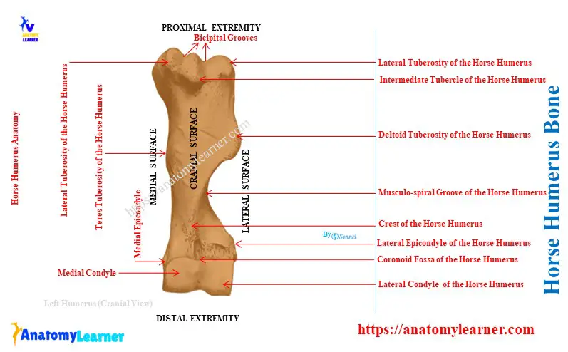
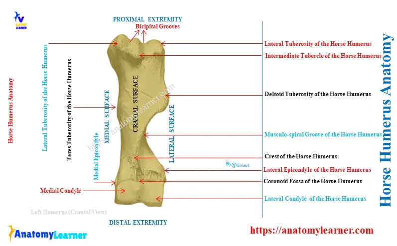
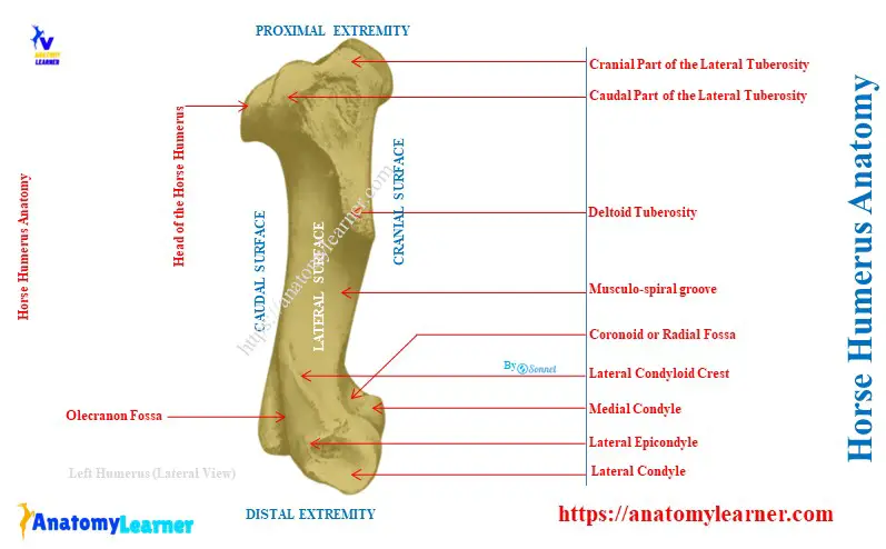
Quick overview: the horse humerus bone consists of an irregular cylindrical shaft and two expanded extremities. A Musculo-spiral groove, deltoid tuberosity, and teres tuberosity are the main osteological features of the shaft. The proximal extremity has a head, neck, and 3 tuberosities, whereas the distal extremity has two condyles and two fossae.
First, I will identify the osteological features in the shaft and extremities of the horse humerus. Then, I will describe these features in detail for each part separately.
What is the humerus in a horse?
The humerus is a long bone in a horse that extends from the shoulder to the elbow joint. It articulates proximally with the scapula bone and distally with the radius and ulna bones.
The direction of the horse’s humerus is obliquely downward and backward. It forms an angle of about fifty-five degrees with the horizontal plane of the body.
Horse humerus bone
For the description purposes, the horse humerus possesses a shaft or body and two extremities. First, let’s identify the below-mentioned osteological features of the humerus from its shaft and extremities.
From the shaft of the equine humerus:
- Lateral, medial, anterior, and posterior surfaces of the shaft,
- Musculo-spiral groove on the lateral surface,
- Teres tuberosity at the medial surface, and
- Crest of the humerus and deltoid tuberosity at the anterior surface,
From the proximal extremity of the equine humerus:
Circular head,
- Well-defined neck,
- Three tuberosities: lateral, intermediate, and medial, and
- Intertuberal or bicipital grooves,
From the distal extremity of the equine humerus:
- Cranially located lateral and medial condyles, and radial fossa,
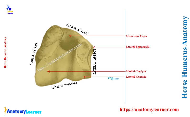
- Posteriorly located olecranon fossa and
- Lateral and medial epicondyles at the corresponding aspects,
All the above-mentioned osteological features are identified in the labeled diagram of the horse humerus bone.
Unique osteological features of the equine humerus
- The Musculo-spiral groove at the lateral surface of the equine humerus is more deep and twisted,
- Prominent deltoid tuberosity is present at the anterior surface of the horse humerus,
- The lateral condyloid crest is more prominent in the equine humerus than that of the ox,
- There is an extra tuberosity (intermediate tuberosity) in between the lateral and medial tuberosities,
- Thus, the bicipital groove divides into two parts by this intermediate tuberosity or tubercle,
- The apex of these tuberosities is at the level of the bicipital groove and
- A nutrient foramen is found at the distal third of the medial surface of the humerus’s shaft.
Now, I will describe all these osteological features from the shaft and extremities separately.
Shaft of the horse humerus anatomy
The shaft of the horse humerus bone is irregular, cylindrical, and twisted in appearance. It practically has four surfaces: lateral, medial, anterior, and posterior.
The lateral surface of the equine humerus is smooth and spiral. It forms the Musculo-spiral groove, which contains the brachialis muscle.
The musculo-spiral groove continues with the posterior surface above. Again, it winds around towards the anterior surface below.
The medial surface is nearly straight and smooth throughout the bone length. It is rounded side by side and blends with the anterior and posterior surfaces.
The teres tuberosity is located just above the middle of the medial surface of the equine humerus. The tendons of the latissimus dorsi muscle and the teres major muscle attach to this teres tuberosity.
You will also find a nutrient foramen at the lower third of the medial surface of the horse humerus.
The anterior surface of the horse humerus divides into two parts: proximal and distal. Here, the proximal part of the anterior surface is smooth, wide, and triangular. Again, the distal part of the anterior surface is narrow and roughened.
A distinct border of the humerus separates the anterior surface from the lateral surface. This is the crest of the humerus bone, which bears the deltoid tuberosity just above its middle portion.
Just below the neck, there are several rough lines at the lateral surface. These lines give the origin to the lateral head of the triceps muscle.
Below the deltoid tuberosity, the crest inclines forward and ends at the coronoid fossa.
The posterior surface of the horse humerus is rounded from side by side and smooth.
Proximal extremity of the equine humerus bone
The proximal extremity of the equine humerus bone consists of a head, neck, two tuberosities, one tubercle, and two intertuberal grooves. Here, the head of the humerus presents an almost circular convex articular surface.
This articular surface is about twice as extensive as the glenoid cavity of the scapula bone. The circular convex head articulates with the distal glenoid cavity of its scapula and forms the glenoidohumeral joint.
There is a fossa in front of the head that contains several foramina. The neck is well-defined and located caudomedial to the head of the humerus.
The lateral tuberosity is larger and located anterior-laterally. It presents two parts: anterior and posterior.
The anterior part of the lateral tuberosity forms the lateral boundary of the intertuberal groove. It also provides attachment to the lateral branch of the supraspinatus muscle.
The posterior part of the lateral tuberosity attaches to the short insertion of the infraspinatus muscle. Again, the outer surface of the posterior part is coated with cartilage.
The medial tuberosity is less developed but also consists of anterior and posterior parts. Here, the anterior part of the medial tuberosity forms the medial boundary of the intertuberal groove. It furnishes insertion to the medial branch of the supraspinatus muscle and deep pectoral muscle.
Again, the posterior part of the medial tuberosity gives attachment to the subscapularis muscle.
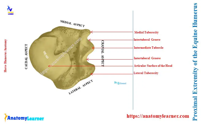
What is a bicipital groove on a humerus bone?
The bicipital or intertuberal groove is the elongated depression between the lateral and medial tuberosities at the proximal end of the horse humerus. It is located in front of the bone and bounded by the anterior parts of the tuberosities.
But the bicipital groove is subdivided into two parts by the intermediate ridge. This ridge is also known as the intermediate tubercle of the horse humerus.
The cartilage covers the bicipital groove of the humerus bone in its fresh condition. The tendon of the biceps brachii muscle attaches to this bicipital groove.
Just below the intermediate tubercle, you will see a fossa with several foramina.
The distal extremity of the horse humerus
The distal extremity of the horse humerus articulates with the proximal end of the radius and ulna bones. This extremity of the humerus consists of two condyles (lateral and medial) of unequal size on its anterior aspect.
Here, the medial condyle of the humerus is larger and crossed by a sagittal ridge. A synovial fossa is on the anterior part of the groove.
This groove continues posteriorly and reaches the olecranon fossa. The olecranon fossa of the humerus is deep and located between the epicondyles. It articulates with the semilunar notch of the olecranon process of the ulna bone.
The lateral condyle of the humerus is small and located somewhat lower and further back. This arrangement gives the distal extremity an oblique appearance. A wide, shallow groove also marks the lateral condyle.
Above the groove of the medial condyle, there is the radial or coronoid fossa. It furnishes the origin to part of the extensor carpi and common digital extensor.
Behind and above each condyle is a thick ridge. These are the epicondyles of the horse humerus bone.
The medial epicondyle of the humerus is well-developed and furnishes the origin of the flexor muscle of the carpus and digits. It also bears a tubercle for the attachment of the medial ligament of the horse’s elbow joint.
The lateral epicondyle possesses the condyloid crest laterally. It forms the outer boundary of the Musculospiral groove and gives the origin of the extensor carpi radialis.
The distal border of this epicondyle gives attachment to the ulnaris lateralis muscle.
Conclusion
So, the horse humerus bone possesses the typical osteological features of the long bone. The most salient features are a deep musculospiral groove at the shaft and an intermediate tubercle at the proximal extremity.