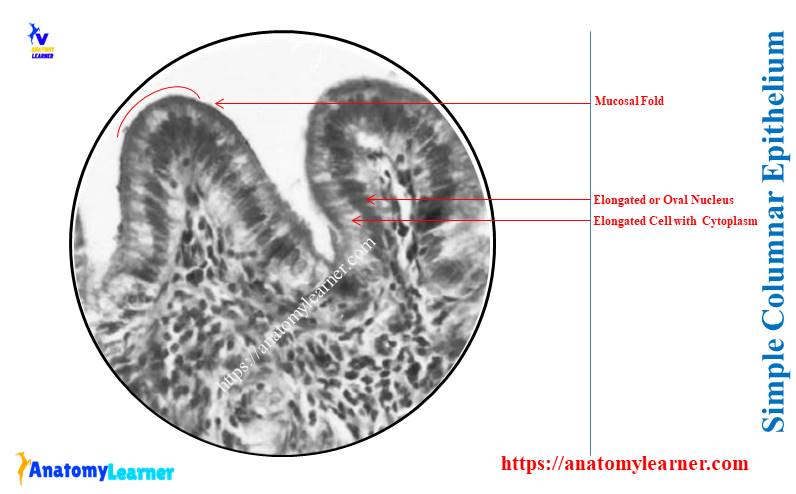
The simple columnar epithelium under the microscope shows a single layer of cells resting on the basement membrane. Here, I will show you the microscopic features of the simple columnar epithelium with their location and labeled diagram.
Quick answer: The simple columnar epithelium under a microscope are the single layer of cells with a greater height than breadth (columnar shape) and an oval basal nucleus. Typically, the simple columnar epithelium locates in the lining of the gastrointestinal tract, gall bladder, respiratory tract, uterine tube, and auditory tube.
So the appearance of the cell and nucleus is essential to identify the simple columnar epithelium. Here, I will enlist the important identifying points for simple columnar epithelium with real microscopic images.
Again, you will get the basic idea of two types of columnar epithelium in this article. Finally, the different labeled diagrams on simple columnar microscope slides will provide here with various magnifications like 4x, 10x, and 400x.
Let’s get started learning the microscopic features of the simple columnar epithelium with the labeled diagram.
Simple columnar epithelium under a microscope
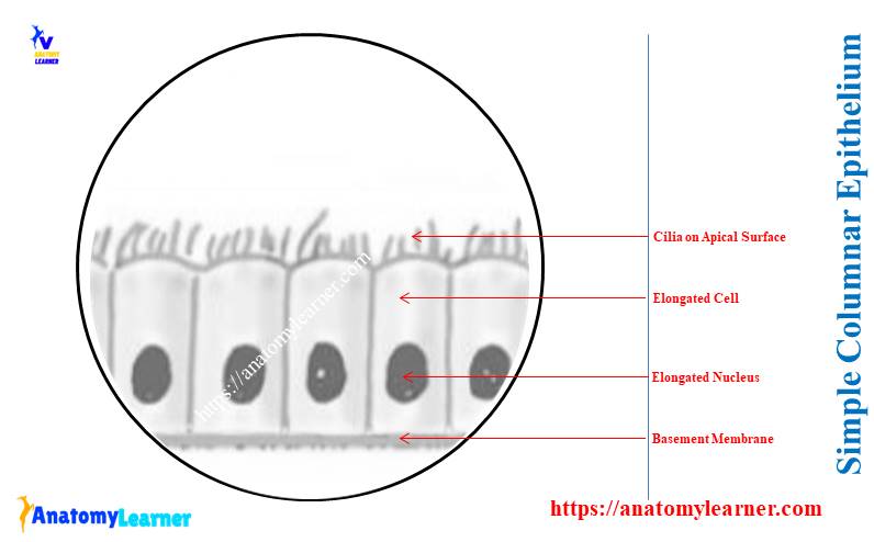
First, let’s try to identify the following features from the simple columnar epithelium under a microscope –
- Tall or elongated narrow cells,
- An oval nucleus that locates at the base of each elongated (column) cell,
- The basement membrane of the elongated or tall cells,
- A unique feature on the surface of the tall columnar cells (the presence of cilia),
The labeled diagram identifies all these features from the simple columnar epithelium. A few simple columnar epithelium microscope slides show the cilia on their apical surfaces.
But, others don’t show any surface specialization; that means you will not find any cilia or microvilli on the apical surface of these simple columnar epithelium. A few simple columnar epithelia (in various organs of the animal’s body) show the microvilli.
You will find the details of various types of simple columnar epithelium in the next section of this article.
Before going to the identification and details description of a simple columnar epithelium microscope or histology slide, let’s see the overview from table 1 –
| Simple columnar epithelium | Features |
| Shape of cells | Tall or elongated |
| Shape of nucleus | Oval |
| Position of nucleus | Base of cell |
| Location of simple columnar | Gastrointestinal tract Gall bladder Respiratory tract Uterine tube, Auditory tube |
| Main functions | Protection Secretion Absorption |
Identification of simple columnar epithelium histology slide
In this part, I will enlist the essential identifying features for the simple columnar epithelium histology slide. Okay, let’s see the identification points of this columnar epithelium histology slide under the light microscope –
- The sample tissue section shows the presence of a single layer of cells that rest on the basement membrane,
- These cells of the provided tissue section show a greater height than their breadth (that is, columnar in shape),
- You will see an oval or elongated nucleus in every single tall or elongated cell of the provided sample tissue section,
- The direction of the nucleus of these columnar cells is similar to the direction of these cells,
- These nuclei of the provided sample columnar cells lie near the base of the cells (same features found in the adjacent cells),
- So, you will find the clear zone of cytoplasm above the nucleus of every columnar cell,
- Sometimes the provided sample tissue section shows the microvilli or cilia on the apical surface of these columnar cells,
So, this is the simple columnar epithelium histology slide.
Typically non-ciliated simple columnar epithelium lines the mucosa membrane of the stomach, gall bladder, and intestine of animals. Again, you will find the ciliated simple columnar epithelium in the mucosa membrane of the respiratory tract, uterine tube, and auditory tube.
The hand drawing and photomicrograph of the simple columnar epithelium are shown in the labeled diagram as a cross-sectional view. Again, if you want to see these simple columnar epithelium as the verticle or transverse section, you will find them rectangular.
So, if you see these columnar from the surface view, you will see the polygonal appearance of simple columnar epithelium.
Types of simple columnar epithelium under a microscope
When you observe the microscopic features of simple columnar epithelium under the light microscope, you may find the followings –
- The apical surface of these columnar represents cilia,
- Some simple columnar show apical microvilli, and
- Another simple columnar doesn’t show any cilia or microvilli,
So, the simple columnar epithelium maybe three (3) types. This is based on the location of this simple columnar epithelium on the different parts or organs of the animal body.
Let’s see the major types of simple columnar based on their location in table 2 –
| Types of simple columnar epithelium | Unique features |
| Simple columnar epithelium | Have no cilia or special features on the cell’s surface |
| Ciliated columnar epithelium | Cell surface bears cilia |
| Columnar with microvilli | Have microvilli on the cell surface |
So, in some locations, the simple columnar epithelium doesn’t show any particular specialization on the apical surface. These are the simple columnar epithelium and lines, typically the gastrointestinal and gall bladder of the animal.
Again, the simple columnar epithelium shows cilia on their apical surface in some locations. These are the ciliated simple columnar epithelium and line the respiratory, uterine, and auditory tubes of animals.
Finally, some of the simple columnar epithelium lines the small intestine, and sometimes the gall bladder shows the striated border on their apical surface. The microvilli are the striated borders on the apical surface of these epithelium cells.
All three types of simple columnar will show in the next section of this article with the labeled diagram.
Location of simple columnar epithelium
So, there are three (3) major types of columnar epithelium in the animal’s body. Here, I will enlist the location of these three types of columnar epithelium –
- #1. Location of the simple columnar epithelium,
- #2. Location of ciliated columnar epithelium, and
- #3. Simple columnar epithelium with microvilli
Here, you will see two forms of microvilli in the simple columnar epithelium. When the microvilli of the simple columnar epithelium are arranged regularly, they have seen as the striated border.
Again, the microvilli of the simple columnar epithelium arrange irregularly and are seen as the brush border. You may clearly see these microvilli from this columnar epithelium under the electron microscope.
First, let’s see the location of the simple columnar epithelium from the animal’s body –
You will see the simple columnar epithelium (with no surface modification or cilia or microvilli) in the mucous membrane of the stomach and large intestine.
The ciliated columnar epithelium may find in the various organs of the animal’s body. Let’s see the exact location of ciliated simple columnar epithelium –
- #1. Mucosa of the respiratory tract,
- #2. Mucosa of the uterus and uterine tube of the animals,
- #3. In the lining of the ductus deference of the animal,
- #4. In the parts of the middle ear and auditory tube,
- #5. Lines in the ependymal lining of the central canal and spinal cord, and
- #6. In the lining of the ventricles of an animal’s brain,
Typically, you will find the microvilli in the simple columnar on the mucosa of the small intestine and gall bladder. You will find the regular-shaped microvilli or striated border on the small intestine’s mucous membrane.
Whereas on the mucous membrane of the gall bladder, you will find the irregularly shaped microvilli or brush border.
Simple columnar epithelium function
The simple columnar epithelium has a wide variety of functions, but the main functions are protection, secretion, and absorption. Here, I will enlist some of the essential functions of the simple columnar epithelium –
- Few columnar epithelia have a secretory function,
- The simple columnar epithelium that lines the large and small intestines secretes mucous (along with goblet cells),
- Few columnar epithelium release enzymes,
- In the uterine tube, the movement of the cilia helps to passage of ova toward the uterus,
- Again, the microvilli (striated and brush borders) increase the surface area for absorption,
In the mucosa of the stomach, you will find secretory vacuoles in the apical part of the mucosa membrane. Here, the columnar secretory cells are scattered and distributed in the mucosa of the stomach.
Along with the simple columnar epithelium, the mucosa of the large intestine shows the typical goblet cells. They secrete mucous that accumulates in the apical part of the cell cytoplasm.
In the respiratory tract, the cilia of the simple columnar epithelium move the mucous in the bronchi and towards the larynx and pharynx.
Simple columnar epithelium under microscope labeled diagram
Now, you will find the different labeled diagrams on the simple columnar epithelium microscope slide. The tall or elongated cells with their elongated nuclei are identified in the simple columnar epithelium labeled diagram.
Here, in both longitudinal and cross sections labeled diagrams, I tried to show all the essential features of simple columnar epithelium.
Now, the labeled diagram of simple columnar epithelium under a microscope shows two types of cells –
- Columnar cells with microvilli or brush border, and
- Oval-shaped goblet cells,
Here, the apical microvilli or brush border from the surface look like the reddish outer layer with longitudinal striation. So, these longitudinal striations on the apical surface of these simple columnar epithelium are the microvilli.
Again, the simple columnar epithelium labeled diagram shows a thin connective tissue basement membrane. The lamina propria made by the loose connective tissue also shows the typical features in the labeled diagram.
You may also know the details of the different layers (including the lamina propria) of a tubular organ from the below-mentioned article of anatomy learner –
In the labeled diagram, you will see the smooth muscle fibers that extend from the lamina propria to the villi.
Let’s find more labeled diagrams on simple columnar epithelium on social media of anatomy learners.
Simple columnar epithelium with goblet cells labeled
You know, the mucous membrane of the intestine sometimes shows the goblet cells along with the simple columnar epithelium. Here, the goblet cells are a pale-stained structure that is interspersed among the simple columnar epithelium.
During the routine histology, the mucus from the goblet cell washed out. Thus, the cytoplasm of these goblet cells appears clear or only lightly stained.
The numerous goblet cells secret mucus that protects the intestinal epithelium from crrosive secretion, which enters the small intestine. You know, some crossive secretion may come from the stomach during digestion toward the intestine.
Simple columnar epithelium under microscope 400x
Now, let’s see the simple columnar epithelium with different magnifications like 4x, 10x, and 400x. Here, the simple columnar epithelium from the gall bladder and stomach with 10x and 400x magnification are shown in the diagrams.
Here, the mucosal fold of the gall bladder and stomach shows the simple columnar epithelium lining. The 400x magnified image of simple columnar epithelium shows oval nuclei of these columnar epithelia that lie perpendicular to the basement membrane.
Thus, the diagram shows the single row of oval nuclei above the basement membrane of the gall bladder and stomach histology slides. Here, the upper part of the cell cytoplasm is eosinophilic and distinct.
Now, let’s see the ciliated columnar epithelium from the uterine or fallopian tube. Here, the labeled diagram of the fallopian tube of an animal shows –
The mucous membrane of the uterine or fallopian tube shows the simple columnar epithelium lining (view with 400x magnification),
On the luminal surface of these simple columnar epithelium cells, you will see the cilia (with 400x magnification),
Frequently asked questions on simple columnar epithelium
Here in this section, you will find the common questions on simple columnar epithelium under the microscope that the histology learners ask. A single layer of oval or elongated cells with a similarly shaped nucleus is the main characteristic feature of the simple columnar epithelium microscope slide.
If you want to get the basic idea of the simple columnar epithelium histology slide, it is recommended to read this full guide. Okay, let’s see the following questions on the simple columnar epithelium with their concise answer –
What does simple columnar epithelium look like under a microscope?
The simple columnar epithelium looks like the elongated or oval structure with the oval nucleus under the microscope. Here, you may find some cilia or microvilli on the apical surface of the simple columnar epithelium microscope slide.
Again, some simple columnar epithelium shows no unique modification or structure on their apical surface. The labeled diagrams provided in this article showed the three major types of columnar epithelium.
How do you identify simple columnar epithelium?
The shape of the cells, nucleus location of the nucleus, and color are the important features to identify the simple columnar epithelium from the microscopic slide. Here, the single layer of elongated or oval shape lies longitudinal to the basement membrane.
Again, the nucleus of the elongated cells also shows the typical oval shape that also lies longitudinal to the basement membrane. The apical surface of the simple columnar epithelium shows different features like cilia or microvilli.
What is the microscopic appearance and location of simple columnar epithelium?
Under the light microscopic view, the simple columnar epithelium appears as an elongated structure with an oval nucleus. You will find the microscopic features of the different types of simple columnar here in this article with the diagrams.
The three major types of simple columnar epithelium find in various organs of animals. You will find these three major types of columnar epithelium in the mucosa of the stomach, gall bladder, intestine (large and small), respiratory tract, uterus, and uterine tubes.
What is the appearance of epithelium cells under a microscope?
If you want to understand the appearance of the epithelium cells under a light microscope, you might have a basic idea of cells and nuclei. You might understand the shape and appearance of the cells and nucleus of various types of epithelium cells under the light microscope.
Here in the below-mentioned article, you will find the overview of different types of epithelium cells with their labeled diagram –
Again, you may learn the specific epithelium cells or tissue from different articles of anatomy learner. You might also know the other basic tissues like connective, muscular, and nervous tissues here.
What is the striated border on simple columnar epithelium?
You will see a thickening area in the various regions of the free surfaces of the simple columnar epithelium cell. Here, on this thickening area of the simple columnar, they are actually vertical striation.
These vertical striations on the columnar epithelium are the striated border (when arranged in a regular pattern). You may find these striated borders on the apical surface of the intestinal mucosa.
Conclusion
So, the simple columnar epithelium under the light microscope shows the elongated cells with the ventrally placed oval nucleus. Three major types of columnar epithelium under a light microscope show their unique features.
All the provided labeled diagrams in this article might help you to understand the fundamental difference and appearance of various types of columnar epithelium under the light microscope. Again, the provided identification features might help you identify this simple columnar epithelium from different animal body organs under a light microscope.
Now, let’s see and identify these three major types of simple columnar epithelium from the real sample at your histology learning laboratory with the help of a microscope.