The stratified squamous epithelium under a microscope shows the multiple layers of cells where the superficial one is flattened. You will also find the basal columnar cells in this stratified squamous epithelium.
Quick summary: in the stratified squamous epithelium under microscope, you will see multiple layers of cells where the basal layer is columnar, and the superficial layer is squamous. There are two types of stratified squamous epithelium under the light microscope in various organs – keratinized and nonkeratinized stratified squamous epithelium.
Here, I will show the microscopic features of keratinized and nonkeratinized stratified squamous epithelium with the labeled diagram. Again, you will find the identification points for these two types of stratified squamous epithelium in this article.
This article will also provide a diagram of the functions and locations of keratinized and nonkeratinized stratified squamous. So, if you want to know the basic of the stratified squamous epithelium and identify it from a real sample with the help of a microscope, let’s continue this article.
Stratified squamous epithelium under a microscope
First, let’s know what structures you might identify from keratinized and nonkeratinized stratified squamous epithelia under the microscope. You should identify the below-mentioned features or parts or cells from the stratified squamous epithelium –
- #1. Multiple layers of cells,
- #2. The basement membrane of the stratified squamous epithelium,
- #3. Basal columnar cells layer and superficial flattened cells layer,
- #4. Absence or presence of keratin intermediate filaments (depends on the type of stratified squamous epithelium),
The labeled diagrams identify all these microscopic features from keratinized and nonkeratinized stratified squamous epithelia. You will find more real microscope pictures and hand-made stratified squamous epithelium labeled diagram below.
Now, let’s see the summary of the stratified squamous epithelium under a microscope from table 1 –
| Stratified Squamous Epithelium | Keratinized | Nonkeratinized |
| Layers of cells | Multiple | Multiple |
| The basal layer of cells | Columnar | Columnar |
| Superficial cells layer | Flattened (squamous) Have keratin layer | Flattened (squamous) No keratin substance |
| Examples (Location) | Epidermis Hard plates Nostril | Esophagus Buccal cavity Laryngopharynx |
| Functions | Protection Prevent water loss | Secretion Protection Prevent water loss |
So, these are the basic difference between the keratinized and nonkeratinized stratified squamous epithelium.
Suggested articles for you to understand the structure of stratified squamous epithelium –
- Simple columnar epithelium under the microscope with labeled diagram, and
- Simple squamous epithelium under a microscope with the labeled diagram,
Now, it will be easy for you to identify both the keratinized and nonkeratinized stratified squamous epithelia with the help of a light microscope.
Identification of stratified squamous epithelium histology slide (Nonkeratinized)
Here, I will enlist the identification points for both the keratinized and nonkeratinized stratified squamous epithelium with the labeled diagrams. First, let’s show the identifying points of nonkeratinized stratified squamous epithelium histology slide –
- The sample tissue section shows the deepest or basal layer of columnar cells,
- In the middle layer of the provided tissue sample, you will find the polyhedral cells layers,
- Again, the superficial layer of the sample shows the increasing degree of flattened squamous cells,
- So, the sample tissue shows oval nuclei in the basal layer, rounded nuclei in the middle layer, and transversely elongated nuclei in the superficial layer,
- There is no superficial keratinized zone in the provided tissue sample,
- Again, the superficial or luminal surface of the provided tissue sample is smooth,
- But, the basal surface of this tissue sample is uneven due to the presence of deep connective tissue papillae of the lamina propria,
Thus, this is the nonkeratinized stratified squamous epithelium histology slide. There are multiple layers of different types of epithelial cells, but they are still called the nonkeratinized stratified squamous epithelium.
This is because of the flattened squamous cells on the most superficial layer of the provided tissue sample. So, to identify the nonkeratinized stratified squamous epithelium microscope slide, you might keep in mind the following essential features that show in table 2 –
| Features | Nonkeratinized stratified squamous epithelium |
| Cell layers | Three layers Basal layer – columnar Middle layer – polyhedral, and Superficial layer – flattened squamous |
| Nucleus | Variable Basal layer – oval (vertically placed) Middle layer – rounded, and Superficial – flattened (transversely) |
| Keratin | No keratin substance on the superficial layer |
Identification of keratinized stratified squamous epithelium
If you want to identify the keratinized stratified squamous epithelium with the help of a light microscope, the following identifying point may help –
- The sample tissue section shows the multiple layers of cells like the nonkeratinized stratified squamous epithelium,
- Here, the basal layer of cells are columnar, and the middle layer possess polyhedral type cells,
- Few cells of the superficial layer of the provided sample are squamous types that possess a flattened nucleus,
- But you will find a clear and non-nucleated keratinized zone on the apical surface of the provided tissue sample,
- This keratinized zone is made of flat, scale-like cells that fill with the keratin substance for the protection of this layer,
- Again, this tough keratin substance becomes firmly embedded in the matrix protein of the provided tissue sample,
- The basal surface of the provided epithelium sample is also uneven due to the presence of epidermal ridge and dermal papillae,
So, this is a keratinized stratified squamous epithelium histology slide. The presence of keratin substances on the superficial layer is the main differentiating point of keratinized stratified squamous from nonkeratinized stratified squamous epithelium.
Thus, before identifying the keratinized stratified squamous epithelium from any tissue sample, you might keep in mind the following features that shown on table 3 –
| Features | Keratinized stratified squamous epithelium under a microscope |
| Cells layers | Three layers like non-keratinized stratified squamous |
| Nucleus | Variable and mainly three types – An oval nucleus in the basal layer, A rounded nucleus in the middle layer, and The flattened nucleus in the superficial layer |
| Keratin | Possess non-nucleated keratinized zone |
| Basal surface | Uneven due to the presence of ridges and papillae, |
| Best example | Epidermis of the skin histology slide |
You may easily identify keratinized and nonkeratinized stratified squamous microscope slides.
The best example of stratified squamous epithelium
You will find the typical features of nonkeratinized stratified squamous epithelium in the lining of internal organs of the animal’s body. I will enlist the example or location of both keratinized and nonkeratinized stratified squamous epithelium in the next section of this article.
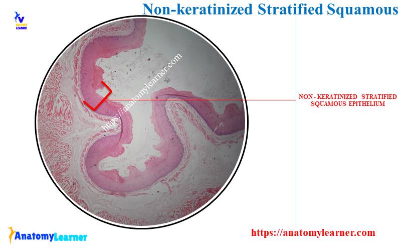
But, the best example of the nonkeratinized stratified squamous epithelium is the esophagus (tunica mucosa layer). For this, you may know more about the lining epithelium (nonkeratinized stratified squamous epithelium) from the below-mentioned article –
- Esophagus histology slide – four different layers description from slide picture and labeled diagram,
The best example of keratinized stratified squamous epithelium is found in the skin histology slide (epidermis of skin). You may learn more about the keratinized stratified squamous epithelium along with other features of the animal’s thick and thin skin from the below-mentioned article of anatomy learner –
Let’s know the details of the location of both keratinized and nonkeratinized stratified squamous epithelium with their diagrams.
Nonkeratinized stratified squamous epithelium under a microscope
Numerous cell layers characterize the nonkeratinized stratified squamous epithelium under a light microscope. In this situation, the outermost layer consists of flattened or squamous cells with nuclei.
Thus, the nonkeratinized stratified squamous epithelium’s outermost or superficial cell layer is alive. This type of stratified squamous epithelium’s superficial surface remains moist and protected from drying.
Again, the thickness of the nonkeratinized stratified squamous epithelium varies in different regions of the animal’s body. For example, you will see the typical features of the nonkeratinized stratified squamous epithelium on the tunica mucosa of the esophagus and also in the oral cavity.
What is the important feature in the nonkeratinized stratified squamous lining of the esophagus? Well, you will see the below-mentioned microscopic features in the lining of the esophagus (nonkeratinized stratified squamous epithelium).
Microscopic features in non-keratinized stratified squamous epithelium
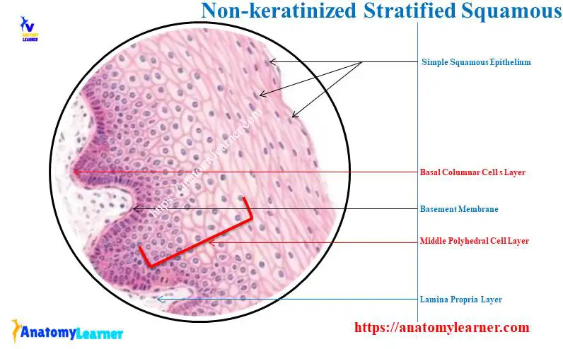
At the base of this type of stratified squamous epithelium, you will find the cuboidal or low columnar epithelium cells. You will see the finely granular cytoplasm of the basal layer of cells.
Again, oval and chromatin-rich nuclei are present on these basal layer cells. The intermediate, second, or middle layer of the non-keratinized stratified squamous epithelium from the esophagus microscope slide also shows the polyhedral layers of cells.
These polyhedral cells of the middle layer possess round or oval nuclei with distinct cell cytoplasm and membrane. You will frequently find mitotic figures in the deeper and basal cell layers of this non-keratinized stratified squamous epithelium.
The cells and their nuclei become progressively flatter in the apical part. These flatter cells migrate toward the luminal or free surface of the epithelium lining (tunica mucosa).
So, above the polyhedral cells layer of this non-keratinized stratified squamous epithelium, you will find the flattened squamous cells. But how is the flattened squamous arranged in the superficial layer of this stratified squamous?
You may find only a few rows (2 – 3) of flattened squamous epithelium on the apical surface. This type of stratified squamous epithelium (non-keratinized) will not possess any keratin substance over these flattened squamous cells.
You will also find a fine basement membrane in the non-keratinized stratified squamous epithelium that separate the cells from underlying connective tissue. And you know, this underlying connective tissue (loose) is the lamina propria.
You may know more about the lamina propria of any tubular organ from the below-mentioned article of anatomy learner –
In this article, you will also learn the microscopic features of other different layers with labeled diagrams.
Other features from the basement membrane
Okay, let’s find some other essential microscopic features from the basement membrane of this non-keratinized stratified squamous epithelium. You know the lower surface of the non-keratinized stratified squamous epithelium (towards the basement membrane) is uneven or wavy.
There are papillae or extensions of connective tissue at the lower surface of the stratified squamous epithelium. And these papillae or extensions of connective tissue give the wavy appearance at the lower surface of stratified squamous.
Again, in this connective tissue, you will find collagen fibers, fibrocytes, capillaries, and arterioles.
Stratified squamous non-keratinized epithelium location
Now, let’s see the location of the stratified squamous non-keratinized epithelium from the animal’s body. Here, I will enlist some of the important organs or parts from the animal body where you will find non-keratinized stratified squamous epithelium –
- #1. Lining epithelium of esophagus,
- #2. Tunica mucosa of the oral or buccal cavity,
- #3. The cornea of animal’s eye,
- #4. Lining on the animal’s tongue,
- #5. Lamina epithelium layer of the oropharynx, and
- #6. Lining epithelium of laryngopharynx
There are other non-keratinized stratified squamous epithelium locations in an animal’s body. You will find the list of non-keratinized stratified squamous epithelium with pictures from animal bodies here.
But, you will find a great variation in the appearance or structure of the non-keratinized stratified squamous between the esophagus and cornea of the eye. In the cornea of an animal’s eye, you will find a comparatively thin layer of stratified squamous epithelium.
Again, there are no connective tissue papillae at the lower aspect (towards the basement) of the cornea epithelium. So, you will see the smooth surface underlying the stratified squamous epithelium of the cornea.
The cornea microscope slide shows a few cell layer thicknesses compared to the esophagus or other organs. But, you will find the same characteristic arrangement of basal columnar, intermediate polyhedral, and superficial flattened squamous cells.
Keratinized stratified squamous epithelium under a microscope
You will also find the typical microscopic features in the keratinized stratified epithelium cells. That means there are several layers of cells, but only the superficial cells have a squamous shape.
The main difference between the keratinized and non-keratinized stratified squamous epithelium is found in their appearance of a superficial layer. In the keratinized stratified squamous epithelium under a microscope, you will find the non-nucleated flattened cells that fill with keratin.
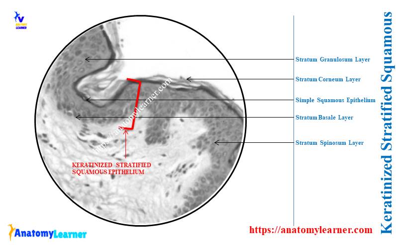
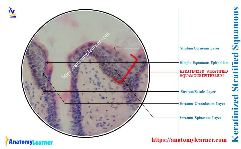
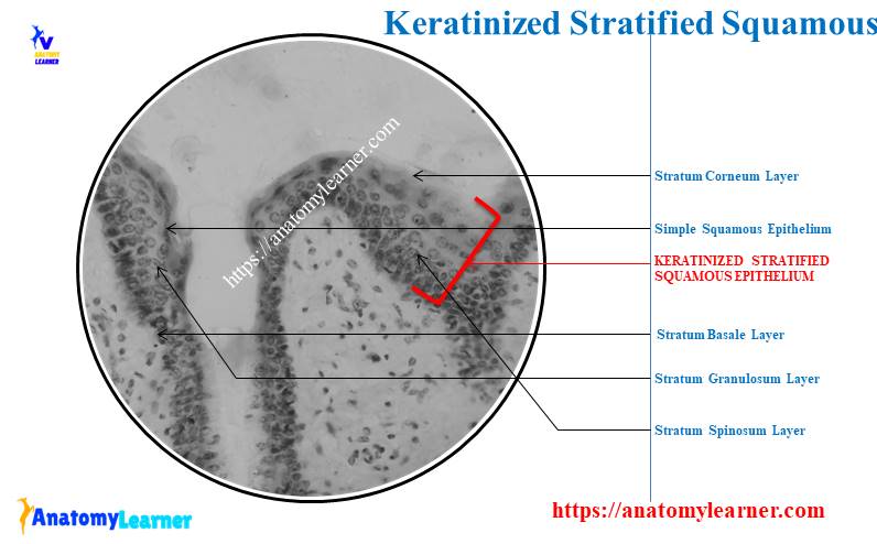
Keratin is a water-resistant protein that forms a protective brarrier against the harmful effect of the environment. You will see three to five distinct cell layers in the microscope slide of keratinized stratified squamous epithelium –
- The deepest layer – stratum basale,
- Next upper layer – stratum spinosum,
- The third layer (toward the surface) – stratum granulosum,
- Next upper layer – stratum lucidum (occurs only in non-hairy skin), and
- Superficial layer – straum corneum,
You will find all these five layers of cells in the epidermis of an animal’s skin. Here, I will describe these five different layers from the keratinized stratified squamous epithelium (with the example of the epidermis of an animal’s skin).
Stratum basale and spinosum of stratified squamous
The stratum basale is the deepest layer of the keratinized stratified squamous epithelium just next to the basement membrane. This is the single cuboidal or low columnar cells layer just near the basement membrane of these stratified epithelia.
Under the light microscope, you may see the mitotic figure in the stratum basale layer of stratified epithelium. This layer gives rise to the new cells that move into the upper layer of the stratified squamous epithelium.
Now, the microscope slide of the skin’s epidermis shows the second layer of polyhedral cells. These polyhedral cells possess rounded or oval nuclei and are adhered to each other by numerous desmosomes.
The electron microscope view between the desmosome of adjacent cells shows –
- Shrinkage between the desmosomal attachment, and
- Small spiny processes radiate from the surface of the adjacent cells,
Thus, this spiny appearance on this cell’s layer gives rise to the name stratum spinosum. You will find the cytokeratin filaments (tonofilaments) in the structure of the spiny process of the adjacent cells.
Again, these tonofilaments of the spiny processes remain to condense at the site of the desmosomes.
Stratum granulosum and lucidum of stratified squamous
The cells from the stratum spinosum layer become more flattened and accumulate keratohyalin and lamellar granules. These cells now move towards the apical surface of the stratified squamous and are termed the stratum granulosum.
This stratum granulosum layer is not found in the nonkeratinized stratified squamous epithelium. Again, you may find this cell layer also in the wall of an animal’s hoof. You may also find this stratum granulosum layer in the cornu of animals.
Now, the next layer of cells (stratum lucidum) only occurs in the nonhairy skin region. This is the intermediate layer of cells between the stratum corneum and granulosum layers.
This keratinized stratified squamous epithelium layer is translucent as it contains eleidin. And you know, eleidin is a protein similar to keratin but has a different affinity for histological stains.
Stratum corneum or keratinized layer of stratified squamous
The stratum corneum is the outermost cell layer of the keratinized stratified squamous epithelium. Here, the flat, scale-like keratinized cells are actively resistant to the environmental irritant.
The cells from the stratum basale to the stratum corneum undergo a series of transformations and form the keratin layer on the apical surface of keratinized stratified squamous epithelium. This process is known as keratinization, where nuclei, Golgi complex, and mitochondria gradually disappear.
These stratified squamous epithelium cells lose their lysosomal activities and accumulate the tonofilaments. The electron microscope view of the stratified squamous reveals that the densely packed tonofilaments are embedded in the dense matrix.
The lamellar granules of the stratum corneum give rise to the intercellular substance between cells. And this intercellular substance from the lamellar granules acts as the barrier component of the stratified squamous epithelium.
I hope you will identify all these different cell layers from the keratinized stratified squamous epithelium.
Keratinized stratified squamous epithelium location
Now, I will show you the location of the keratinized stratified squamous epithelium from the animal’s body. Let’s enlist the location or examples of the keratinized stratified squamous epithelium –
- #1. Epidermis of the skin of the whole body (best example for keratinized stratified squamous epithelium),
- #2. The lining of the hard plates of animals,
- #3. The epithelial lining of the nostril,
- #4. In the wall of the hoof and cornu of animals,
- #5. The lining of the palm and soles of the animals,
You might find a little difference in the layers of epidermis between an animal’s thick and thin skin. The typical features of different layers of cells will find in the thick skin of the animal’s back, palm, and sole regions.
Keratinized and the nonkeratinized stratified squamous epithelium is located over the animal’s body surfaces that are subject to friction. Due to the friction, the most superficial layer of cells constantly remove.
So, the removed cells are replaced by the proliferation of cells from the stratum basale of the stratified squamous epithelium. Thus, both keratinized and nonkeratinized stratified squamous epithelium show the mitotic figures.
Stratified squamous epithelium function
The main functions of the stratified squamous epithelium are protection, secretion, and prevent water loss. Let’s enlist the functions of the stratified squamous epithelium to form various locations of the animal’s body –
- The superficial keratin layer of the stratified keratinized squamous epithelium prevents dehydration of the underlying tissue,
- Again, the superficial layer of the stratified squamous epithelium make a protective layer that prevents the harmful substance from the environment,
- The specific stratified squamous epithelium in various regions of the animal body also participate in secretion and other protection,
Stratified squamous epithelium under a microscope labeled
Now, let’s see the features of stratified squamous epithelium under a microscope with a labeled diagram. Here, I will provide both hand drawings and real microscope figures of stratified squamous epithelium (keratinized and nonkeratinized).
First, the labeled diagram of keratinized stratified squamous epithelium shows the five different types of cells. The stratum basale, spinosum, granulosum, lucidum, and corneum from the keratinized stratified squamous epithelium are identified in the labeled diagram.
Again, the papillae or extension of connective tissue from the lamina propria are also identified in the labeled diagram. Here, the keratin layer of the keratinized stratified squamous epithelium did not show any nucules.
Let’s see the second labeled diagram where I tried to show the three major cell layers from the nonkeratinized stratified squamous epithelium. The labeled diagram identifies the basal columnar or oval and intermediate rounded nucleus.
Again, this labeled diagram identifies the flattened or squamous cells with flattened nuclei from their superficial layer.
Now, let’s draw both the keratinized and nonkeratinized stratified squamous epithelium with the help of provided samples. You will find all the characteristic features of both keratinized and nonkeratinized stratified squamous epithelium in the provided hand-drawing diagram.
If you want to get more labeled diagrams on both keratinized and nonkeratinized stratified squamous epithelium, let’s find them here on the social media of anatomy learners.
Frequently asked questions on stratified squamous epithelium
Here, I will enlist some important questions on the stratified squamous epithelium that histology learners frequently ask. But, it is suggested to know the details of the keratinized and nonkeratinized stratified squamous epithelium features with the labeled diagrams.
Okay, let’s see the common questions on the keratinized and nonkeratinized stratified squamous epithelium with their concise answer –
Where is stratified squamous epithelium found?
You will find the stratified squamous epithelium in the area of the body where the frictions are more. Depending on the functions and structure, you may find two types of stratified squamous epithelium in various regions of the animal’s body.
The keratinized stratified squamous epithelium covers the skin (both thin and thick) of the whole animal’s body. Typically, the epidermis of the thick and thin skin possesses keratinized stratified squamous epithelium.
Again, you may also find the keratinized stratified squamous epithelium in the wall of the hoof and cornua of the animals.
On the other hand, the nonkeratinized stratified squamous epithelium covers the wet surface and is exposed to wear and tear. Typically, you will find the nonkeratinized stratified squamous in the lining of the esophagus, tongue, oropharynx, and laryngopharynx.
What is the function of stratified squamous epithelium?
Protection, secretion, and prevention of water loss are the three essential functions of the stratified squamous epithelium. The mechanism behind the protection and prevention of water loss are described in the previous section of this article.
The tall columnar or low cuboidal cells of the stratified squamous epithelium have secretory functions.
What does stratified squamous epithelium look like under a microscope?
The stratified squamous epithelium looks like a combination of columnar, polyhedral, and squamous cells under the light microscope. But, depending on the various organs, you may find two types of stratified squamous epithelia under a light microscope.
How would you recognize squamous epithelium?
To recognize the squamous epithelium from the stratified squamous, you might notice the appearance of the superficial cell layer. Here, the superficial cells layer of the stratified squamous shows the typical features of the squamous epithelium.
And that is, the cells look flatter and possess a flattened or elongated (transversely) nucleus.
What does stratified squamous keratinized epithelium look like?
The stratified squamous keratinized epithelium looks translucent on its apical surface. And you know this is due to the presence of the keratin layer over the apical surface of the stratified squamous keratinized epithelium.
Conclusion
So, the stratified squamous epithelium under a microscope shows three to five different layers of cells. According to the appearance of cell layers, they may divide into keratinized and nonkeratinized stratified squamous epithelium.
The labeled diagrams help you identify the keratinized and nonkeratinized stratified squamous epithelium under the light microscope. You will see the significant difference in the cell layers and the presence or absence of keratin in the superficial layer between keratinized and nonkeratinized stratified squamous epithelium.