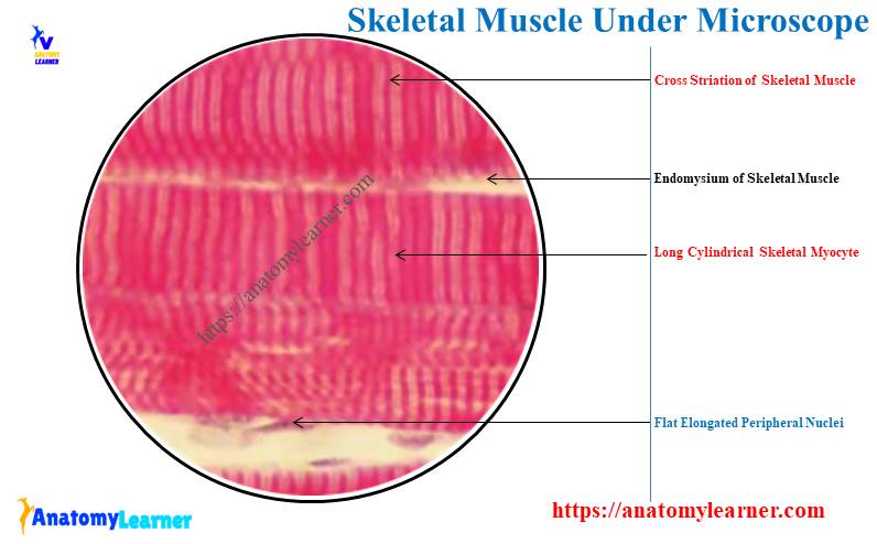
The skeletal muscle under a microscope shows long, cylindrical, unbranched fibers and flat nuclei. Each skeletal muscle fiber comprises compactly packed long myofibrils that arrange parallel to the long axis.
This article might help you to understand the microscopic features of the skeletal muscle with the labeled diagram. Again, you will find the complete list of the identifying points for the skeletal muscle microscope slide in this article.
Quick summary: The skeletal muscle under a microscope are long, parallel, cylindrical muscle fibers without branching. Numerous flat peripheral nuclei and unique cross-striation are found in the skeletal muscle microscope slide.
Finally, I will share the main differentiating points of the skeletal muscle microscope slide from smooth and cardiac muscles. So, if you want to learn the basic microscopic features of the skeletal muscle histology slide and differentiate it from smooth and cardiac muscles, let’s continue this article till the end.
The skeletal muscle under a microscope
In the structure of skeletal muscle fibers under the light microscope, you will find elongated myocytes that vary in diameter and length. These skeletal muscle fibers are derived from the prenatal fusion of numerous mononuclear myoblasts.
Thus, you may find multiple nuclei in the single skeletal muscle fiber in the peripheral aspect. Individual skeletal muscle fibers possess numerous myofibrils, which form the dot appearance in the cross-section of that fiber at the light microscope.
Again, the individual myofibril of the skeletal muscle fiber comprises thick and thin myofilaments. These myofilaments of the skeletal muscle fibers are responsible for contraction.
You will know the details of these structures from the skeletal muscle microscope slide in the description section. Now, let’s see what structure you should identify from the skeletal muscle fibers at the light microscope view –
- Individual skeletal muscle fibers from both longitudinal and cross sections,
- Flattened and peripheral nuclei from the skeletal muscle fibers,
- Cross-striation (dark and light bands) of skeletal muscle fibers,
- Endomysium, perimysium, and epimysium of the skeletal muscle,
- Fibroblast in the endomysium of skeletal muscle,
- Individual muscle fascicle,
- Myofibrils of the skeletal muscle, and
- Larger blood vessels and capillaries from the skeletal muscle,
The provided labeled diagrams identify all these microscopic features from the skeletal muscle histology slide. You might see these microscopic features of skeletal muscle both from the longitudinal and cross (transverse) sections.
Summary of the skeletal muscle microscope slide
While identifying the skeletal muscle histology slide with the help of a light microscope, you might identify the cell or muscle fiber, nuclei, and other associated structures. The followings are the essential features (in table 1) that will provide the summary of the skeletal muscle microscope slide and help you to identify it at the light microscope –
| Features | The skeletal muscle under a microscope |
| Muscle fiber or cell | Long and cylindrical muscle fiber |
| Branching in fiber | No branching |
| Appearance of fibers | Parallel to each other |
| Nucleus | Elongated or flattened |
| Location of nucleus | Peripheral (near the sarcolemma) |
| Number of nuclei | Multiple |
| Cross striation | Present in skeletal muscle |
I hope you got the basic idea of the shape of an individual skeletal muscle cell or fiber, its nucleus, and other essential features. Now, let’s see the important identifying characteristics of the skeletal muscle histology slide.
Identifying points of skeletal muscle microscope slide
If you are asked to identify the skeletal muscle microscope slide, the following identification point might help you. Here, I will only enlist the essential identification points for the skeletal muscle microscope slide with the diagram –
- The provided tissue sample shows the elongated, cylindrical, parallel muscles fibers that don’t possess any branch,
- There are numerous flattened nuclei present at the periphery of the individual muscle fiber,
- The muscle fibers can easily distinguish as they show unique transverse cross striations,
- The individual muscle fiber is separated from others by loose connective tissue,
So, this is the longitudinal section of the skeletal muscle histology slide. Here in the longitudinal section of the skeletal muscle, 2 important features you might remember –
- #1. Presence of multiple peripheral nuclei in the single or individual muscle fiber, and
- #2. Each muscle fiber of the bundle shows cross or transverse striations,
Now, let’s see the identifying points from the cross or transverse section of skeletal muscle –
- The provided muscle tissue shows an irregular oval-shaped structure,
- Individual muscle fiber shows the peripheral oval or elongated multiple nuclei,
- Numerous muscle fibers form a single fasciculus,
- The provided muscle tissue section shows numerous fasciculi under the microscope view,
Again, the higher magnification of the provided muscle tissue shows dot-like myofibrils within the individual fiber,
So, this is the transverse section of the skeletal muscle histology slide. You will also find 3 important features in the transverse section of skeletal muscle – endomysium, perimysium, and epimysium.
These are the connective tissue layer that covers the individual muscle fiber, fascicle, and skeletal muscle bundle. You will learn more about these connective tissue layers (endomysium, perimysium, and epimysium) in the next section of this article.
The skeletal muscle under microscope description
So, the skeletal muscle fibers under a light microscope are long cylindrical, multinucleated cells with peripheral nuclei. The elongated and flattened nuclei of the skeletal muscle fibers are normally seen under the cell membrane sarcolemma.
Again, adjacent to these nuclei of skeletal muscle, you will see the thin cytoplasm (known as the sarcoplasm) with other organelles. The cytoplasm of the skeletal muscle cells fills with numerous longitudinal myofibrils.
In the transverse section of skeletal muscle fiber with the routine stain, you will find the groups of myofibrils. These myofibrils arrange parallel and distribute uniformly throughout the skeletal muscle fiber.
Under the electron microscope, you will see the numerous microfilaments in the structure of individual myofibrils. You will learn the details of the microfilaments or finer structure of the myofibril with a diagram in the next section of this article.
You will also see the cross or transverse striation in the skeletal muscle fibers microscope slide. These cross striations are of the alternate dark (A) and light (I) bands with a Z line that intersects with the I band.
You may easily observe this cross striation in the skeletal muscle histology slide that stains with Hematoxylin and Eosin. But, you might know the finer structure to get a clear idea of the myofibril.
The Dark (A) and light (I) bands and Z and M lines will be described in the electron microscopic features of the skeletal muscle fiber.
The sarcoplasm (SC) of the skeletal muscle fiber contains various cell organelles that tend to aggregate near the multiple nuclei. You will also find numerous mitochondria and glycogen that provide energy for skeletal muscle contraction.
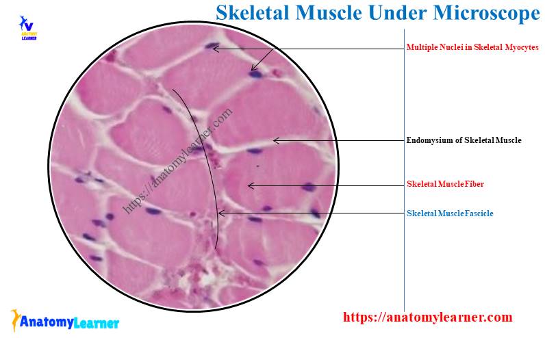
Cross section of skeletal muscle in microscope slide
The organization of the skeletal muscle fibers in the cross or transverse section is somewhat different. Individual muscle fibers or skeletal myocytes are bound together to form a primary bundle.
This primary bundle of the skeletal myocytes is known as the fascicle. Here, in the cross-section of the skeletal muscle fibers, you will see the followings –
- Endomysium – an individual myocyte within the fascicle surrounds by the reticular fibers and forms the endomysium,
- Perimysium – each fascicle of the skeletal muscle surrounded by the dense irregular connective tissue; termed as the perimysium,
- Epimysium – the entire skeletal muscle or bundle is surrounded by the irregular connective tissue layer known as the epimysium,
Various blood vessels, nerves, and muscle stretch receptors (muscle spindles) are present in the perimysium layer. All these connective tissues of the skeletal muscle fibers are interconnected and provide contractile force. These contractile forces created by the connective tissue transmit to each other tissue.
The number of fascicules in a skeletal muscle, and the number of fibers in each fascicule, are highly variable. Some of the smaller skeletal muscles possess delicate and fewer fascicules. In comparison, the larger skeletal muscle fiber possesses coarse and numerous fascicules.
Ultrastructure of skeletal muscle under the electron microscope
If you want to see the ultrastructure of the skeletal muscle, use the electron microscope. Under the electron microscope, you will see the below-mentioned features from the skeletal muscle fibers –
- Sarcolemma and sarcoplasma,
- Sarcoplasmic reticulum, and sarcosome,
- Different organelles in the skeletal myocytes,
- Individual structure of the myofibrils (different bands, zones, and lines),
The individual skeletal muscle fiber is covered by the plasma membrane, also known as the sarcolemma. Again, this skeletal muscle fiber sarcolemma also covers the basement membrane of the outer connective tissue.
This connective tissue basement membrane of the skeletal muscle makes a connection between the muscle fiber and the endomysium’s fiber. The cytoplasm of the skeletal muscle cell possesses numerous organelles and pushes the nuclei to a peripheral location.
Now, let’s know what the sarcoplasmic reticulum and sarcosome in the skeletal muscle are. You will see the elaborate system of a membrane-lined tube in between the myofibrils of the skeletal muscle. This elaborate system of the membrane-lined tube is the sarcoplasmic reticulum.
Again, the elongated mitochondria and cluster of glycogen scattered distribute in the myofibrils of skeletal muscle. Here, the elongated mitochondria of skeletal muscle are known as the sarcosome.
You will also find other organelles like Golgi bodies, ribosomes, lysosomes, and lipid droplets in the cytoplasm of an individual skeletal muscle cell. The individual skeletal myocyte labeled diagram shows all the organelles that are provided earlier.
Structure of skeletal myofibrils
The electron microscope reveals that the skeletal myofibrils comprise numerous fine (both thick and thin) myofilaments. These myofibrils have alternative darker A band (anisotropic) and lighter I (isotropic) bands.
When the thick and thin myofilaments of the skeletal myofibrils overlap, they form the darker (A) bands. Again, you will only find the thin myofilaments in the lighter I bands.
In the structure of the individual myofibril of the skeletal muscle, you will find the followings –
- Bands and I bands,
- Z bands and H zones, and
- The sarcomere,
From the labeled diagram, let’s see these bands and zones from the individual myofibril of the skeletal muscle. As you have seen before, the darker (showed) bands are A, whereas the lighter areas are the I bands.
In between the lighter bands (I), you will find a cross-striated thin dark line. This cross-striated and thin dark line is the zwischenschiebe (Z) band.
The middle part of the darker bands (A) is traversed by a lighter band which is known as the H –band. This is also called the Hensen band of the skeletal muscle.
Now, within the H – band, you will find a thin dark line. This thin dark line in H – the band is known as the Mittleschiebe or M band.
Now, the part of the skeletal myofibril between two consecutive Z bands is known as the sarcomere.
All these bands that have been described above run transversely across the skeletal muscle fiber. Again, the adjacent myofibrils of the skeletal muscle lie in the same alignment with one another. Thus, their orderly arrangement in the skeletal muscle shows unique cross-striation features.
Why is a skeletal muscle called striated muscle?
The contractile thin protein actin and thick protein myosin form the myofilament of the skeletal muscle. You will see the regular arrangement of the actin and myosin in the sarcoplasm of the skeletal muscle fiber.
This regular arrangement of these skeletal muscle proteins forms the cross-striation pattern. Because of these cross-striation patterns, the skeletal muscle is also called the striated muscle.
Structure of myofilaments
The contractile myofilaments of the skeletal muscle fibers are primarily actin or myosin II. Again, you will find another type of myofilament that contain protein involved in either binding the primary filament together or regulating actin-myosin interaction.
So, you will find two types of myofilaments in skeletal muscle at the electron microscope –
- #1. Thin myofilaments of the skeletal muscle – consist of actin, troponin, and tropomyosin, and
- #2. Thick myofilaments of the skeletal muscle – consist of myosin II,
In the structure of the thin myofilaments of the skeletal muscle, you will find globular molecules (G-actin). These G-actin or globular molecules polymerize to form thin filamentous strands. These thin filamentous strands are known as F-actin.
Again, each globular molecule has the binding site for myosin – II. Here, two filamentous strands of F-actin twist together to form the double helix.
You will also find the filamentous tropomyosin molecule that lies in the groove between two twisted strands of F-actin (filamentous strands). Again, the filamentous tropomyosin covers the myosin II binding sites on the actin filament.
The myofilament structure also shows the triple globular unit of troponin that lies in the regular internal of the tropomyosin.
On the other hand, the thick myofilaments are composed of myosin II, where you will find two heavy chains and four light chains of amino acids. Here, the two heavy chains twist together to form a rodlike tail with two globular heads.
You will also find the two light chains associated with each globular head of the myofilaments. These globular heads have binding sites for actin and adenosine triphosphate.
Other proteins in the skeletal muscle
You will also find several other proteins in the structure of the skeletal myofibrils other than actin and myosin. Let’s see other different proteins from the skeletal myofibrils –
- Nebulin,
- Desmin,
- Dystrophin,
- Actinin,
- Titin, and
- Myomesin protein,
The nebulin protein of the skeletal muscle is associated with actin. This protein may regulate the assembly and length of the actin filaments.
Desmin is the intermediate filament that locates at the Z line of the myofibrils. This desmin links the adjacent myofibrils together side by side. They also attach the myofibrils to the cell membrane, known as the costamere.
Dystrophin is the transmembrane complex of protein that links the skeletal myocyte to the surrounding basement membrane. Again, this dystrophin also makes a complex link with the actin-myosin filaments.
The actin is present in the region of the z bands and binds the tail ends of the actin filaments with the bands. Again, titin links the head ends of the myosin filaments to the z bands.
The myomesin present in the region of M bands binds the tail end of the myosin filament with the bands.
Types of skeletal muscle fibers under a microscope
Based on the speed of contraction, gross anatomic appearance, and fatigue resistance, the skeletal muscle fibers may be divided into two types –
- #1. Red skeletal muscle fibers, and
- #2. White skeletal muscle fibers,
The red skeletal muscle fibers contain a large amount of myoglobin, contributing to their red color. You know that myoglobin is an oxygen-carrying protein that is similar to hemoglobin.
The structure of the red skeletal muscle shows numerous densely packed mitochondria under the sarcolemma. These red muscle fibers contract and fatigue slowly and are termed slow-twitch fibers.
The red muscle fibers are narrower than white fibers and numerous in the skeletal myofilaments. Thus, the striation on the red skeletal muscle is less distinct than those of the white skeletal muscle fibers.
In contrast, the white skeletal muscle fibers have less myoglobin, and thus they become lighter. You will find fewer mitochondria in the white skeletal muscle fibers that remain clustered between the myofibrils.
There is extensive smooth endoplasmic reticulum in white fibers compared to the red skeletal muscle fibers. These extensive smooth endoplasmic reticula allow them to release calcium and initiate the contraction rapidly.
The contraction and fatigue of the white skeletal muscle fibers are rapid compared to the red fibers. Thus, the white skeletal muscle is also known as the fast twitch fibers.
You will also find the intermediate type of muscle fibers that have the combined characteristic of red and white skeletal fibers.
Difference between white and red skeletal muscle fibers
From table 2, you will find the main difference between the white and red skeletal muscle fibers. Okay, let’s see the differentiating points of the white skeletal muscle fibers from red fibers –
| Features | Red skeletal muscle | White skeletal muscle |
| Myoglobin | More | Less |
| Diameter | Narrow | Wider |
| Striation | Less | More |
| Nuclei | Not always at the periphery | At periphery |
| Myofibrils | Less | More |
| Mitochondria | Numerous | Less |
| Contraction | Slow and continue | Rapid |
Skeletal muscle under microscope labeled diagram
In this article section, I will provide more labeled diagrams of skeletal muscle under a microscope. Here, I will identify all the microscopic features from both longitudinal and cross (transverse) sectional views of the skeletal muscle.
Here, the longitudinal section of skeletal muscle histology slides (low magnification) the long cylindrical muscle fibers with multiple peripheral nuclei. Again, the provided diagram (low magnification) shows the cross-striation of the skeletal muscle fibers.
The diagram also shows the dark (A) and light (I) bands in the cross striation. Sometimes, you may find the Z discs that bisect the light I bands of the cross striation.
You can view the typical structural features of the skeletal muscle histology slide in the cross or transverse section. Here, the cross-section of the skeletal muscle histology slide shows the fascicule.
Again, the epimysium, perimysium, and endomysium are clearly identified from the cross-sectional view of the skeletal muscle. In between various skeletal muscle fascicules, the diagram shows numerous blood vessels.
The sarcoplasm of the skeletal muscle fibers shows a granular appearance in the provided sample. And you know, this occurs due to the cross-section of the skeletal muscle’s myofibrils.
You will find more labeled diagrams on the skeletal muscle fibers (both longitudinal and cross-section) here on social media of anatomy learners.
Skeletal muscle under microscope 10x, 40x, and 400x
Here, you will find various labeled diagrams of skeletal muscle histology slide with magnification of 10x, 40x, and 400x. The skeletal muscle microscope slide with 10x magnification is shown in the labeled diagram.
The 40x and 400x magnified labeled diagrams of the skeletal muscle microscope with all important features are also shown here. Multiple nuclei with the skeletal myofibers and the cross striation are easily observed in the 40x and 400x magnified labeled diagrams.
Frequently asked questions on skeletal muscle microscope slide
Here, you will find the questions on the skeletal muscle microscope slide that the histology learners ask. I will enlist the most commonly asked question on the skeletal muscle microscope slide with their concise answer.
Let’s see the questions and concise answers of the skeletal muscles microscope slide –
What does skeletal muscle look like under a microscope?
The skeletal muscle under a microscope looks like long cylindrical fibers with multiple nuclei at the periphery. You will find the typical identifying features in the skeletal muscle on the microscope slide.
The most identifying feature of the skeletal muscle is the presence of cross-striation in the long cylindrical muscle fibers.
But, the cross-section of the skeletal muscle is somewhat different than these of the longitudinal section. The skeletal or striated muscle cross-section shows numerous muscle fascicules with numerous myofibrils.
What is the microscopic structure of a skeletal muscle?
Microscopically, you will find the myofibrils in the skeletal muscle fibers. Again, the myofibrils of the skeletal muscle fibers contain numerous myofilaments that contain various proteins.
Microscopic features of the skeletal muscle also show the multiple peripheral nuclei and cross striation in the longitudinal fibers.
What are the differences between smooth muscle and skeletal muscle under a microscope?
You may easily differentiate the smooth muscle fibers from the skeletal muscle fibers under the light microscope. The main differentiating point between the smooth and skeletal muscles is the cell shape and nuclei variation.
You know, the skeletal muscle cells are cylindrical, whereas the smooth muscle possesses fusiform cells. Individual skeletal muscle fiber contains multiple peripheral nuclei, but the smooth muscle contains only one centrally placed elongated nucleus.
You will find more differences between the smooth muscle and skeletal muscle fibers in table 3 –
| Features | Skeletal muscle | Smooth muscle |
| Cells | Long cylindrical | Fusiform |
| Nucleus | Flat, multiple | Single elongated |
| Location of nucleus | Peripheral | Central |
| Striation | Striated | Nonstriated |
| Contraction | Quick | Slow |
With their microscope features, you will easily differentiate the smooth muscle fibers from the skeletal muscle fibers.
Conclusion
The skeletal muscle myocytes under the light microscope are long elongated cells that arise from the parental fusion of numerous mononuclear myoblasts. You might know the typical features of the skeletal muscle histology slide to identify it quickly at your laboratory.
The provided labeled diagrams help you to understand every single feature from the skeletal muscle histology slide. Now, you will easily differentiate the skeletal muscle microscopic slide from smooth muscle.