The ligamenta flava is one of the important ligaments of the inter neural articulation. In this guide, I will provide the exact answer to the question “What is ligamenta flava?” with proper diagrams.
Quick answer: The ligamenta flava is the ligament that connects the arches of the adjacent vertebrae of the interneural articulation.
I will also try to inform you why this ligamenta flava is important in an animal’s interneural articulation.
What is ligamenta flava?
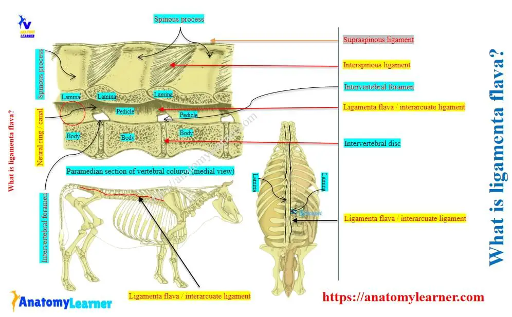
The ligamenta flava fill the dorsal gaps between the adjacent vertebral arches along the vertebral column. They are the membranous ligament and consist largely of yellow elastic tissue.
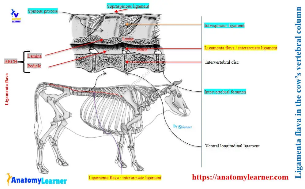
Here, the figure shows the attachment and extension of the ligamenta flava in the cow’s vertebral column (in the inter neural articulation).
Thus, to understand the ligamenta flava clearly, you might have the knowledge of the followings –
- Different parts of a typical vertebra (including body, arches, and processes),
- Joints of the animals’ vertebral column (especially the intercentral, interneural, and intertransverse articulation),
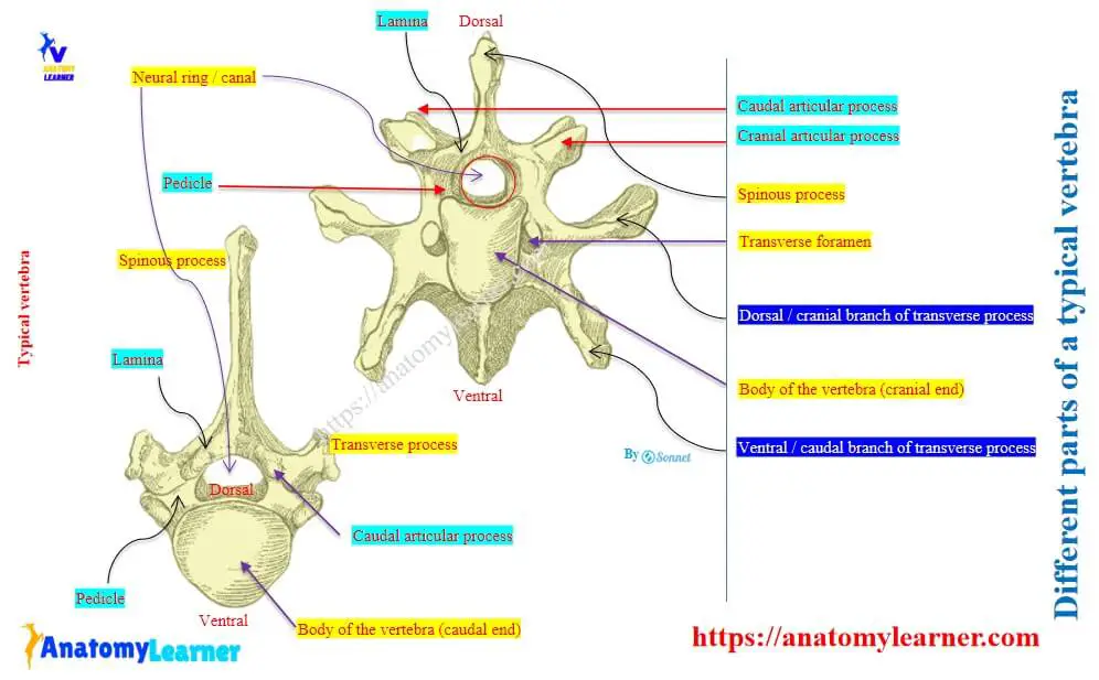
Parts of a typical vertebra of animals
The typical vertebra of an ox shows 3 different parts –
- A cylindrical body: it consists of cranial convex and caudal concave extremities. You will also find the dorsal surface that forms the floor of the neural ring / canal and the ventral surface that possess ill-developed spine.
- Right and left lateral arches: both right and left lateral arches consist of the ventral pedicle and dorsal lamina. Here, the pedicles bear the notches on both cranial and caudal aspects that help to form inter-vertebral foramen with adjacent vertebrae.
- Processes of a vertebra: There are three processes in a typical vertebra. The spinous process is single and directed upward. Whereas the transverse processes are present on either side and projected outward from the lateral aspect of the arch. Finally, the articular processes are divided into paired cranial and paired caudal processes.
Here, the figure shows the 3 different parts (body, arches, and processes) of the cow’s typical vertebra.
Joints of the vertebral column of animals
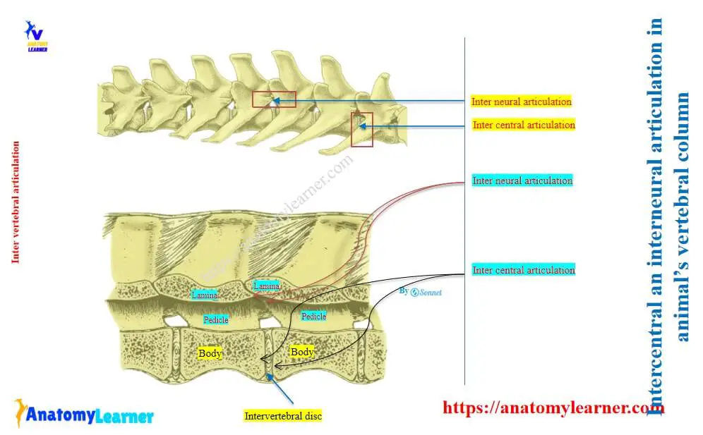
The bodies, arches, and processes of the vertebral bones are serially articulated and form the intercentral and inter neural joints. In most cases, they are termed the common vertebral articulation in animals, with very few exceptions.
Intercentral articulation: the cranial convex and caudal concave extremities of the bodies of two adjacent vertebrae articulate together to form an inter central articulation. A fibrocartilaginious convexo concave disc is placed between the bodies of these adjacent vertebrae.
Inter neural articulation: the arches and processes of the adjacent vertebrae articulate together and form the inter neural articulation.
Here, the figure shows both inter central and inter neural articulation from the animal’s vertebral column.
In both inter central and inter neural articulation of the animal’s vertebral column, you will find different ligaments. In most cases, these joints are articulated by some of common / common variety of ligaments.
Again, a few joints of the vertebral column are not articulated by the common ligaments. These joints of the vertebral column are the special vertebral articulations.
What are the ligaments of the inter neural articulation?
You will find the followings ligaments in the inter neural articulation in any animal’s vertebral articulation –
- Capsular ligament: encloses the articular process and is well-developed in the cervical region.
- Yellow ligament / ligamenta flava: connects the arches of the adjacent vertebrae.
- Intertransverse ligament: connects the adjacent transverse processes of the vertebrae.
- Interspinous ligament: connects the adjacent border of the supraspinous processes.
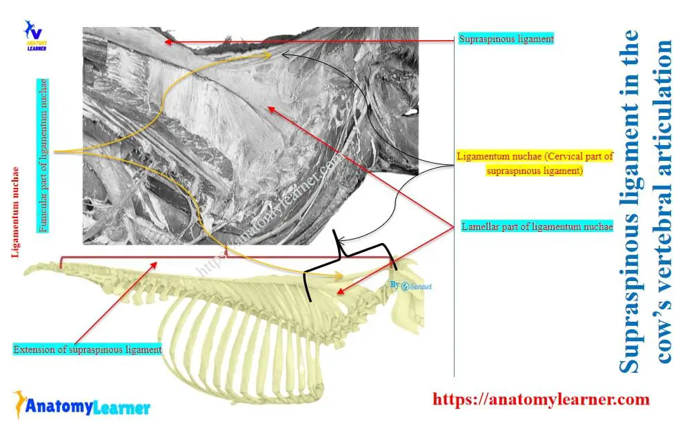
- Supraspinous ligament: It is a very strong ligament and extends from the occipital condyle to the sacrum in animals.
The supraspinous ligament in animals divides into the thoracolumbar and cervical parts. Here, the cervical part of this ligament is known as the ligamentum nuchae.
Again, the ligamentum nuchae is further divided into the lamellar and the funicular parts. The figure shows the supraspinous ligament in a cow (including the ligamentum nuchae).
Ligamenta flava anatomy and diagram
So, the dorsal gaps between the animal’s vertebral arches are filled with the ligamenta flava. These ligamenta flava are thin and made up of yellow elastic tissue.
Another name of ligamenta flava:
- Ligamentum flavum,
- Interarcuate ligament, and
- Yellow elastic ligament,
Why is ligamenta flava important in animals?
The ligamenta flava are clinically important in animals, as they are pierced / punctured during caudal epidural blocking. This procedure involves infiltration of blocking materials into the epidural space through the yellow ligamenta flava.
Best sites for blocking: at the cranial lumbar region between L1 and L2, at the lumbosacral region between L6 and S1, at the tail region between CD1 and CD2 vertebrae. (Here, L = lumbar vertebrae, S = Sacral vertebrae, and CD = caudal vertebrae).
Conclusion
So, the exact answer to the question “What is ligamenta flava?” is the yellow elastic ligament that connects arches of the adjacent vertebrae. It is a membranous ligament that binds the lamina and pedicle perfectly and completes the inter neural articulation of the vertebrae.