The ligamentum nuchae in an ox is better developed than that of the horse. It is part of the supraspinous ligament and plays a major role in intra-neural articulation.
I will show you the location and anatomical features of the ligamentum nuchae in bovine. You will also understand the exact extension of this structure in the ox neck from this article.
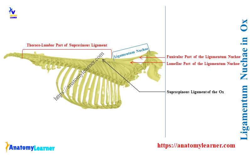
Ligamentum nuchae in ox is the cervical part of the supraspinous ligament that divides into lamellar and funicular parts. It extends from the occipital protuberance to the second or third cervical vertebrae of an ox.
I will also enlist the unique features of the ligamentum nuchae from different animals like horses, dogs, and pigs. First, let’s know the detailed structure of the bovine ligamentum nuchae with the labeled diagram.
Ligamentum nuchae in ox
The supraspinous ligament of intra-neural articulation becomes remarkably modified at the neck region. It becomes wide and flat and forms the ligamentum nuchae.
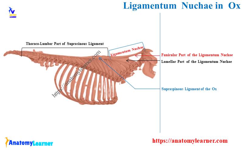
Here, the diagram shows the cervical and thoracolumbar parts of the supraspinous ligament of an ox. The cervical part of this ligament shows major modification, which is known as the ligamentum nuchae of ox.
Again, the diagram shows the dorsal rounded bundle and ventral flattened parts of the ligamentum nuchae. Thus, the bovine ligamentum nuchae divides into the following 2 parts –
- Funicular part – dorsally, and
- Lamellar parts – ventrally,
Let’s discuss the funicular and lamellae parts of the bovine ligamentum nuchae with a diagram.
Funicular part of the cattle ligamentum nuchae
The funicular part of the cattle ligamentum nuchae arises from the external occipital protuberance. It inserts into the summits of the vertebral spine at the level of 2nd or 3rd thoracic vertebrae.
You may know the details of ox thoracic vertebrae anatomy from the below-mentioned article –
- Thoracic vertebrae of ox with diagram,
The funicular part form of a rounded bundle becomes wide and flat as it goes caudally. Finally, it continues with the thoracolumbar part of the ligamentum nuchae at the withers.
The funicular part of the bovine ligamentum nuchae clearly divides into two lateral halves. Both are rounded in their occipital attachment, but from the axis backward, they become wider and flat.
The wide part of the two lateral halves of the funicular part is almost sagittal. It lies on either side of the vertebral spine of the ox. Again, the wide parts of the funicular part are covered by the trapezius and rhomboideus muscles.
Suggested article on bovine muscles –
- Cow muscle anatomy – bovine muscles identification with diagram (neck region),
At the level of the third thoracic spine, the funicular part gradually diminishes in size. Finally, this funicular part fades out and continues with the thoracolumbar part of the ligamentum nuchae.
The lamellar part of the bovine ligamentum nuchae
The lamellar part of the bovine ligamentum nuchae arises from the wider funicular part. It consists of two laminae separated medially by a layer of loose connective tissue.
Each lamina directs downward and forward in a radiating manner. They end on the spine of the cervical vertebrae of the ox except the first.
Each of the lamellar parts of the bovine ligamentum nuchae is stronger than the horses. They are thick and consist of two portions –
- Anterior portion of the lamellar ligamentum nuchae, and
- Posterior portion of the lamellar ligamentum nuchae,
Here, the anterior portion is thin and double in cattle. The fiber of the anterior portion terminates on the spine of the 2nd, 3rd, and 4th cervical vertebrae.
Again, the posterior portion of the lamellar ligamentum nuchae is single. Its fiber extends from the first thoracic spine to the 5th, 6th, and 7th cervical vertebral spines.
Let’s know the details of ox cervical vertebrae from the below-mentioned articles –
- 7th cervical vertebra of an ox with labeled diagram,
- Axis of an ox – second cervical vertebra with diagram,
Which bones are deep to a ligamentum nuchae in ox?
Quick answer: the occipital protuberance and the spines of cervical and thoracic vertebrae are deep to the ligamentum nuchae. However, the spine of the first cervical vertebra is not deep to the ligamentum nuchae in the bovine.
Here, the diagram shows the spines of the vertebral bones that are deep into the ligamentum nuchae. It shows the spine of the 2nd to 7th cervical and first 3 thoracic vertebrae attached with this nuchae.
Function of the ligamentum nuchae in animals
The ligamentum nuchae of an animal is a powerful elastic apparatus. It is a very strong part of the supraspinous ligament.
It attaches the summit of the spines of the cervical (2 – 7) and the first 3 thoracic vertebrae. Let’s see the major function of the ligamentum nuchae in animals, especially grazing animals like cows –
It assists the extensor muscles of the head and neck of animals like cows and horses,
This ligament is well-developed in cows due to their grazing habit of extending the head toward the ground,
Where are ligamentum nuchae located in horses?
Quick answer: the ligamentum nuchae of the horse extends from the occipital protuberance to the withers. It is directly continued with the lumbo-dorsal segment of the horse supraspinous ligament.
The parts of the ligamentum nuchae in a horse are almost similar, with a little exception. You will find both the funicular and lamellar parts in the horse ligamentum nuchae.
Here, the diagram shows the extension and structure of the ligamentum nuchae of the horses. Let’s find the video on ligamentum nuchae of animals, where I explained everything about it. You may also find more labeled diagrams of the animal’s ligamentum nuchae here.
Ligament nuchal in horses – exceptional features
The ligament nuchal in horses is a little different than in other animals. You will find a few bursae below the supraspinous ligament.
Typically, these bursae are found below the cervical part of this ligament. There are 2 bursae under the ligament nuchal in the older horses –
- Atlantal bursa – lies between a funicular part of the ligament and the dorsal arch of the atlas and
- Supraspinous bursa – over the third and fourth thoracic vertebral spines,
However, this supraspinous bursa may remain over the second thoracic spine and extend up to the fifth.
There may be another bursa present at the spine of the horse axis vertebra. This bursa lies between the funicular part and the large digitation attached to the axis.
It is believed that infection of these atlantal and supraspinous bursae leads to poll evil and fistula withers. However, the fistula withers in a cow may rarely occur due to the absence of these types of bursae.
In the funicular part of the horse ligamentum nuchae, you will find two bands closely attached. Near and at the withers, it broadens greatly and forms an expanded portion.
Again, in this area of the funicular part, you will see the covering of the trapezius and rhomboideus muscles. Behind the 4th thoracic spine, it becomes thinner and narrower.
In the structure of this ligament nuchal in horses, you will find the fat and elastic tissue. It varies greatly in the stallion of draft breeds.
What is the supraspinous ligament of the ox?
Quick answer: the supraspinous ligament of the ox is a very strong and long ligament of intra-neural articulation. It extends medially from the occipital protuberance of the ox skull to the sacrum bone.
The supraspinous ligament of the ox divides into –
- Thoracolumbar part – starts from the sacral spine and attaches to the summits of the spines of lumbar and thoracic vertebrae and
- The cervical part – is also known as the ligamentum nuchae in ox,
But, there is no practical demarcation between the cervical and thoracolumbar parts of the ligamentum nuchae. You will also find other various ligaments except the supraspinous ligament in the intra-neural joint.
The intra-neural joints are the articulations of vertebral arches and processes. You will find the below-mentioned ligaments that connect in the spines, processes, and laminae of the vertebrae –
- Capsular ligament – well-developed in the cervical region and encapsulates the articulation between the articular processes,
- Ligaments of the arches – also known as the ligament flava and connect the arches of adjacent vertebrae,
- Interspinous ligament – thin and connect adjacent transverse processes of the vertebrae,
- The transverse ligament – is well-developed in the lumbar region and connects the adjacent transverse processes and
- Supraspinous ligament – very strong and longest ligament of the vertebral column,
Frequently asked questions on ligamentum nuchae in ox
Here, you will find the list of questions on the ligamentum nuchae in ox or other animals. But to understand the extension and structure of the ligamentum nuchae, you might go through the full guide.
Let’s see the questions on ligamentum nuchae of animals with their concise answer –
What is the ligamentum nuchae formed by in bovine?
Quick answer: the ligamentum nuchae is formed by the yellow elastic tissue in the bovine. The elastic tissue is more in the bovine ligamentum nuchae due to the extension of the head toward the ground during grazing.
Where does the nuchal ligament attach in dogs?
Quick answer: the nuchal ligament attaches to the spine of the axis and anterior thoracic spine in dogs. The dog’s nuchal ligament consists of small fibrous bands.
Sometimes, it may be regarded as a small fibrous raphe between the right and left muscles of the dog’s neck. So, the ligamentum nuchae is less developed in dogs compared to cows or horses.
What is special about the nuchal ligament of a pig?
Quick answer: the nuchal ligament of the pig consists of a fibrous raphe and a thin layer of elastic tissue. This ligament extends between the cervical spines of the pigs.
Conclusion
So, the ligamentum nuchae in ox consists of funicular and lamellar parts. The starting part of the funicular part is rounded and clearly divides into two lateral halves.
However, the lamellar part of the bovine ligamentum nuchae is thick and consists of anterior and posterior parts. The anterior part is double, and the posterior part is single and attaches with corresponding spines of the vertebrae.