The conjunctiva is an important accessory organ of the animal’s eye. Here, in this article, I will describe the animal eye conjunctiva anatomy along with their practical identification.
Quick answer: the animal eye conjunctiva is a thin transparent membrane that covers the front part of the sclera and cornea and consists of 3 divisions – palpebral, bulbar, and fornix.
So, let’s get started to learn and identify three divisions or parts of the conjunctiva from the animal’s eye. Here, I will describe and identify the conjunctiva using both cow and goat eyes.
Animal eye conjunctiva anatomy
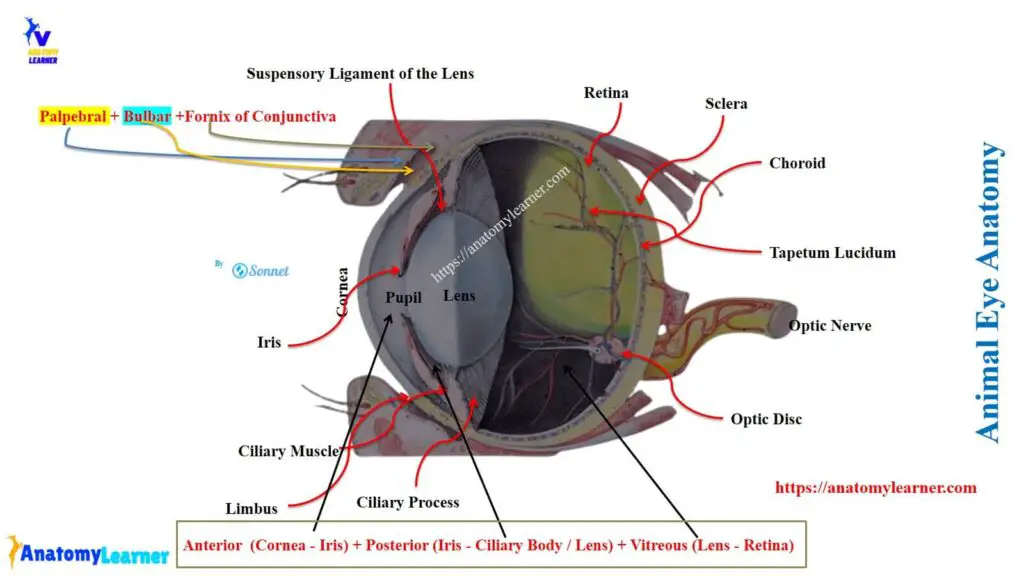
Before describing the animal eye conjunctiva anatomy, you might have basic knowledge of the animal eye structure. The animal eye consists of three basic organs –
- The eyeball or globe of the eye,
- Optic nerve element, and
- The certain accessory organs (which consist of orbital fascia, orbital muscle, eyelids, conjunctiva, and lacrimal apparatus).
Here, the picture shows the different parts of the animal eye.
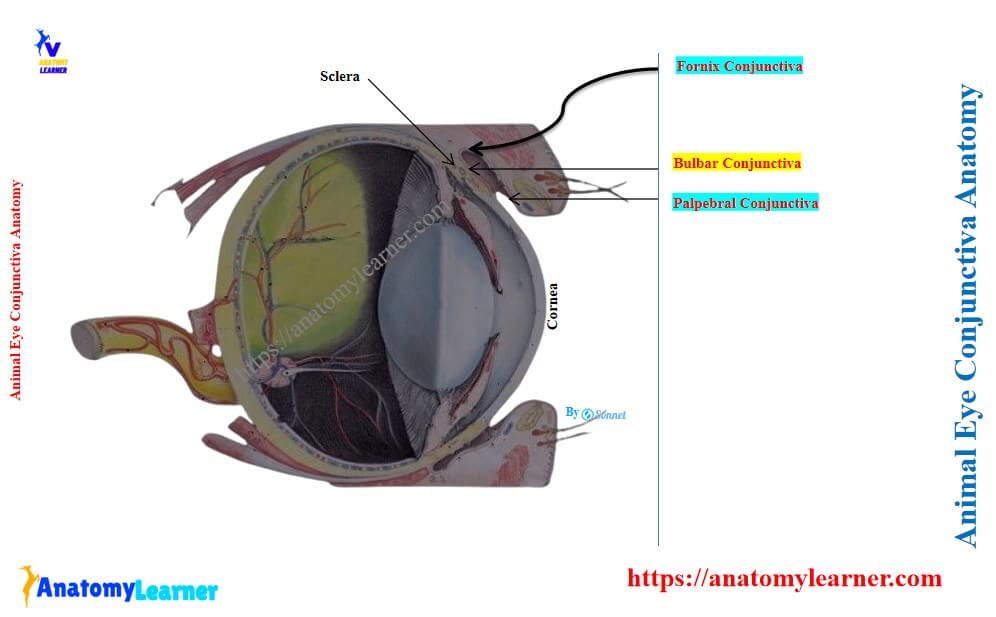
Now, let’s know what the conjunctiva of the animal eye is. The conjunctiva of the animal eye is a thin, transparent mucous membrane that covers the front portion of the sclera and cornea. It reflects the inner surface of the palpebra at the palpebral fissure.
Palpebral fissure: it is the interval between the upper and lower eyelids. The ends of the palpebral fissure are known as the canthi or angle. Thus, there are lateral (also known as nasal angle) and medial (also known as temporal) angles or canthus in the palpebral fissure.
So, the key features of the animal conjunctiva are shown in Table 1 –
| Features | Description of the specific anatomical features |
| Type of structure | Thin transparent mucous membrane |
| Covering | Covers the front part of the sclera and the pigmented part of the corneo-scleral junction |
| Reflection or attachment | Reflects the inner surface of the palpebra and the palpebral fissure (palpebral part of the conjunctiva) Also reflects from the upper and lower eyelids and forming folds (fornix of the conjunctiva) Finally, continue to the globe of the eye and attach near the limbus or corneoscleral junction (bulbar part of the conjunctiva) |
Parts of the animal eye conjunctiva
The conjunctiva of animals (cow, goat, sheep) has 3 parts –
- Palpebral conjunctiva,
- Fornix conjunctiva, and
- Bulbar conjunctiva
Now, let’s know the anatomical features of these 3 parts of the animal conjunctiva and identify them from the labeled diagrams.
What is the palpebral conjunctiva?
The part of the conjunctiva that lines the inner part of the eyelids or palpebra is known as the palpebral conjunctiva. Thus, the key anatomical features of the palpebral conjunctiva of animals are –
- Attaches the eyelids (palpebra) to the globe of the animal eye, and
- It extends from the innermost surface (identified in the picture) of the eyelids (upper and lower)
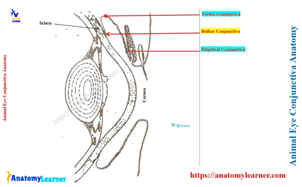
Here, the labeled diagram shows the palpebral part of the animal eye conjunctiva (cow). The palpebral part of the conjunctiva is closely adherent to the tarsus glands and highly vascular. But it is loosely attached farther back.
Again, it is papillated and is covered with the stratified cylindrical epithelium. Within this epithelium, you will find many goblet cells (histological features).
What is fornix conjunctiva of the animal eye?
Fornix conjunctiva of the animal eye is the continuation of the palpebral part, which reflects from the upper and lower eyelids and forming folds. This part of the animal eye conjunctiva consists of tubular glands. Near the vicinity of the fornix conjunctiva, there are numerous lymph nodules.
Here, the diagram also shows the fornix conjunctiva from the cow eye anatomy.
What is the bulbar conjunctiva of an animal’s eye?
The bulbar conjunctiva of the animal eye continues from the upper and lower fornices to the globe. This part attaches near the limbs or corneoscleral junction of the animal eye.
The bulbar part of the animal eye conjunctiva is loosely attached to the anterior part of the sclera. It is pigmented in the vicinity of the corneoscleral junction of the animal eye. On the cornea, it is presented by a stratified epithelium (seen on histological features).
Here, the diagram shows the bulbar part of the animal eye conjunctiva (cow and goat eyes).
Cow or animal eye conjunctival sac
The conjunctiva of the animal eye encloses the capillary space between the eyelids and the eyeball. This enclosed space is known as the animal eye conjunctival sac.
The conjunctiva of the lateral part of a cow or goat’s eyelid is pierced near the fornix by the orifice of the excretory ducts of the lacrimal glands.
Other structures related to the conjunctiva of a cow’s and a goat’s eye
You will find the third eyelid in a cow or goat’s eye that derives from the conjunctiva. It is a semilunar fold of the conjunctiva that covers and encloses a curved plate of hyaline cartilage. The third eyelid of a cow or goat is located at the temporal or medial angle of the eye and moves over the medial portion of the bulb of the eye quite freely.
Branches of ophthalmic and facial arteries chiefly supply the eyelids and conjunctiva of the animal’s eye (cow, sheep, goat). Again, the sensory nerves that branch from the ophthalmic and maxillary divisions of the cow’s trigeminal nerve innervate the conjunctiva of cow and goat eyes.
Cow eye conjunctiva diagram and pictures
Now, it is time to identify the different parts of the cow eye conjunctiva from the real sample. Here, the picture shows the actual cow eye conjunctiva (palpebral, fornix, and bulbar parts). The inner thin transparent mucous membrane that lines the eyelid is the palpebral part of the cow eye conjunctiva.
Again, the mucosal fold in the upper and lower eyelids is the fornix conjunctivae. Finally, the bulbar part of the cow eye conjunctiva reflects upon the anterior portion of the eyeball (sclera).
Conclusion
So, the conjunctiva is a thin, transparent mucous membrane and constitutes the major accessory structure of the animal eye. Three parts of the animal eye conjunctiva (palpebral, fornix, and bulbar) are easily identifiable from the actual gross sample.