The dog eye anatomy includes eyeball, orbit, eyelid, and lacrimal apparatus. This article will help you to know the details of these structures from the dog eye with labeled diagrams.
Quick overview: The eyeball of a dog eye is located in the orbital cavity and consists of 3 tunics – fibrous, vascular, and nervous. You will find the conjunctiva in the inner aspect of the eyelids that help to form 3rd eyelid.
Again, the dog’s lacrimal apparatus includes these structures responsible for producing, dispersing, and disposing tears. You will also see the intraocular and extraocular muscles in the structure of the dog eye.
I have examined the dog’s eye structure several times. Thus, I may help you to learn the structure of eyeball, orbit, eyelid, and lacrimal apparatus perfectly.
I will also show you the dog eye’s muscles, innervation, and vasculature with the labeled diagram. So, let’s learn the details of eye structure from the dogs.
Dog eye anatomy
The dog eye is the organ of sight, consisting of the eyeball and its accessory structures. Light passes through the transparent structure of the eye to reach the receptor part of the retina.
Here, the transparent structures of the dog eyes are cornea, aqueous humor, pupil, lens, and vitreous humor. The receptor structures of the retina are rods and cones.
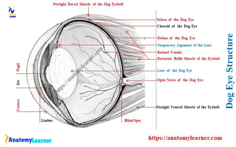
First, I would like to show you the following parts of the dog eye anatomy with a labeled diagram –
- Sclera – the white of the eye,
- Cornea – transperant cranial part,
- Limbus – junction between sclera and cornea,
- Choroid – thin, dark, highly vascular membrane,
- Ciliary body – the thickest part between the choroid and iris,
- Suspensory ligament – between lens to ciliary body,
- Iris–colored, doughnut-shaped that surrounds the pupil,
- Pupil – central opening of the iris that lets light into the dog eye,
- Retina – inner coat of the eye,
- Optic disc – form optic nerve,
- Lens – transparent, biconcave structure behind the iris,
- Anterior chamber – between cornea and iris,
- Posterior chamber – between iris and lens,
- Vitreous chamber – located between lens and retina and contains vitreous humor and
- Vitreous body – a substance that fills the vitreous chamber of the dog eye,
All these parts from the dog eye are identified in the labeled diagram.
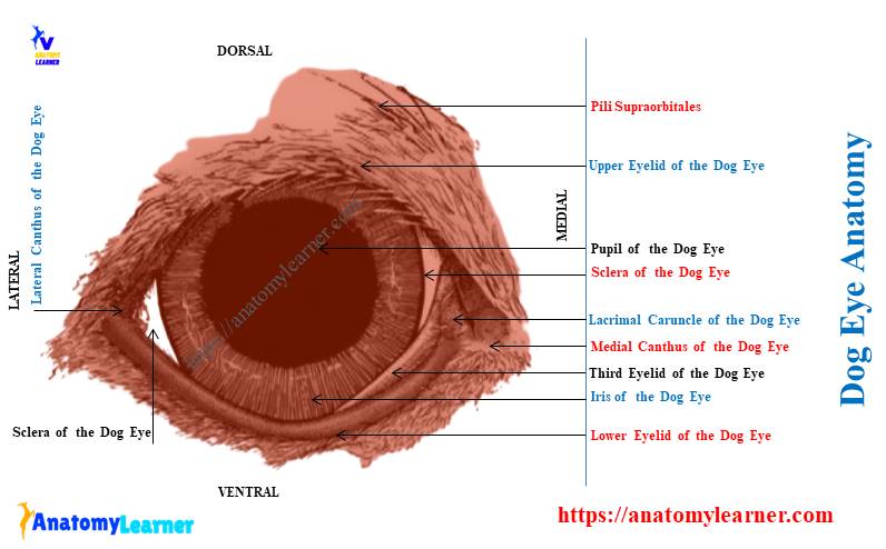
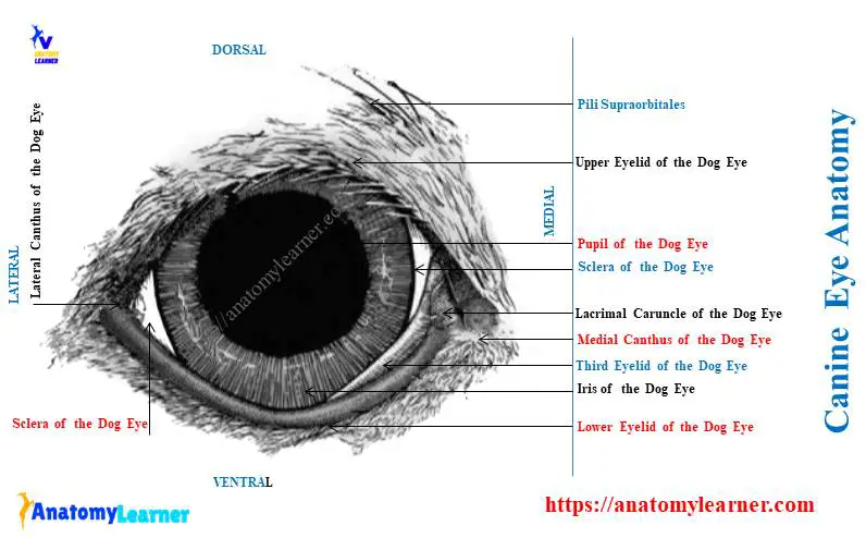
Now, let’s see and identify the following accessory structures from the dog eye –
- Upper and lower eyelids of the dog eye,
- Lateral and medial canthus,
- Lacrimal caruncle and puncta,
- Conjunctiva – a special mucous membrane that lines the eyelids and eyeball,
- Palpebral conjunctiva – inner surface of the eyelid,
- Bulbar conjunctiva – a reflection of palpebral conjunctiva onto the eyeball,
- Third eyelid – between eyelid’s medial angle to eyeball and
- Different muscles of the eyeball,
Again, all these accessory structures are identified from the dog eye-labeled diagram.
What are the parts of the dog eye?
You may know the different primary parts of the dog eye with their subdivisions. Let’s see the summary of the different parts of the dog eyeball from Table 1 –
| Primary parts of the dog eyeball | Subdivision or contents |
| Outer fibrous tunic | Sclera – caudal part of the fibrous coat, Cornea – transparent anterior part |
| Middle vascular tunic | Choroid – pigmented layer, Ciliary body – consists of ciliary process and muscles, Zonule – suspensory apparatus, Iris – the most anterior segment of the vascular tunic Pupil – central opening of the iris |
| Inner nervous tunic | Retina – has three distinct areas, Lens – soft, transparent, and biconcave structure |
So, the above 9 structures are the primary components of the dog eyeball. You will also see three different chambers in the structure of the dog eyeball.
Table 2 shows the three chambers from the dog eyeball with their location –
| Chambers of the dog eyeball | Location |
| Anterior chamber | Space bounded by the cornea cranially and iris and anterior lens surface caudally |
| Posterior chamber | Is the space bounded cranially and caudally by the lens capsule and cranial face of the vitreous |
| Vitreous chamber | It is the largest space lying between the lens and retina that fills with vitreous humor |
You will know the details of these chambers of the dog eyeball in the next section of this article. Again, the structures and parts of the conjunctiva and muscles from the eyeball will be discussed separately.
What is unique about the dog’s eye?
Before going to discuss the detailed anatomical features of the dog eye, let’s see the special about it –
- The eyeball of the dog is almost spherical and relatively large compared to the other animals,
- You will see the thick sclera in the ciliary region that contains a well-developed venous plexus,
- The cornea of the dog eye is almost circular,
- You will find richly pigmented choroid that present a well-defined tapetum cellulosum,
- The dog eye’s tapetum cellulosum is triangular in outline and has a metallic luster,
- A well-developed ciliary muscle present in the ciliary body of the dog eye,
- The iris of the dog eye is commonly light or brown-yellow,
- The pupil of the dog eye is round and bears minute round prominence,
- A round central area present in the retina of the dog eye,
- The surface of the lens is not strongly curved in the dog compared to the other animals,
- You will find the flat lacrimal gland that lies chiefly under the orbital ligament,
- The conjunctival epithelium is cylindrical with many goblet cells, and
- You will not find any distinct cilia in the lower eyelid of the dog eye,
These are the most unique features of the dog eye compared to the cow. Let’s find the detailed features of these structures of the dog eye in the next section.
Canine eye anatomy description
The location of the eye, the size of the orbit, size and shape of
the palpebral opening may vary in dog breeds. However, the basic structure of the dog eye is almost similar in various breeds.
To describe the anatomy of the canine eye, you might cover the following –
- Location of the eye within the orbital cavity,
- Parts of the eyeball with their associated structures,
- Structure of the eyelid (including various types of conjunctiva),
- Features of the lacrimal apparatus,
- Major muscles of the dog eyeball,
- List of nerves innervate the canine eye, and
- List of vessels that supply the canine eye,
Let’s discuss the structures above from the canine eye with the labeled diagram.
What is an orbit in a dog?
The dog’s orbit is an incomplete conical cavity that contains the eyeball. Most parts of the dog’s eyeball are enclosed with the orbital cavity.
It starts from the base of the orbit and projects a variable distance from the rostral to the orbital margin.
The bones form the four-fifth part of the dog’s orbital margin. The caudolateral one–fifth part is completed by the orbital ligament.
You will find the frontal bone dorsally, maxilla cranially, and zygomatic ventrally of the structure of the canine orbital margin. Let’s know the details of these bones that form the incomplete orbital margin in the dogs –
Orbital ligament of the dog eye
The orbital ligament is the thick fibrous band that completes the caudolateral part of the orbital margin. It unites the zygomatic process of the dog’s frontal bone with the frontal process (fp) of the zygomatic bone.
The orbicularis oculi and lateral palpebral ligament are attached to an orbital ligament. In the brachycephalic breed, you will find more thickened orbital ligaments.
How the orbital margin is formed in a dog?
The frontal bone forms the dorsal and medial part of the dog’s orbital margin. A part of the lacrimal bone forms the small portion of the ventromedial aspect of the orbital margin.
However, in some brachycephalic breeds, the lacrimal bone is confined to the medial orbital wall. It does not contribute to the orbital margin in these dog breeds.
Here, the maxillary bone forms the medioventral aspect of the orbital margin. You will find the zygomatic bone in the ventrolateral orbital margin of the brachycephalic breeds.
The lateral wall and floor of the dog’s orbital wall consist of soft tissue. Thus, you will not find this structure after skeletal preparation.
The orbital segment of the frontal bone forms the medial wall of the dog’s orbit. Here, the wing of the presphenoid bone forms the caudal part of the medial wall. Within this wall, you will find the optic canal.
The lacrimal bone forms the small part of the rostroventral medial wall of the dog’s orbit. It contains the fossa for the lacrimal sac and the caudal orifice of the nasolacrimal canal.
The medial wall of the dog’s orbit also possesses 5 foramina. You will find an optic canal and orbital fissure at the apex of the dog’s orbit.
- Optic canal – the optic nerve and internal ophthalmic artery pass the cranial cavity through this canal,
- Orbital fissure – is a fissure that forms between the basisphenoid and presphenoid bones. It passes to the oculomotor, trochlear, abducent, and ophthalmic nerves. You will also find the external ophthalmic artery and ophthalmic venous plexus within this fissure.
Two small ethmoidal foramina – rostrodorsal to the orbital fissure. Within these foramina, the external ethmoidal artery and the ethmoidal nerve pass.
What is the ventral orbital crest?
The ventral orbital crest is the dorsally convex crest of the frontal bone. It demarcates the boundary between the orbit and the pterygopalatine fossa.
The medial pterygoid muscle of the dog arises from this ventral orbital crest. This muscle forms the medial third of the orbital floor in a dog.
Again, the zygomatic salivary gland of the dogs remains on the dorsolateral surface of the pterygoid muscle. The maxillary artery and nerve also cross the orbital floor.
You will also find the pterygopalatine ganglion on the dorsal surface of the orbital floor.
The zygomatic process of the frontal bone makes the medial part of the roof of the canine orbit. You will see very small foramen in the midorbital process through which small arteries pass.
The ventral aspect of the zygomatic process of the frontal bone possesses fossa for the lacrimal gland. It is located just at the origin of the dog’s orbital ligament.
The medial surface of the temporalis muscle and orbital ligament forms the dorsolateral boundary of the orbit.
Now, let’s summarize the boundary of the dog’s orbital margin in Table 3 –
| Surfaces of the orbital cavity | Boundary of the dog orbital margin |
| Lateral wall and floor | Soft tissue |
| The medial wall of the orbit | Orbital part of the frontal bone |
| Medial aspect of orbital roof | Zygomatic process of the frontal bone |
| Dorsolateral and lateral | Temporalis muscle, and Orbital ligament, |
Dog eyeball structure
The dog eyeball comprises three concentric coats – fibrous, vascular, and nervous tunics. You will find a nearly spherical-shaped eyeball in a dog. It differs little in its sagittal, transverse, and verticle diameter.
The size of the dog’s eyeball varies in different breeds. However, the average diameter of the dog eyeball varies from 19 – 21 millimeters.
A transparent cornea forms the anterior one-fourth of the dog’s eyeball. Let’s discuss the features of three various tunics of the dog eyeball.
Fibrous tunic of the dog eye anatomy
This is the outer tunic of the dog eye structure. This fibrous outer tunic of the dog eye is responsible for the shape of an eye.
Again, it protects the eye from the external environment and conduction with the refraction of light rays. Here, the cornea of the fibrous tunic is responsible for this work. The sclera is the site for inserting the extraocular muscles in the dog eye.
You will find 2 parts in the fibrous tunic of the dog eye –
- A transparent cornea cranially and
- The opaque sclera that encloses the caudal three-fourths of the globe,
Again, you will find the limbus corneae between the junction of the cornea and sclera of the dog eye. Let’s see the features of the sclera and cornea of the dog eye structure.
Sclera of the dog eye
The sclera of the dog eye anatomy comprises a dense meshwork of collagen and elastic fibers. Externally, the sclera of the dog eye is white and covered in front by the conjunctival mucous membrane.
Again, the internal surface of the sclera is brown and possesses various grooves. Caudally, the sclera is pierced by the optic nerve, ciliary vessels, and ciliary nerves.
The dog’s optic nerve pierced the sclera just medial to the caudal pole at the optic disc or blind spot. You will find the cribriform plate-like appearance in this area, also known as lamina cribrosa sclerae.
The thickness of the dog sclera varies in different areas. You will find the most significant thickness of the sclera at the region just caudal to the corneoscleral junction.
In this area, you will also find the insertion of the straight and oblique eye muscles. Again, this area contains the scleral venous plexus.
The ciliary muscle attaches to the small ridge of the fibrous tissue. It forms a ring in the inner surface of the sclera, just caudal to the iridocorneal angle.
Let’s see the micrometry of the dog sclera from Table 4 –
| Sclera of the dog eye | Measurement |
| Scleral surface | 12 – 15 cm2 |
| Thickness of the sclera | 0.25 – 0.45 millimeter |
The sclera is sievelike, where the optic nerve leaves the dog’s eyeball. Here, you will find numerous bundles of collagen, elastic, and reticular fibers.
The short ciliary nerve and ciliary vessel enter the dog eyeball through the foramina of the sclear. These foramina are located at the periphery of the area of cribrosa.
The cornea of the canine eye
The cornea forms the cranial segment of the fibrous tunic of the dog eyeball. It is a transparent membrane that is convex in front and concave behind.
The thickness of the dog cornea may vary in different breeds. You may find the average thickness of the dog eye’s cornea 500 – 600 micrometers.
This thickness may increase with the age of the dogs. Again, the central part of the dog cornea is thinner than the peripheral part.
The cornea of a dog eye is one of the refracting media and consists of the following –
- The anterior epithelium,
- Bowman layer or anterior limiting lamina,
- The substantia propria,
- A posterior limiting lamina and
- The caudal epithelium,
You will see these layers of the cornea under the light microscope. Here, the anterior epithelium of the dog cornea consists of three distinct cell layers.
Why is the cornea transparent?
The dog cornea is transparent for the following three reasons –
- Uniform size and distribution of the collagen fibers (consists of extracellular matrix),
- The distribution of media of various refractive indices within the stroma of the cornea and
- Expression of the crystallins by the stromal cells of the dog cornea,
Again, the lack of pigment, velles, and many myelinated fibers also causes the cornea’s transparency. However, any disruption of the collagen fibers may result in the loss of transparency of the dog cornea.
So, the dog’s cornea is avascular and nourished by the capillary loop at the sclerocorneal junction (limbus). The branches of the ciliary nerves innervate the cornea.
These ciliary nerves arise from the ophthalmic nerves, the branch of the trigeminal nerve. The trigeminal nerve innervation for the dog cornea is essential to maintain homeostasis.
What is the iridocorneal angle in a dog’s eye?
The iridocorneal angle in a dog’s eye includes the following –
- The most anterior internal aspects of the sclera,
- The most posterior internal aspect of the cornea,
- An anterior external aspect of the ciliary body,
- The root of the iris, and
- All intervening tissue associated with these structures,
Vascular tunic of the dog eye
The vascular tunic is the middle, thick part of the dog’s eyeball. This tunic remains between the retina and the sclera of the dog’s eye.
The vascular tunic of the dog eyeball consists of the following distinct parts –
- Posterior choroid,
- The middle ciliary body and
- Anterior iris,
You will also find the suspensory ligaments of the lens that extend from the ciliary process to the lens capsule. Let’s see the overall functions of the vascular tunic of the dog eye –
- Regulating the amount of light entering the eye through the pupil,
- Production of the aqueous humor that maintains the intraocular pressure,
- Suspensory ligament positioning the lens of the eye,
- The ciliary muscle helps to change the visual focus,
- Tapetum lucidum of the choroid increases the photic stimulation of the retina under low light and
- The ciliary body and choroid provide nutrition to the structure within the dog eye,
Now, let’s discuss the features of the three main structures of the vascular tunic of the dog eye.
Iris of the vascular tunic
The iris of the dog eye is the most anterior segment of the vascular tunic. It is a thin, circular diaphragm-like structure of the dog eye.
The iris remains suspended in the aqueous humor behind the cornea and front of the lens. Structurally, it consists of radiating and circular muscle fibers, pigment cells, vessels, and a layer of endothelial covering.
The pigment cells in the iris are responsible for the color of the dog’s eye.
A central aperture is present in the dog’s iris, known as the pupil. Here, the pupil of the dog eye is circular.
The size of the dog’s pupil is variable in different breeds. It regulates the amount of light reaching the retina of the dog’s eye.
You will find the small diameter of the pupil in the greater intensity of illumination. The size or diameter of the dog eye’s pupil is regulated by the contraction of radiating and circulating muscle fibers.
Histologically, a discontinuous layer of flat fibrocyte is found in the anterior surface of the iris. You will also find the intercellular space between the anterior chamber and the underlying stroma of the dog eye.
Various anatomists found melanin as the only identified pigment in the dog’s iris. But blue-eye dogs lack the pigment restricted to the caudal pigment of the iris.
Thus, the blue color of the dog’s eye is caused by the light’s various absorption and selective reflection. It is caused by the iris tissue itself and the caudally located melanin of the dog eye iris.
Which muscles regulate the diameter of the dog eye’s pupil?
Two antagonistic muscles called sphincter papillae and dilator papillae regulate the diameter of the dog eye’s pupil. These muscles are derived from the outer layer of the neuroepithelial part of the retina.
The sphincter papillae is the most significant muscle that consists of a sheet of circumferentially arranged smooth muscle. On the other hand, the dilator papillae consist of radially arranged smooth muscle fibers.
It forms a meshwork by which the collagen bundle of the iris stroma is bounded. Again, this dilator muscle is located caudal to the sphincter muscle.
Both sphincter and dilator papillae are well-developed in the dog eye compared to other animals. Now, let’s see the innervation and blood supply to these antagonistic muscles of the dog eye’s pupil.
Both parasympathetic and sympathetic nerves innervate to the sphincter and dilator muscles. The postganglionic parasympathetic axon innervates the sphincter papillae muscle. Again, the preganglionic axon innervates the sphincter muscle through the oculomotor nerve.
The blood supply of the iris arises primarily from the two long caudal ciliary arteries.
The ciliary body of the eye
The ciliary body is the thickened middle part of the vascular tunic of the dog eyeball. It is located between the iris and choroid segments of the vascular tunic.
The ciliary body of the dog eye consists of the following –
- A ciliary ring – is the caudal flat part of the ciliary body. It locates the cranial border of the retina and continues with the choroid.
- The ciliary crown – is the raised part of the ciliary body. It is located cranial to the ciliary ring and adjacent to the iris.
You will find the well-developed ciliary process in the structure of the ciliary crown. Cranial to the retina, you will see numerous small, flat, parallel, and regular processes at the ciliary crown.
These processes increase rapidly in their height and merge as they pass cranially. They form tall, thin folds rising from the base plate of the ciliary body.
These folds lose their outer attachment to the sclera through ciliary body muscles at the root of the iris. They arc towards the lens as short, blunt, and free ciliary processes.
Why is the ciliary body thicken in a dog eye?
Due to the presence of numerous major ciliary processes in the ciliary body, it becomes thickened. You will find approximately 75 – 80 major ciliary processes in the ciliary body of the dog eye.
You will also find the minor ciliary folds and processes between the major ciliary processes. The ciliary zonule almost entirely covers the caudal surface of the ciliary process.
Both the major and minor ciliary processes form the ciliary crown in the dog eye.
Other features of the ciliary body
The ciliary body of the dog eye is highly vascularized. You will find the radial ciliary arteries that supply to the ciliary body. It directly comes from the caudal margin of the greater arterial circle of the dog iris.
The ciliary body musculature consists of various smooth muscle fascicles. These muscles are located in the outer part of the ciliary body.
In the case of other animals, the ciliary muscles consist of circumferential and meridional fibers. But in the dog, you will only find the meridional fibers in their ciliary muscles.
Here, the meridional fibers arise from the scleral ring on the inner surface of the sclera. This muscle inserts in the stroma of the ciliary body.
Choroid of the dog eye
The choroid is the dark-colored, thin vascular membrane of the dog eyeball. It comprises capillary plexus, small blood vessels, and loose connective tissue containing pigmented cells.
It is another one of the refractive media of the dog eye and continuous with the ciliary body anteriorly. Except for a few areas, the choroid envelops the caudal hemisphere of the eyeball.
The choroid of a dog eyeball divides into the following –
- Suprachoroid layer,
- A vascular layer (lamina vasculosa),
- The reflective layer, or tapetum lucidum,
- A choriocapillaris layer and
- The basal lamina of the choroid,
However, the basal lamina is not well-developed in the dog eye. The dog eye’s vascular layer of the choroid is a plexus of arteries, arterioles, veins, and venules.
The collagenous and elastic stroma supports this vascular layer of the choroid. It traditionally subdivides into the outer large-sized vessels layer and inner middle-sized vessel layer.
Here, the outer large-sized vessels are the terminal branches of the ciliary arteries. The vascular layer of the choroid and retinal pigment layer of the dog eye is darkly pigmented in most dogs.
What is tapetum in a dog eye?
The tapetum is the specialized reflective layer of the choroid of a dog eye. It increases the ability of the retina to function under low light levels.
You will find tapetum in most domestic mammals except for the pig eye. It is cellular in the canine eye.
The tapetum occupies one-third of the superior area of the choroid. You will find tapetum cells in the central area of the dog tapetum.
However, the number of tapetum cells decreases peripherally and near the optic nerve. The shape and size of the dog tapetum vary according to the various breed and size of the dog’s eyes.
Zonule or suspensory ligament of the lens
The zonule is a group of fine fibers that arise from the groove between the ciliary process. This is also known as the suspensory ligament extending to the lens capsule.
The lens of the dog eye is fixed in its position with the help of this suspensory ligament or zonule. Here, the zonule consists of a highly ordered array of zonular fibrils.
This zonule lies caudal to the iris and ciliary body of the dog eye. It also separates the posterior chamber of the dog eye from the vitreous body.
Most zonular fibers arise from the pars ciliaris retinae, just cranial to the ora serrate. They pass cranially and closely adherent to the surface of the ciliary body.
The suspensory apparatus that attaches to the caudal part of the lens capsule is not well-developed.
Internal nervous tunic of the dog eye
The nervous tunic is the innermost tunic of the dog eyeball. This very thin and delicate membrane consists of a retina with its pigmented epithelium.
The dog eye’s retina is a membranous expansion of the optic nerve elements. You know, the optic nerve is a central nervous system tract with characteristics of central nervous tissue.
You may know the details of the dog nervous system from the below-mentioned article –
The meninges of the brain (covering the brain and spinal cord) continue along the optic nerve to the eyeball. Here, the optical part of the retina ends just caudal to the ciliary body.
It is serrated and known as the ora serrate. The ora serrate is the boundary between the visual and nonvisual parts of the dog retina.
The intraocular myelinated part of the optic nerve forms the optic disc. It is round to oval or triangular to quadrilateral in shape.
However, the shape of the optic disc varies significantly in different breeds of the dogs. It is pink or white but also may contain pigment.
You will find a central depression in the optic disc of the dog eye. There are no photoreceptors in this disc; thus, it is termed as the blind spot of the eye.
Areas of the dog eye retina
You will see three distinct areas in the dog eye retina –
- A pars iridicus retinae,
- The pars ciliaris retinae, and
- A pars optica retinae,
But, the pars optic retinae is the only photosensitive area of the dog retina. The parts iridicus and ciliaries together form the nonvisual part of the dog retina.
Here, the pars iridicus retinae is the bilayered epithelium of the retina. It covers the caudal surface of the iris.
Its outer surface gives rise to the muscle of the dog’s iris. Again, the inner layer of this pars iridicus is adjacent to the lens and contains pigmented epithelium.
The parts iridicus retinae reflect onto the ciliary body and become the pars ciliary retinae. This is the bilayred cuboidal epithelium that produces aqueous humor.
You will find heavy pigmentation in the outer layer of the pars ciliary retinae. But the inner layer of the pars ciliary is unpigmented.
The pars optica retinae is responsible for transducing the photic energy to the visual energy. In this optic retinae of the retina, you will find 10 layers. Let’s find the detailed guide on the appearance of these 10 layers from the retina histology slide –
Lens of the Dog Eye Anatomy
The lens of the dog eye is a soft, transparent, protein-rich, biconcave structure. It suspends in contact with the posterior face of the iris and the anterior face of the vitreous body.
The primary function of the lens is to bring an image into critical focus on the photoreceptor layer of the retina. It is the chief refractory medium of the dog eyeball and consists of concentric layers of modified cells.
The posterior part of the dog eye lens is more convex. At an older age, it becomes opaque, and cataract occurs.
The eye becomes hypertrophic when the lens is surgically removed during the cataract. If you perform a transverse section of the lens, it becomes circular. But in the sagittal or dorsal section, it looks slightly elliptical.
The entire lens of the dog eye is covered with the lens capsule. It is the basement membrane secreted by the cells of the lenticular epithelium. This lens capsule is highly refractile and elastic.
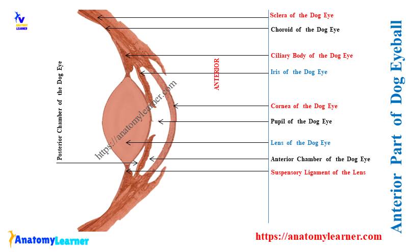
How many chambers does a dog eye have?
There are three chambers within the dog eyeball – anterior, posterior, and vitreous. Let’s see the boundary and contents of these 3 chambers of the dog eyeball –
- The anterior chamber of the dog eye – is the space bounded cranially by the cornea and the iris and cranial surface of the lens caudally. This anterior chamber is filled with aqueous humor.
- The anterior chamber of the dog eyeball has direct communication with the posterior chamber. Between these two chambers, you will find the aperture in the iris.
Again, the periphery of the chamber continues with the space of the iridocorneal angle. Now, let’s see the boundary and content of the posterior chamber of the dog eyeball.
Posterior chamber of the dog eyeball – it is a smaller chamber than the anterior and cranially bounded by the iris. The lens of the capsule and anterior face of the vitreous bounds the posterior chamber posteriorly.
Again, the suspensory apparatus and ciliary epithelium bound the posterior chamber peripherally. You will also find the aqueous humor in the posterior chamber of the dog eyeball.
What is aqueous humor in the dog eyeball?
Aqueous humor is the clear and colorless fluid that fills the cranial and posterior chambers of the dog eyeball. It is secreted by the active ciliary processes of the ciliary body and deposited in the posterior chamber.
Now, the aqueous humor passes to the anterior chamber of the dog eyeball through the pupil. A small amount of aqueous humor escapes from the iridocorneal angle to the anterior ciliary vein through sinus venosus sclerae.
The aqueous humor consists of water, glucose, amino acids, and ascorbic acids. It also serves as a refractive medium for the dog eye.
If there occurs any interference in the circulation of aqueous humor, it may increase the intraocular pressure. Thus, it leads to glaucoma in the dog’s eye.
What are the vitreous chamber and vitreous body?
The vitreous chamber is the largest of the 3 chambers of the dog eyeball. It possesses almost 80% of the volume of the eyeball.
The suspensory apparatus and posterior lens capsule form the anterior limit of the vitreous chamber. The rest of the area is covered with the retina of the dog’s eye.
The vitreous body remains in the vitreous chamber of the dog’s eyeball. It is a soft, clear gel that maintains the chamber’s shape.
The posterior face of the lens covers the anterior face of the vitreous body. This vitreous body of the dog eyeball is almost acellular.
But, the bulk of the vitreous body consists of liquid components. You will find water, protein, and carbohydrates in the components of the vitreous body.
The membrane vitrea limits the anterior face of the dog’s vitreous body. Again, it is tightly adherent to the pars ciliary retinae.
Dog eyelid anatomy
The dog eyelids are two fibrous sheets attached to the periphery of the orbital margin. It covers the anterior part of the eyeball (when it is closed).
The eyelid also maintains the light and protects the cornea. An opening between the two eyelids is the palpebral fissure. The palpebral fissure’s orientation and size greatly vary in dog breeds.
The external angle of the palpebral fissure is thick, and the inner angle is rounded. You will see the cilia or eye lashes at the margin of the eyelids.
However, the cilia or long hair do not present in the inferior eyelid of most of the dogs. Numerous long hairs are projecting from the superior eyelid of the dogs.
The superior and inferior eyelids join at the medial and lateral commissures. They together form the lateral and medial canthi.
You will see a triangular prominence in the medial angle of the dog’s eye. This is the lacrimal caruncle, which may or may not be pigmented.
From the lacrimal caruncle, a small, fine haris projects. You will find the sebaceous, ciliary, and tarsal glands in the eyelid of the dogs.
Here, the sebaceous glands open into the follicles of the cilia on the superior eyelid of the dogs. Again, the ciliary glands secret into the hair follicles or the eyelid margin.
Tarsal glands are the specially modified compound sebaceous glands. They present both in the superior and inferior eyelids of the dogs.
The medial and lateral palpebral ligaments stabilize the commissures of the eyelids. Here, the lateral ligament arises from the zygomatic arch and ventral end of the orbital ligament. The medial ligament is a fibrous band that arises from the periosteum of the frontal bone.
Dog eye anatomy conjunctiva
The conjunctiva is the thin transparent membrane of the dog eye anatomy. It covers the front part of the sclera and cornea of the dog’s eyes.
This membrane is loosely connected over the sclera and continuous as the corneal epithelium over the dog’s cornea. You will find two types of conjunctiva in the dog eyelids –
- Palpebral conjunctiva – inner aspect of the eyelids and lines with special mucous membrane and
- Bulbar conjunctiva – is the part of palpebral conjunctiva that reflects onto the surface of the globe at the level of the orbital rim,
The area where this reflection occurs is known as the conjunctival fornix. Again, the space between the two eyelids and the eyeball is the conjunctival sac. You will find the mucous and fluid within the conjunctival sac of the dog eye.
The histological section of the dog conjunctiva shows the stratified squamous epithelium on its palpebral margin. Again, this becomes the cuboidal epithelium towards the fornix of the conjunctiva.
The conjunctival epithelium rests on the loose connective tissue stroma. You may get the complete guide on the loose connective tissue from the below-mentioned article –
The dog eye conjunctiva is highly vascularized and supplied by dorsal and ventral palpebral artery branches. Again, the branches of malar arteries and terminal branches of the ciliary arteries also supply the conjunctiva.
Dog third eyelid
The third eyelid of a dog is the semilunar fold of the conjunctiva. It is well-developed in dogs and arises as a fold from the ventro-medial aspect of the conjunctiva.
The free concave edge of the third eyelid faces superiolaterally and makes contact with the eyeball. It is highly mobile and sufficient in extent to cover the entire anterior face of the cornea.
In the normal position of the dog eye, the third eyelid is hidden within the orbit. However, the free edge of the third eyelid is visible in the ventromedial aspect of the palpebral fissure.
The T-shaped hyaline cartilage reinforces the body of the dog eye’s third eyelid. The superficial glands of the third eyelids surround this structure.
This is the pink, tear-drop shape, mixed seromucous gland. Again, this gland is also known as the harder’s gland or nictitating gland.
Lacrimal apparatus
The dog lacrimal apparatus consists of the following –
- Lacrimal glands – pink, oval, lobulated glands,
- Carancula lacrimalis – small rounded body helps in the flow of the tear in the proper direction,
- Puncta lacrimalis – minute orifice of the of the lacrimal canal at the lacrimal papillae,
- Lacus lacrimalis – triangular space at the medial angle of the dog eye,
- Lacrimal canals – two in number and conmect the lacus lacrimalis to the lacrimal sac,
- Lacrimal sac – This is the sac-like structure logged in a fossa at the front end of the medial wall of the orbit,
- Nasolacrimal ducts – start from the lacrimal sac and pass through the nasolacrimal canal. Finally, it opens on the external wall of the dog’s nostril.
Let’s find the full guide on the dog nostril from the below-mentioned article –
Dog eye muscle anatomy
The dog eye muscle anatomy is essential to the function of the visual apparatus. You will find the three groups of muscles in the dog eye structure – intraocular, extraocular, and palpebral muscles.
The intraocular muscle of the canine eye – lies entirely internal to the sclera. It regulates the pupillary diameter and shape of the lens.
Extraocular muscles of the canine eye – these are the seven muscles that insert on the eye’s sclera. They help in the rotation and retraction of the whole eyeball.
Palpebral muscles of the dog eye – these are the group of muscles of the eyelids. They regulate the shape and position of the palpebral fissure.
What are the intraocular muscles in the dog eye?
The dilator and sphincter muscles of the dog’s iris and ciliary muscles are the intraocular muscles in the dog eye. They lie entirely within the eyeball and consist of smooth muscle fibers.
- Iris muscles (dilator and sphincter) – regulate the amount of light that enters the retina and
- Ciliary muscles – contraction of these muscle fibers loses the suspensory ligament, allowing the dog’s lens to become more convex.
What are the extraocular muscles in the dog eye?
Four straight, two oblique, and one retractor muscles are the dog eye’s extraocular muscles. The straight muscles are dorsalis, ventralis, medialis, and lateralis in the dog eyeball.
Again, the two oblique extraocular muscles of the dog eye are dorsal and ventral. Here, Table 5 shows the extraocular muscles from the dog eye structure –
| Dog eye muscles | Name of the dog’s eye muscles |
| Four Straight muscles | Straight dorsalis muscle Ventralis straight muscle Straight medialis muscle, and Straight lateralis muscle, |
| Two oblique muscles | Obliquus dorsalis muscle and Obliqqus ventralis muscle, |
| One retractor muscle | Retractor bulbi muscle |
Now, let’s know the functions of these extraocular muscles of the dog eye. You will also know why the dog eye rotates 90 degrees of the arc of the dorsal plane.
All these extraocular muscles of the dog eye help to rotate the globe in three perpendicular axes –
- The dorsal and ventral straight muscles rotate the globe around the medial-to-lateral axes,
- The medial and lateral straight muscles of the dog eye rotate the globe superior to inferior axes,
- Again, two oblique muscles of the dog eye rotate the eyeball around the axis bulbi,
The retractor bulbi muscle can retract the dog eyeball into the orbit along the optic axis. Again, two or more muscles of the eyeball provide the oblique movement.
Thus, the dog can rotate the eye 90 degrees of the arc in the dorsal plane. Again, they can rotate the eye 60 degrees in a sagittal plane.
The dog eye’s oblique muscle also helps fix the eye against the caudal pull of the straight muscle. Now, let’s know the anatomy of these three extraocular muscles from the canine eye.
Straight muscles of the dog’s eyeball
The dorsal straight muscle of the dog eye arises from the optic canal and orbital fissure. The rest of the straight muscle arises from the ventral part of the orbital fissure.
These respective muscles are inserted on the dog eyeball’s dorsal, ventral, medial, and lateral aspects. In the transverse section, you will find the oval bellies of these straight muscles of the dog eyeball.
Here, the medial straight muscle of the dog eyeball is slightly larger than the other muscles. A flat tendon arises from this muscle and is inserted on the anterior sclera of the dog eye.
The tendon from the ventral straight muscle passes deep to the ventral oblique muscle. The oculomotor nerve supplies the dorsal, medial, and ventral straight muscles of the dog eye. In contrast, the abducent nerve innervates the lateral straight muscle.
Obliqqus muscles of the canine eyeball
The obliquus dorsalis muscle of the dog eyeball arises from the dorsomedial margin of the orbit. This muscle runs cranially and passes between the dorsal and medial straight muscles.
The obliquus dorsalis muscle gives rise to the small, thin, round tendon that passes over the small cartilaginous structure.
The obliquus ventralis muscle of the dog eyeball arises from the apex of the orbit. This muscle courses dorsomedially and passes ventral to the ventral straight muscle.
You will find two short tendon that arises from the obliquus ventralis muscle. Here, the short tendon inserts deep into the lateral straight muscle. The longer tendon arises from the ventralis muscle and passes lateral to the lateral straight muscle.
The branch from the malar artery supplies the dog eyeball’s obliquus ventralis muscle. Again, the oculomotor nerve innervates this obliquus ventralis muscle of the dog eye.
Retractor bulbi muscle of the dog eye
This is only one retractor muscle of the dog eye that arises from the periosteum of the orbit. This muscle passes laterally between the dorsal and lateral rectus muscles of the eye.
It divides into two parts – dorsal and ventral that pass to the optic nerve. The retractor bulbi muscle of the dog eye pulls the eyeball deeper into the orbit. This retractor muscle also plays an essential role in the rotatory movement of the dog’s eyeball.
The muscular branch of the ophthalmic artery supplies the retractor bulbi muscle of the dog eye. Again, the branches from the abducent nerve innervate the retractor bulbi muscle of the dog eye.
What are the palpebral muscles of the dog eyelid?
The palpebral muscles of the dog eyelid include the following –
- Orbicularis oculi muscle,
- Levator palpebrae superioris muscle,
- Levator anguli oculi medialis muscle,
- Retractor anguli oculi lateralis muscle,
- Tarsalis superior and inferior muscles, and
- Pars palpebralis muscle,
The orbicularis oculi muscle of the palpebral helps to close the palpebral fissure. It arises from the medial palpebral ligament and encircle the palpebral fissue.
It looks like wedge-shaped in the transverse section. You will find two parts in the orbicularis oculis muscles – pars orbitalis and pars palpebralis. Here, the pars palpebralis part of the orbicularis muscle is well-developed in the superior eyelid of the dog.
The levator palpebrae superioris muscle of the dog retracts the superior eyelid. It arises deep into the orbit, dorsal to the optic canal.
Cranially, this muscle of the palpebrae becomes progressively wider and flatter. The oculomotor nerve innervates this levator muscle of the palpebrae.
A small, flat retractor anguli oculi lateralis muscle arises from the temporozygomatic fissure. This muscle is parallel and superficial to the lateral palpebral ligament.
The dog eye’s superior and inferior tarsal muscles arise from the periorbital orbitalis muscle. It consists of smooth muscle fibers and maintains the position of the dog’s eyelids.
The pars palpebralis arise from the ventral midline of the orbit. It consists of delicate muscle fibers and acts as a depressor of the inferior eyelid.
How are the dog eyes innervated?
The dog eye and its associated structures are innervated by the following –
- Optic nerve (cranial nerve II) – occupies the center of the cone within the orbit formed by the extraocular muscle,
- The oculomotor nerve (cranial nerve III) – is the primary general somatic efferent innervation to the muscles of the dog eye,
- Trochlear nerve (cranial nerve IV) – It reaches the orbit through the orbital fissure and innervates the dorsal oblique muscle,
- Trigeminal nerve (cranial nerve V) – it innervates the eye and orbit of the dog,
- Abducent nerve (cranial nerve VI) – supplies general somatic efferent axon to the lateral straight muscle and retractor bulbi muscle,
- The facial nerve (cranial nerve VII) – supplies somatic efferent innervation to the eyelid muscles. Again, it provides the parasympathetic innervation to the lacrimal gland.
Different branches of the ophthalmic nerve also innervate the various parts or structures of the dog eye. Let’s see the main three branches of the ophthalmic nerve that innervate the dog eye –
- Frontal nerve – is a small nerve that innervates the superior eyelid of the dog eye. It also innervates the dorsal straight muscle of the dog’s eyeball.
- The lacrimal nerve – is a very small branch that innervates the lateral edge of the straight dorsal muscle and lacrimal gland.
- Nasociliary nerve – passes between the dorsal and ventral rami of the oculomotor nerve. It divides into long ciliary, infratrochlear, and ethmoidal nerves.
The long ciliary innervates the dilator muscle of the pupil. Again, the infratrochlear passes rostrodorsally along the medial edge of the dorsal straight muscle.
The ethmoidal nerve innervates the nasal mucosa and skin of the muzzle.
What is the main artery that supplies blood to the dog’s eye?
The external carotid artery branches are the main source of blood supply to the dog eye and its associated structure. Maxiallry and ophthalmic are two main branches of the outer carotid artery (ecr) that supply blood to the dog eye.
Again, the superficial temporal and malar arteries supply blood to the eye and associated structures. The malar artery supplies blood to the third eyelid, ventral oblique muscle, and nasolacrimal duct.
The maxillary artery of the dog eye gives rise to various branches before entering the caudal alar foramen. You will find the external ophthalmic artery from the maxillary artery that passes dorsally and enters the periorbita.
The external ethmoidal artery arises from the external ophthalmic artery and consists of two muscular branches. The ventral muscular branch of the external ethmoidal artery runs rostrally toward the eyeball. It supplies the dog’s eyeball muscles and the third eyelid’s superior gland.
The muscular branches of the ethmoidal artery continue as the anterior ciliary arteries. Again, the dorsal muscular branch supplies the dorsal straight, oblique, and levator palpebral superior muscles.
You will also see a lacrimal artery from the dorsal muscular branch and supply lacrimal gland. The internal ophthalmic artery arises from the rostral cerebral artery and is distributed in the eyeball.
Two long ciliary arteries arise from the internal and external ophthalmic and supply the anterior part of the eyeball. At the posterior aspect of the eyeball, you will find a variable number of short ciliary arteries.
Which veins drain blood from the dog eye?
The angular, dorsal, and ventral ophthalmic veins help drain the blood from the dog’s eye. Here, the angular vein continues with the facial vein dorsally and caudally.
The angular vein of the dog eye arises from the medial commissure of the eyelids. Again, it passes caudally over the superficial surface of the medial palpebral ligament. It also passes cuadodorsally medial to the commissure of the eyelids.
The dorsal external ophthalmic vein is the most prominent intraorbital vein in the dog eye. It passes medially deep to the dorsal oblique muscle and joins with the ventral external ophthalmic vein.
A lacrimal vein drains blood from the lacrimal gland of the dog eye. It joins with the dorsal external ophthalmic vein at the apex of the orbit.
The ventral ophthalmic vein of the dog eye lies within the periorbita. It passes between the extraocular and pterygoid muscles of the dog eye.
Conclusion
So, the dog eye anatomy possesses an eyeball and its associated structures. Here, the associated structures of the dog eye consist of the eyelid, muscles, lacrimal apparatus, nerves, and vessels.
The eyelids consist of conjunctiva, a vital accessory structure of the dog eye. Again, the 7 muscles of the dog eye structure are essential to understand the various movements.