The dog lung anatomy consists of different lobes, surfaces, borders, apex, and base. There are right and left lungs in a dog which are considered the main organ of respiration.
These two lungs of a dog occupy most of the parts of the thoracic cavity and are separated by the mediastinum. Again, each of these canine lungs is covered by the visceral layer of pleura.
This short guide might help you learn the anatomical facts of the dog lung with the labeled diagrams. Here, I will describe every single lobe from both the right and left lungs of a dog.
After completing this guide, you will quickly identify the right lung from the left one based on its peculiar anatomical features. Again, you will also learn the surface anatomy of the dog lung so that you may determine the exact location externally.
Finally, I will show you some of the significant anatomical differences between the dog, horse, and ruminant lungs at the end of this article.
Dog lung anatomy
The lung of an older dog is soft and spongy in appearance. In the fresh condition, the canine lung shows a pink color. Again, the older dog’s lung crepitates to touch and float on water.
But, the still-born puppy’s lung is solid and does not float on water. Here, the dog’s right lung is larger than the left. Both these right and left lungs are conical in shape and separated by a fissure (mediastinum).
In the anatomy of the dog lung (right and left), you will find the followings –
- Two surfaces of both the right and left lungs,
- Two borders in both lungs,
- A base (concave; towards diaphragm) and apex (larger and rounded extremity), and
- Lobes in the dog’s lungs – (4 in the right lung, and 3 in the left lung),
In the right lung structure, you will find the cranial (apical), caudal (diaphragmatic), middle (cardiac), and accessory lobes. But, you will not find any accessory lobe in the left lung of a dog.
Before going to the details of the dog lung structure, you might know the lumber and anatomical location. Let’s see where the lung is located in a dog.
How many lungs do dogs have?
From the previous discussion and labeled diagram, you can easily understand there are 2 lungs in the dog’s thoracic cavity. These 2 lungs (right and left) have many anatomical features.
But, the dog’s right lung is larger than the left. Again, you will find 4 different lobes in the right lung and 3 in the left lung.
The base of the right lung is more concave, whereas the base is less concave in the left lung. Here, the apical lobe of the dog’s right lung is larger but smaller than the left lung.
There is a large cardiac notch in the left lung as most of the portion of the canine heart is located left of the mediastinum. So, you will see a smaller cardiac notch in the canine right lung.
But, the cardiac impression of the right lung is more deep compared to the left lung. Again, you will see an accessory (intermediate) lobe in the right lung of a dog.
The difference between the dog’s right and left lungs is given in Table 1 –
| Features | Right Lung | Left Lung |
| Size | Larger | Smaller |
| Lobes | 4 | 3 |
| Intermediate lobe | Present | Absent |
| Base | More concave | Less concave |
| Cardiac notch | Narrow | Indistinct |
| Cardiac impression | More deep | Shallow |
So, if you want to know these anatomical features both from the right and left lungs of a dog, you may continue this article till the end. You will quickly understand these terms that I have used in the table to compare the features between the canine right and left lungs.
Okay, let’s know the exact location of the dog’s right and left lungs so that you may identify them externally from a live dog.
Where is the lung located in a dog?
The lung is located in the thoracic cavity, extending from the 1st to the 10th rib. A thin transparent serous membrane known as the pleura encloses the lung and lines the thoracic cavity of the dog.
You know there are 2 lungs (right and left) in a dog, so that you might know the exact location of these lungs in the thoracic cavity. The dog’s right lung is clinically used for auscultation. You may easily identify the area of this lung by following the below-mentioned procedure –
You may draw a triangular area on the dog’s thoracic cage or cavity to identify the location of the right lung. For this, you should draw 3 lines as shown in the picture.
- Let’s draw a line from the caudal border (angle) of the dog’s scapula to the tuber coxae of the hip bone,
- Then, a second line should be drawn from the caudal angle of the scapula to the olecranon process of ulna bone,
- Finally, let’s draw the third line from the olecranon process to the last rib of the thorax,
So, you will find a triangular area in the dog’s thorax where the dog’s lung is located. Unfortunately, this triangular area does not represent the exact location of the right or left lung perfectly.
So, what should you follow to identify the exact location of the dog’s right or left lungs from its thorax? Well, you should know the extension (longitudinal) of both the right and left lungs in the dog’s thorax.
Dog lung location – right
First, let’s see the location of the right lung of the dog in its thoracic cavity. The long axis of the dog’s right lung extends from the first rib to the tenth rib (2nd intercostal space – 9th intercostal space).
The dorsal border of the right lung is located at the ventral half of the thoracic cavity, whereas the ventral border lies opposite the sternum. I hope the diagram below might help you understand the exact location of the dog’s right lung.
You may precisely locate the dog’s right lung from its thoracic cage, as shown in the diagram. Okay, now, let’s see the exact location of the left lung of the dog.
Where is the dog’s left lung?
You know the dog’s left lung is smaller compared to the right. The left lung extends from the 2nd to 10th rib and lies left of the median plane.
The extension for the dorsal and ventral border of the dog’s left lung is similar to the right lung. But, the left lung is unsuitable for auscultation as most of the part of the heart is located on the left side (occupied in the left lung).
I hope this information is enough to locate the exact location of the dog’s right and left lungs. Finally, you may tell that the dog’s lungs (right and left) are located in the middle of the thoracic cavity, which lies right and left of the median plane. Again, they extend from the thoracic cage’s 1st to the 10th ribs.
Special features of the canine lung anatomy
Now, I will show you the most important features (unique) of the dog lung anatomy. I will describe the base, apex, surfaces, and borders of the dog’s lungs shortly. Let’s see the unique features of the dog’s right and left lungs together in a minute.
First, you should identify the base and apex of the dog’s lung. The apex is the cranial part of the dog’s lung that lies in the thoracic inlet. It is more pointed in the dog’s right lung and extends further cranially than the apex of the left lung.
Again, the base is the caudal part of the dog’s lung that is more or less concave in both lungs. Here, the base of the left lung extends further caudally (up to the 10th rib) than the base of the right lung (ends in the 9th intercostal space).
In both the right and left lungs of the dog, you will find 3 distinct surfaces –
- The lateral or costal surface of the lungs,
- Medial or vertebral or mediastinal surfaces, and
- Caudal or diaphragmatic surfaces,
The lateral surface of both lungs is convex, known as the costal surface, as it faces towards the ribs. So, you will find the impression of the ribs on the coastal surface of the dog’s lungs.
The flattened surface that faces the mediastinum (space between right and left pleural sacs) is known as the medial surface. This medial surface of the dog’s lung divides into two parts –
Vertebral part – the vertebral bodies protrude ventrally from the dorsal thoracic wall and intervene between two lungs; this portion of the medial surface is the vertebral part for both the right and left lung.
Mediastinal part – remaining ventral part of each medial surface faces the mediastinum and is known as the mediastinal part.
Where is a diaphragmatic surface on a dog’s lungs?
Each dog’s lungs (right and left) possess the diaphragmatic surface. It is concave in both lungs as it lies against the convex surface of the dog’s diaphragm.
Here, the diaphragmatic surface of the right lung is approximately one–third larger than that of the dog’s left lung. But why is the diaphragmatic surface of the right lung larger?
The accessory lobe of the right lung extends ventrally and to the left of the median plane. Again, this lobe ends in a process at the apex of the dog’s heart.
Border or margin of the dog’s lungs
As I told you before, there are two distinct borders or margins in both the right and left lungs of the dogs – dorsal and ventral. But, you may also find the caudal or basal margin and the acute margin in the dog’s lungs.
So, there are total 4 margins or borders in both the right and left lungs of the dog.
Now, let’s see the extension (location) of these margins or borders from the dog’s lungs. Here, the border along the vertebral part of the dog’s lung is known as the dorsal margin. This dorsal margin of the dog’s lungs (both right and left) extends from the apex to the base of the lung.
Now, the convex lateral costal surface communicates with the medial surface ventrally and forms a margin. This is the ventral margin of the dog’s lung and also extends from the apex to the base of the lungs.
Again, the ventral margin of the dog’s lungs meets with the dorsal margin along the caudal aspect of the caudal lobe. Thus, the caudal or the basal margin is formed in the dog’s lungs. That means the costal and diaphragmatic surfaces communicate at the basal margin.
Finally, the ventral and basal margins form an indistinct acute margin in the dog’s lungs.
Cardiac notch and impression in the dog’s lung
As the dog’s heart is located in between the right and left lungs (along the middle of the middle mediastinum) and left of the median plane, you will find two essential features of the lungs –
- Cardiac notch in the dog’s lungs, and
- Cardiac impressions on the dog’s lungs,
Let’s see where these cardiac notch and impressions are located.
The cardiac impression means the depression made by the heart on the dog’s lungs. The medial surface of each lung is deeply indented by the heart over an area between the third and sixth ribs. This is the cardiac impression on the dog’s lungs.
The ventrally diverging borders of the cranial and middle lobes of the dog’s lung form the cardiac notch in the dog’s lung. Here, the cardiac notch of the dog’s right lung is V-shaped, with the apex located dorsally.
The cardiac notch of the dog’s lungs is located opposite the beginning of the distal fourth of the 4th rib of the thoracic cage.
Where are the dog’s lungs, hilus?
You know the area of each lung receives the principal bronchi and furnishes passages for the pulmonary and bronchial vessels and nerves. This is the hilus of the dog’s lung, and you will find this structure on the medial surface.
This is also known as the root of the dog’s lung, which helps to maintain its position. From this hilus of the lung, you may easily identify the different lobes.
But, you know, the lung lobes are determined by the branching pattern of the principal bronchi. Externally you will see the interlobar fissure that indicates the different lobes.
You will find mainly 2 interlobar fissures in both the right and left lungs of the dog –
- Caudal interlobar fissure, and
- Cranial interlobar fissure,
In the case of a dog’s right lung, you will see the caudal interlobar fissure in between the middle and caudal lobes. Again, you will see the caudal interlobar fissure in between the cranial and caudal lobes of the left lungs.
Again, the cranial interlobar fissure lies between the cranial and middle lobes of the dog’s right lung. You will also find the cranial interlobar fissure in between the cranial and middle lobes of the left lungs.
Now, you can easily understand the basic features of the dog’s lungs with the above-mentioned information. But, you should also know the structure of the pleura and the mediastinum space that are closely related to the dog lung anatomy.
So, let’s know what the pleura, pleural cavity or sacs, and mediastinum are.
What are pleura and pleural sacs in a dog’s lungs?
Each lung of the dog covers with a thin transparent serous membrane which is known as the pleura. As the pleura covers the different parts of the dog’s lung, so you will find the following segments of the pleura –
- Costal pleura of the dog’s lung,
- Mediastinal or medial pleura of the dog’s lung, and
- Diaphragmatic pleura of the dog’s lung,
These parts of the lung’s pleura attach to the concerned wall of the thoracic cavity. If you see the structure of the lung’s pleura, you will find two parts –
- Visceral pleura – attaches to the lung surface, and
- Parietal pleura – connects with the different segments of the thoracic wall,
Here, the visceral pleura invest into the lung surface and continue with the parietal pleura at the root of the dog’s lung. The two parts of the pleura (visceral and parietal) form the pleural sac. Thus, you will find two pleural sacs (one for the right lung and another for the left lung) in the dog.
In the pleural sac, you will find the fluid (known as the liquor pleurae). This liquor pleura helps to remain the pleural sac moist always. Thus, this fluid of the pleural sac provides a frictionless environment during inspiration.
The visceral pleura of the dog’s lung passes into the interlobar fissure. Now, this structure has blended intimately with the connective tissue framework of the dog’s lungs.
The pleural sac of a dog’s lungs receives blood supply from intercostal and bronchial arteries. Whereas the vagus, sympathetic, and intercostal nerves innervate to the lung’s pleura.
Histologically, you will find the simple squamous epithelium lining on the external surface of the pleura. Again, there are also numerous microvilli, and few cilia are present on the free surface of these cells.
How is mediastinum form in a dog’s thoracic cavity?
You know the right and left pleural sacs of the corresponding lungs are separated by an interpleural space. This intrapleural space is the mediastinum of the dog’s thoracic cavity.
So, you will find this mediastinum from the thoracic inlet to the diaphragm along the midline of the median plane of the dog’s thorax. Again, this space (mediastinum) extends vertically from the vertebral column to the dog’s sternum.
In the dog’s thoracic cavity, the mediastinum may divides into three (practically, there is no clear division of mediastinum) parts –
- The cranial part of the mediastinum,
- The middle part of the mediastinum, and
- Caudal part of the mediastinum,
Now, let’s see the area of these three parts of the dog’s mediastinum. Here, the cranial mediastinum locates in front of the dog’s heart.
Thus, the cranial mediastinum consists of the great vessel, vagus, pherenic, and cardiac nerves. The middle mediastinum of the dog’s lung consists of the heart, origin of the great vessels, esophagus, bifurcating trachea, vagus, and pherenic nerves.
Finally, the caudal mediastinum is the interpleural space located behind the heart. This space (caudal mediastinum) accommodates the abdominal aorta, vena cava, thoracic part of the esophagus, vagus, and pherenic nerves.
Now, with the labeled diagrams, you will learn the specific features of the different lobes from both the right and left lungs. Let’s start with the anatomical details of the dog’s right lung lobes.
Dog right lung lobe anatomy
You have already understood the number of lobes in the dog’s right lung. You will find 4 distinct lobes in the dog right lung anatomy. The lobes from the dog’s right lung are –
- A cranial lobe of the dog’s right lung (apical lobe),
- Middle lobe of the dog’s right lung (cardiac lobe),
- An accessory lobe of the dog’s right lung (intermediate lobe), and
- A caudal lobe of the dog’s right lung (diaphragmatic lobe),
Again, you will find two interlobar fissures among the different lobes of the dog’s right lung. Here, the cranial interlobar fissure of the dog’s right lung separates the cranial and middle lobes. Whereas the caudal interlobar fissure of the dog’s right lung separates the right middle and caudal lobes.
Now, I will describe all these 4 lobes from the dog’s right lung.
Right cranial lung lobe dog
The right cranial lung lobe of the dog is also known as the apical lobe. It extends from the dorsal part of the cranial interlobar fissure cranially and ventrally to the right of the median plane.
You will find the gentle curved convex border in the cranial and ventral parts of the dog’s right lung. These curve form the acute, cranial, and dorsal margins will form a slightly convex surface cranial to the dog’s heart.
Now, this portion of the dog’s right cranial lung lobe extends across the median plane to the left side. Again, the most cranioventral part of a dog’s right cranial lung lobe lies adjacent to the caudal portion of the apex of the left lung.
The cranioventral part of the lung’s cranial lobe extends across the midline to the right side. You will also find the cranial mediastinum space between the right and left lung’s cranial lobe.
You will also find the cranial interlobar fissure that has a great relationship with the right cranial lung lobe of the dogs. This fissure separates the caudoventral margin of the cranial lobe of the dog’s right lung from the craniodorsal part of the middle lobe.
Middle lobe of the dog’s right lung
The middle lobe of the dog’s right lung is also known as the cardiac lobe. This lobe begins at the cranial interlobar fissure.
You will see a broad coastal surface in the middle lobe of the dog’s right lung. Again, the ventral extremity of the middle lobe tapers to a narrow, pyramid shape.
Here, the ventral extremity of the middle lobe of the dog’s right lung lies caudal to the apex of the heart. On the medial surface of the middle lobe of the dog’s right lung, you will find a depression (deep) formed by the heart. This is the cardiac impression on the middle lobe of the dog’s right lung.
Here, you will also find a cardiac notch between the middle and cranial lobe of the dog’s right lung. The middle lobe (cardiac lobe) of the dog’s right lung is comparatively small compared to the other lobes.
The middle lobe again forms an acute angle between the caudal border of this lobe and the cranioventral border of the caudal lobe. You will find a pulmonary vein that enters into the middle lobe of the dog’s right lung.
A caudal lobe of the dog’s right lung
The caudal lobe of the dog’s right lung is also known as the diaphragmatic lobe. It is pyramidal shaped and wholly separated from the middle and caudal part of the cranial lobe of the dog’s right lung.
This caudal lobe of the dog’s right lung is smaller than the left caudal lobe and lies to the right of the median plane of the body. It extends from the 5th intercostal space to the 10th rib of the dog’s thoracic cavity.
The diaphragmatic surface of the caudal lobe of the dog’s right lung is irregular. It forms a small concave surface on its caudal aspect.
On the medial surface of the caudal or diaphragmatic lobe of the canine’s right lung, you will find the attachment of the accessory lobe. Now, let’s learn the anatomical features of the accessory lobe from the dog’s right lung.
An accessory lobe of the dog lung anatomy
The accessory lobe of the dog’s right lung is also known as the intermediate lobe. It is the most irregular lobe in the dog lung, and you will find this lobe in the right lung only.
You will easily find this accessory lobe in the dog lung from the medial surface. It lies in contact with the apex of the heart and the adjacent area of the right caudal lobe cranially. Again, this lobe of the dog’s lung is molded against the diaphragm caudally.
The caudal mediastinum of the dog’s thoracic cavity separates the accessory lobe of the right lung from the left caudal lobe. In the anatomy of the accessory lobe of the dog’s right lung, you will find the followings –
- A thicken middle portion of the accessory lobe, and
- Three processes of the accessory lobe – dorsal, ventral, and right lateral,
Here, the dorsal process of the accessory lobe is a sharp-pointed pyramid-shaped structure. It extends caudally and makes contact with the caudoventral face of the dorsomedial part of the caudal lobe of the right lung.
Now, the ventral process of the accessory lobe of the dog’s right lung is wedge-shaped that lies in the space between the diaphragm and the apex of the heart. This process of the accessory lobe runs almost directly ventral to the dorsal surface of the sixth sternebra.
Finally, you will find a notch between the dorsal and right lateral process of the accessory lobe of the right lung. In this notch, the caudal vena cava and right pherenic nerves pass.
Now, you will learn the anatomical facts of the left lung lobes of the dog with the labeled diagrams.
Dog left lung lobe anatomy
Like the right lung, you will also find different lobes in the left lung of a dog. You know these lung lobes are named for the branching of the principal bronchi into the lobar bronchi.
You will find two lobar bronchi in the left principal bronchus of the dog – cranial and caudal. Again, the cranial lobar bronchus immediately divides into two segmental bronchi, which serve the cranial and caudal parts of the left cranial bronchi.
How many lobes does a dog’s left lung have?
So, while studying the anatomy of the dog left lung, you might know the details of the lobes. You will find 3 lobes in the dog’s left lung –
- Cranial part of the left cranial lobe or apical lobe,
- The caudal part of the cranial lobe of the left lung of the dog or cardiac lobe, and
- A caudal lobe of the left lung or diaphragmatic lobe,
Here, the accessory lobe is absent from the dog’s left lung. Now, with the labeled diagrams, let’s know the anatomical facts of the apical, cardiac, and diaphragmatic lobes from the dog’s left lung.
The cranial part of the left cranial lobe of the dog’s lung
This is the apical lobe of the dog’s left lung that extends from the thoracic inlet to the dorsal part of the fifth rib. The apex of this cranial part of the left cranial lobe of the dog’s lung lies cranial to the transverse plane through the first rib and the right of the median plane.
This lobe is the largest in the dog’s left lung and is compressed between the heart and the lateral thoracic wall. You will find the cranial fissure between the cranial and caudal part of the left cranial lobe of the dog’s lung.
A cardiac lobe of the dog’s left lung
This is the caudal part of the cranial lobe of the dog’s left lung. On its cranial extremity, you will find a convex border, whereas the caudal extremity possesses a concave caudal border.
The convex dorsocranial border of the cardiac lobe of the dog’s left lung overlies the caudal thickened portion of the cranial part of the cranial lobe. Again, the caudal concave border of the cardiac lobe overlies the cranial border of the caudal lobe of the dog’s left lung.
You will also see the ventral margin of the caudal part of the cranial lobe of the dog’s left lung. This margin of the cardiac lobe lies nearly in a dorsal plane from the midventral line of the body.
You will not find practically any cardiac notch in the left middle or cardiac lobe in the dog’s lung. But, if you see the left lung of any ruminant, you will find a wide cardiac notch in the left cardiac or middle lobe.
A caudal lobe of the dog’s left lung
The caudal lobe of the dog’s left lung is also known as the diaphragmatic lobe. It forms the dorsal, ventral, and diaphragmatic margins of the dog lung.
The caudal lobe of the dog left lung is pyramidal in shape and completely separated from the cranially lying caudal part of the cranial lobe. Here, you will find a caudal interlobar fissure between the caudal and caudal part of the cranial lobe (cardiac or middle lobe) of the dogs.
The caudal fissure between the caudal and middle lobe extends from the sixth rib to the seventh costochondral joint.
The basal or diaphragmatic margin of the caudal lobe of the dog’s left lung is concave. The dorsal end of the diaphragmatic or caudal lobe lies in the 9th intercostal space. In comparison, the ventral end of the caudal lobe or basal margin lies just upper to the 7th costochondral joint.
Relationship of other organs to dog’s lung
If you read this article from the beginning, you may easily understand the relationship of the other different organs with the dog’s lung. Mostly you will find a close relationship between the following organs with the dog’s lungs (Table 2) –
| Organs or Structures | Relationship |
| Heart | Form cardiac impression on the medial surface of each lung |
| Caudal vena cava | It lies between the dorsal and right lateral process of the accessory lobe |
| Diaphragm | Lies caudal to the caudal lobes of the dog’s lung |
| Pulmonary vessels | Enter into the hilus of both right and left lungs |
| Ribs | Form costal impression on the coastal surfaces |
So table 2 shows the primary organs or structures that closely relate to the dog’s lungs are the heart, caudal vena cava, diaphragm, ribs, and pulmonary vessels.
Heart and dog’s lung
The heart of the dog locates between the right and left lung (ventral half of the mediastinum) and forms significant impressions on the medial surface. This is known as the cardiac impression, which is deeper in the medial surface of the right cranial lobe.
You will also find the impression of the heart on the middle lobe laterally and accessory lobe caudally. There is a V-shaped notch located between the cranial and middle lobes of the dog’s right lung. This is the cardiac notch on the dog’s lung structure.
You will also see a significant relationship between the dog’s lung and caudal vena cava. There is a notch for the caudal vena cava on the dorsal and right lateral process of the dog lung’s accessory lobe.
Close to this caudal vena cava, you will also see the right phrenic nerve in the notch that innervates to the diaphragm of the dogs. Now, the caudal surface of the dog’s right and left lungs is closely related to the diaphragm.
Again, the medial surface of both the right and left lungs closely relates to the mediastinal spaces. The thymus and mediastinal lymph nodes may also be closely connected to the lungs.
Other structures related to the dog’s lungs
You will also find the impression of the different ribs (not so distinct as the ruminant’s lung) on the lateral surfaces of both the right and left lungs. So, the dog’s lungs have an excellent relationship with the thoracic wall (muscles and ribs).
In the ventral part of the dog’s lung, you will find the sternum, whereas the dorsal portion will find the vertebral bodies. Again, on the medial surface of each lung, you will find some essential structures like the pulmonary veins, artery, bronchus, and bronchi (within the hilus).
Dog lung pulmonary anatomy
From the dog lung pulmonary anatomy, I will discuss the followings –
- Pulmonary trunk – consists of right and left pulmonary arteries, and
- Pulmonary veins – the number varies within the species,
You know, the pulmonary artery of the dog lung carries the deoxygenated blood from the heart to the lung for gaseous exchange. Whereas the pulmonary vein returns the oxygenated blood from the lung to the left atrium of the dog’s heart.
The dog’s pulmonary trunk arises from the fibrous pulmonary ring of the right atrium of the heart. It bifurcates (pulmonary trunk) into right and left pulmonary arteries, which enter the right and left lungs, respectively.
Now, let’s learn a little about the right and left pulmonary arteries of the dog’s lung.
Left pulmonary artery of the dog’s lung
The left pulmonary artery for the dog’s left lung is large compared to these of the right one. This artery curves dorsally cranial to the vein from the cranial part of the cranial lobe. It also crosses the lobar bronchus of the cranial part of the cranial lobe of the dog’s left lung.
After this crossing, the dog’s left pulmonary artery bifurcates into 2 segments –
- Larger terminal branch, and
- Branch to the caudal part of the left cranial lobe,
Here, the large terminal branch of the dog’s left pulmonary artery runs cranially as the main vessel to the cranial part of the left cranial lobe. Again, another branch of the left pulmonary artery (branch to the caudal part of the left cranial lobe) lies cranial to the bronchus and caudal to the large vein.
Right pulmonary artery for the dog’s right lung
Here, the right pulmonary artery for the dog’s right lung is comparatively shorter than the left one. This right pulmonary artery runs caudolaterally across the base of the dog’s heart from the left to the right.
Again, this structure passes ventral to the left lobar bronchi and dorsal to the large left lobar veins. You will see 2 unequal branches of the right pulmonary artery –
- A small branch of the right pulmonary artery, and
- A large branch of the right pulmonary artery,
Here, the small branch of the right pulmonary artery enters the right cranial lobe of the dog’s right lung. Again, the larger branch of the right pulmonary artery courses caudally and enters the right caudal lobe of the right lung.
You may also find the small right middle lobar artery near the origin of the large branch of the right pulmonary artery. It actually arises from the right cranial lobar artery and runs laterally, and finally enters into the dorsal third of the middle lobe.
You will also see the pulmonary lobar artery that enters the accessory lobe of the dog’s right lung. It enters the thickened middle of the accessory lobe and divides into three parts.
These three segments of the pulmonary lobar arteries enter into the three processes of the accessory lobe of the dog’s right lung.
Pulmonary veins that exist in the dog’s lungs
The number of pulmonary veins in the dog’s lungs may vary. You may find 8 – 9 pulmonary veins in the structure of the dog’s lungs.
You know the pulmonary veins return the blood distributed to the bronchial tree to the dog’s heart. But, the blood from the limited area near the hilus of the lung is drained by the bronchial veins.
You will find 1 pulmonary vein for each lobe of the dog’s lung (both right and left). But, there may present 2 veins in the right cranial lobe of the dog’s right lung.
The single trunk drains the blood from the dog’s right caudal and accessory lobes. This single trunk lies to the right of the similar trunk from the left caudal lobe of the dog’s left lung.
The pulmonary lobar veins from the left lung open individually into the dorsal part of the dog heart’s right atrium. Again, the pulmonary veins from the right caudal lobe of the dog’s right lung run parallel to the bronchus.
The bronchial arteries and veins will also find in the dog’s lung structure. The bronchial arteries of the dog’s lung are smaller and arise from the bronchoesophageal artery at the right fifth intercostal artery.
Near the hilus of the dog’s lungs, you will also find different smaller bronchial veins. There is also some pulmonary lymphatic in the structure of the dog’s lungs.
Dog lung anatomy labeled diagram and video
Finally, I will again show you the different labeled diagrams on the dog lung anatomy. I will also add a video here so that you may learn the summary of the anatomical features both from the right and left lungs of the dogs.
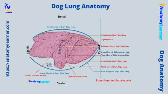
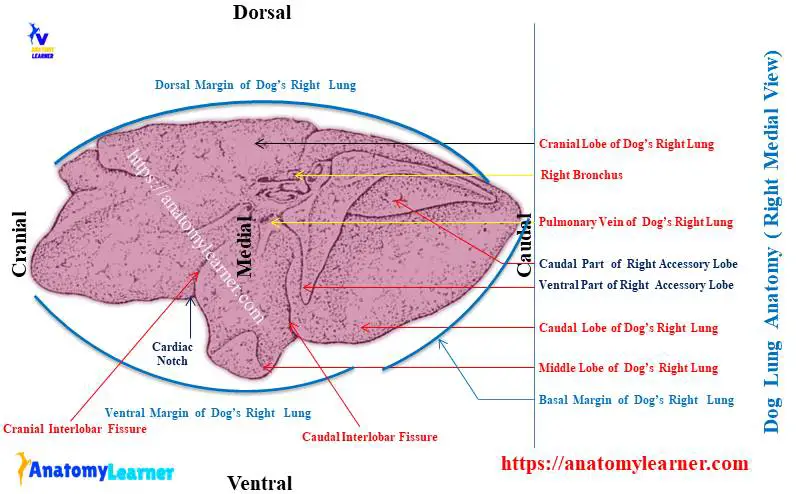
From the dog’s right lung, I tried to show you the 4 distinct lobes from the medial surface in the labeled diagram. Again, this diagram shows different margins (borders) and other different structures from the medial surface of the right lung.
Again, the cranial and caudal interlobar fissures from the dog’s right lung are also identified in the labeled diagram.
Now, the 3 distinct lobes (cranial, middle, and caudal) are identified from the lateral surface of the dog’s left lung. Here, you will also find the cranial and caudal interlobar fissure (shown in the diagram) from the lateral or coastal view.
Again, the 3 different margins from the dog’s left lung are also identified in the labeled diagram. The structures from the hilus are also identified from the medial surface of the left lung.
Finally, you will get the summary (with the peculiar anatomical features) of both the right and left lungs of the dogs from this video.
Horse, ox, and pig lungs compare to the dogs.
You will find a great variation in the structure of the lung’s lobes among the horse, ox, and pig compared to the dogs. Here, I will show you some of these essential features of the lungs of horses, ox, and pigs.
You will find more variation in the ox lung structure compared to the dogs. The apical lobe of the ox lung is larger compared to the left lung. You will see a wide cardiac notch in the left lung of the ox and a more deep cardiac impression on the medial surface of the right lung.
Again, in the horse lung, the interlobar fissures are not so deep; thus, the lobes are not so prominent. The apical lobe of the horse’s right lung is not as large as found in the ox or dog. You will not find any apical bronchus in the horse lung.
The pig’s right lung possesses 4 distinct lobes, but in the left lung, you will only find 2 lobes. Here, the apical bronchus is present in the pig’s right apical lobe at the level of the 3rd rib.
Common inquiries on dog lung anatomy
In this part of the article, you will find some common questions on dog lung structure that veterinary students ask. Here, I will enlist some of these questions (on dog lungs) and try to provide the perfect information.
How are the lungs of a dog divided?
You know there are 2 lungs (right and left) in the dog’s thoracic cavity. These 2 lungs of the dogs are separated from each other by the mediastinum space.
Again, you may also find the division of both the right and left lungs of the dog. The interlobar fissures form this division of the dog’s lungs.
You will find cranial and caudal interlobar fissures in both lungs. These 2 interlobar fissure divides the dog’s right lungs into 4 parts or lobes – cranial, middle, caudal, and accessory.
Again, the cranial and caudal interlobar fissure divides the dog’s left lung into 3 lobes.
I hope you can understand the division pattern of the dog’s lungs.
How many lobes does a dog lung have?
The dog lung has total 7 lobes (all together in the right and left lungs). In the right lung of a dog, you will find 4 distinct lobes – cranial (apical), middle (intermediate), caudal (diaphragmatic), and intermediate (accessory) lobes.
But, the accessory lobe of the dog’s right lung has 3 processes – dorsal, cranial, and caudal.
Whereas in the left lung of a dog, you will find 3 distinct lobes – cranial, middle, and caudal. There are no intermediate or accessory lobes in the dog’s left lung.
Thus, there are total 7 lobes present in the dog lung.
Conclusion
The dog lung anatomy comprises the features of 7 lobes, surfaces, borders, and hilus. Here, the right lung of the dog possesses 4 distinct lobes with a typical anatomical feature, whereas the left has 3 lobes.
The dog’s right lung is larger compared to the left one. You will find the more concave base in the right lung anatomy, whereas the left lung possesses the less concave base.
The cardiac impression is more distinct and deep in the medial surface of the dog right lung anatomy. Now, you should identify all the anatomical features from the different lobes of both the right and left lungs from the actual sample with the help of a labeled diagram.