The dog hip anatomy consists of bones, muscles, and joint. Some of the nerves and vessels supply to the hip region of a dog and pass it along.
In this guide, I will tell (show) you the complete anatomical features of the dog’s hip with the different labeled diagrams. Here, I will discuss the main anatomical facts of the bones, muscles, vessels, and nerves directly or indirectly related to the dog’s hip.
You will find a detailed guide on a dog’s hip joint formation in this article. Finally, I will briefly discuss some common problems in the canine hip.
So, if you want to know the details about the canine hip joint anatomy, let’s continue this article till the end. I hope you will enjoy the short and detailed guide on the dog hip with the labeled diagrams.
Dog hip anatomy
The dog hip extends from the coxal tuber to the ischiatic tubercle. So, the hip bones from both sides together form the hip region in a dog.
The most important features of the dog hip anatomy are the formation of joint along with muscles, vessels, and nerves that passes over this area. Here, I will focus on these structures involved in a dog’s hip joint formation.
The anatomy of the canine hip joint is clinically important as different problems like hip dysplasia commonly occur. Again, the muscles (like gluteus and biceps femoris) and nerves (like ischiatic) are clinically significant structures from the dog hip.
Before knowing the details of the different structures from the canine hip, ensure you have a piece of good knowledge about the different bones and muscles from the pelvic limb. Again, you should know the different joints of the dog’s pelvic limb and the bone’s involvement.
I hope the below-mentioned articles might help you to know these (bones and muscles) from the dog’s pelvic limb –
- Dog leg anatomy – bones, muscles, and vessels with the labeled diagrams,
Again, the following article will also help you to get a basic idea of the different joints from the dog’s pelvic limb –
- Dog joints anatomy with the labeled diagram,
Now, let’s discuss the anatomical facts (main) of the followings –
- Dog hip joint formation,
- Muscles involved in the hip joint and hip area,
- Vessels of the dog’s hip, and
- Nerves that supply to the dog’s hip region,
Where is a dog’s hip located?
The dog’s hip is located at the caudal part of the body that extends from the coxal tuber to the ischiatic tuberosity. Again, the hip joint is located between the articular surface between the cotyloid cavity of os coxae (acetabulum) and the head of the femur bone.
The below-mentioned diagram shows the different joints from the dog’s pelvic limb. From this diagram, you will easily understand the location of the dog’s hip with its bony involvement.
Summary of dog hip
So, in the hip area of a dog, you will find the involvement of two important bones – the hip or os coxae (ilium, ischium, and pubis) and the femur bone. I will describe all the features from the hip bone and femur from the dog pelvis.
Here, the dog hip joint is a ball and socket type of joint in the pelvic limb. So, you will find the polyaxial movement in the dog’s hip joint.
- Type and movement of dog hip joint: ball and socket joint; polyaxial movement,
- Bone involvement in the dog hip joint: cotyloid cavity of the os coxae (acetabulum) and head of the dog’s femur bone,
- Ligaments of the dog’s hip joint: transverse acetabular, joint capsule or capsular ligament, iliofemoral ligament, ischiofemoral ligament, and ligament of the head of the femur (round ligament),
The transverse ligament of the dog hip is also known as the cotyloid ligament. Compared to the canine, you will find an accessory ligament of the femur (cheek ligament) in the horse hip joint anatomy.
There are different muscles in the dog’s hip that I will describe in the canine hip muscle anatomy section. The most important muscles or tendons that attach to the hip joint are –
- Tendon of the psoas minor muscle,
- Iliacus muscle of the dog hip,
- Gluteus medius, deep and superficialis,
- Tensor fascia latae and sartorius muscles,
- Gemelli muscle of the hip joint,
- Internal and external obturator muscles,
- Articularis coxae muscle, and
- Different parts of the quadratus femoris muscle,
The caudal gluteal and branches of the deep femoral arteries (which arise from the external iliac) supply the hip region and joint. Again, the branches of the ischiatic nerve (cranial and caudal gluteal) and caudal cutaneous femoral nerves supply to the hip joint area.
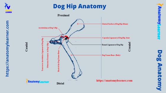
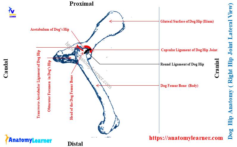
Dog hip joint anatomy labeled diagram
Now, I would like to show you the different labeled diagrams on the dog hip joint anatomy. These might help you to understand the complete structure of the dog’s hip.
In the canine hip labeled diagram, I tried to show you the different muscles that have a direct attachment to the hip joint.
Again, the other muscles located in the hip area are also identified in the labeled diagram. You will also find some other muscles that are not directly related to the hip (showing muscles from the thigh region of a dog).
Now, let’s see the formation of the dog hip joint in between the articular surface of the acetabulum (cotyloid) and the head of the femur. In the diagram, I tried to show all the ligaments from the canine hip joint (capsular, transverse, iliofemoral, and round ligaments).
Different arteries that supply the hip joint and area are also identified in the labeled diagram. Here, I only identified the major vessels (arteries) from the hip area of the dog.
Branches of the external iliac artery (caudal, cranial gluteal, and deep femoral artery with different branches) are identified in the dog leg arteries labeled diagram. Again, you may also find the diagram on the different veins supplied in a dog’s hip joint area here.
Finally, I tried to show the different nerves (like the pelvic, obturator, pudendal, perineal, cranial and caudal gluteal, and the main ischiatic nerve) from the hip region of a dog. The main ischiatic nerve (longest peripheral nerve) of the dog passes caudomedial to the hip joint (shown in the diagram).
Dog hip bone anatomy
To describe the anatomical facts of the canine hip, you might have a good piece of knowledge on the different features of the hip bones. Here, the main bone of the dog hip is os coxae which consist of the ilium, ischium, and pubis.
Again, another important bone of the canine hip is the femur bone that forms the hip joint with the acetabulum (cotyloid) of the os coxae. The below-mentioned article (guide) might help you to know the details of these two bones from the canine hip –
- Dog femur bone anatomy with the labeled diagram, and
- Dog pelvic bone anatomical features with the labeled diagram,
Again, this article (related to animal hip bone anatomy) might give you a basic idea of different features (showing all the features in a video).
But, you may also find the important features of the dog hip and femur bones in the followings –
- Osteological features of dog’s hip bone (os coxae), and
- Osteological features of the head of the dog’s femur bone,
Let’s continue the article to learn the basic structure of the two important bones (hip and femur) that form the canine hip joint.
Hip or os coxae of a dog
The dog hip or os coxae consists of the ilium, ischium, and pubis bone. Together these bones of the hip form a socket that receives the head of the femur bone.
This socket is deep and known as the cotyloid cavity of the dog hip. You may also be called this cotyloid cavity, the acetabulum.
Here, I will not describe all the anatomical features in detail from the three bones of the dog’s hip. The previously suggested article will help you know the features of these three bones from the canine hip. So, now, I will enlist some of the important features you might know to describe hip anatomy.
You know the ilium of the dog hip is the largest and most cranial bone with a wing and body. Here, the wing is most cranial and forms a concave cavity on its lateral aspect.
The body of the dog’s ilium is narrow and more irregular caudal. The body expands in its caudal end and forms the cranial two-fifth of the acetabular cavity.
Again, the ischium is the second large bone in the dog hip, comprising the body, ramus, table, and tuberosity. This bone forms the caudal third of the dog’s hip. Again, this bone also forms the caudal part of the acetabulum, obturator foramen, and pelvic symphysis (ischial symphysis).
Finally, the pubis is a small triangular bone in the dog’s hip. It extends from the lateral aspect of the ilium and ischium bone to the pubis symphysis medially. The caudal border of the dog’s pubis bone from the cranial part of the obturator foramen.
Osteological features of the canine hip bone
Overall, you should know the below-mentioned osteological features from the three bones (ilium, ischium, and pubis) of the dog hip –
- Crest of ilium and cranial dorsal iliac spine,
- Wing of ilium and gluteal surface of ilium,
- Caudal dorsal iliac spine,
- Sacropelvic surface (iliac tuberosity and auricular surface),
- Body of the ilium and arcuate line,
- Greater and lesser ischiatic notch,
- The ischiatic spine of the dog’s hip,
- Acetabular cavity (acetabulum) with acetabular fossa and lunate surface,
- Obturator foramen of the dog hip,
- Ischiatic table, tuberosity and ischiatic arch,
- Iliopubic eminence and pecten of pubic bone,
- Pubic tubercle, and
- Pelvic symphysis (ischiatic and pubis symphysis part),
The labelled diagram identifies all the above-mentioned osteological features from the dog hip bone (os coxae). But, it would help if you focused on the osteological features of the acetabulum of the dog as it receives the head of the femur and forms the hip joint.
So, let’s see how the acetabulum forms and contribute to forming the dog’s hip joint.
Dog acetabulum structure
The cotyloid cavity of the dog hip is formed by the union of corresponding angles of ilium, ischium, and pubis bones. You will find the following essential osteological features in the dog acetabulum –
- Acetabular notch,
- The outer smooth articular (lunate) surface of the acetabular cavity,
- Non-articular part in the middle of the fossa, and
- Acetabular notch in the acetabulum,
There is an outer smooth articular part in the acetabulum of the dog hip. You will see a notch at the posteromedial aspect of the acetabulum. This acetabular notch is wider than other animals like a goat, cows, or sheep.
There is a deep acetabular fossa present in the acetabular cavity. Again, there is a non-articular part in the middle and deep area of the fossa. This non-articular part is connected with the acetabular notch.
You will also find a secondary notch at the anteromedial border of the cotyloid cavity (acetabulum).
Dog femur head anatomy
In this part, I will show you only the osteological features from the head of a dog femur. The articular surface of the femoral head articulates with the lunate surface of the acetabulum and forms the major part of the dog hip anatomy.
But, you may know more about the different parts (proximal and distal extremities and body) of the dog’s femur from the below-mentioned article –
- Dog femur bone anatomy with the labelled diagram,
You will find the head at the proximal end of the femur bone. It is smooth and nearly hemispherical and caps the dorsocaudal and medial parts of the constricted neck.
There is a small indistinct pit on the medial aspect of the head. You will also see a moderately rough, depressed, nonarticular strip from the fovea capitis femoris to the nonarticular margin.
The ligament of the head of the (dog) femur (round ligament) attaches to the fovea capitis femoris of the femur head.
How is the dog’s hip joint formed?
So, the dog hip joint is formed by the head of the femur articulated with the acetabulum of the os coxae. Here, the femur (head part) and acetabulum meet at the hip joint at a 90-degree angle.
It is a ball and socket (multiaxial) joint in the dog’s pelvic limb, allowing a great range of limb movement. But, the main movement of this joint is extension and flexion.
A different muscle that directly or indirectly attaches to the hip joint restricts the hip movement in a great range. Different ligaments provide the stability of the dog hip joint. Let’sLet’s see the common ligaments from the canine hip joint –
- Capsular ligament or joint capsule,
- Round ligament or ligament of the head of dog’s femur bone,
- Transverse or cotyloid ligament of the dog hip,
- An iliofemoral ligament of the canine hip, and
- The ischiofemoral ligament of the dog’s hip joint,
Capsular ligament of the dog’s hip
The joint capsule or the capsular ligament of the dog hip is capacious. It extends from the margin of the acetabulum (cotyloid cavity) to the border around the head of the femur.
You will find two parts in the capsular ligament of the dog’s hip – fibrous coat and synovial membrane. Here, the fibrous coat possesses various thickening but doesn’t contribute to forming the main ligament.
The dorsal part of the fibrous coat is thicker and causes a nearly horizontal bulging of the synovial membrane. Here, the synovial membrane of the joint capsule reflects on the round ligament.
The horizontal bulging of the synovial membrane is the orbicular zone. This structure arches from the cranial to the caudal border across the dorsal surface of the neck. It parallels the dorsal part of the cotyloid cavity’s margin and the dorsal part of the head-neck junction.
Cotyloid or transverse ligament of canine hip joint
This is the fibrocartilaginous structure that attaches along the rim of the cotyloid cavity to make it deeper. A small part of the cotyloid ligament makes a bridge over the acetabular notch. This structure is the transverse ligament of the dog’s hip joint.
You will also find two other small ligaments in the dog hip joint – iliofemoral and ischiofemoral ligaments. The iliofemoral ligament occurs cranially and runs between the part of the ilium and femoral head. Again, the ischiofemoral ligament occurs caudally and runs between the ischium and femur bones.
Round ligament of dog hip anatomy
The round ligament is the ligament of the head of the dog’s femur bone. It is a short, thick, and flattened structure that extends from the fovea capitis femoris of the canine femur bone to the acetabular fossa.
This structure of the dog hip is intraarticular and not weight-bearing. You will find the synovial membrane covering the round ligament of the dog.
Again, you will see the wide acetabular attachment that bends with the periosteum of the acetabular fossa. It also attaches to the transverse acetabular ligament of the dog hip.
You will find the more wide part on the round ligament that attaches to the head of the femur. Again, in the acetabular fossa, you will see a small amount of fat around the round ligament.
So, you see, there are fewer ligaments in the structure of the dog hip joint compared to the other different joints of the body, like the shoulder or elbow joint. Therefore different surrounding muscles with their tendons help to keep the joint in its position and provide great stability.
Now, I will show you some of the muscles that greatly contribute to the formation of the canine hip anatomy. Let’sLet’s continue this article to know the details of these selected muscles from the dog’s hip area.
Dog hip anatomy muscles
You will find different muscles in the dog hip that may be divided into three major groups – lumbar hypaxial, lateral pelvic, and medial pelvic groups. Here, the lumbar hypaxial muscle lies on the ventral surface of the lumbar vertebrae and ilium.
Again, the lateral pelvic muscles lie on the lateral side of the dog’s pelvis. Finally, the medial pelvic muscles are deep and partly inside of the dog’s pelvis.
If you wish to know the details of anatomical features of the dog pelvis, you may read the below-mentioned article from anatomy learner –
- Dog pelvis anatomy with the labeled diagram,
From these hip muscles, I will describe the most important muscles of the hip. Again, I will try to point out the important muscle that has a direct relationship with the hip joint structure (provide stability to the hip joint).
But, first, let’s enlist the major muscles from the hypaxial, lateral pelvis, and medial pelvic regions of the dogs –
The major muscles from the dog’s lumbar hypaxial are –
- Psoas minor muscle (have a direct contribution to hip joint formation),
- Psoas major muscle,
- Quadratus lumborum muscle of the hip,
Again, the medial pelvic muscles of the dog hip area include the following –
- Obturator extenrus and internus muscle of the dog hip,
- Gemelli muscle of the hip,
- Quadratus femoris muscle,
Both these muscles directly contribute to the formation of the canine hip anatomy. Now, let’s see the lateral muscles from the dog’s hip –
- Gluteus medius, superficialis, and profundus,
- Piriformis muscle of the canine hip, and
- Tensor fascia latae muscle of canine hip,
All the muscles mentioned above have a great contribution to providing the stability of the dog’s hip structure. You will find all these muscles from the dog hip in the below-mentioned labeled diagram.
Psoas minor and major muscle of the dog hip
These two are the lumbar hypaxial muscles that arise from the ventral surface of the caudal thoracic and lumbar vertebrae. Finally, they inserted the os coxae and femur bone of the dogs.
Here, the psoas minor muscle runs towards the pelvis ventromedially and lies between the iliac fascia and peritoneum. This muscle of the dog arises from the tendinous fascia of the quadratus lumborum muscle at the level of the last thoracic vertebra.
Again, this muscle runs to the arcuate line and inserts an iliopubic eminence of the pubis bone on this line. It flexes the lumbar part of the vertebral column of the dogs.
Again, the psoas major muscle lies ventral to the quadratus lumborum and dorsal to the psoas minor muscle. It arises from the transverse process (3) of the second and third lumbar vertebrae.
This muscle receives fibre from the iliacus along the cranioventral border of the ilium bone. The ilicus and psoas major muscles contribute to the dog hip anatomy. The main function (action) of this muscle is to draw the pelvic limb cranially by flexion of the hip joint.
Tensor fascia latae muscle
The tensor fascia latae is the triangular lateral pelvic muscle in the dog. This muscle of the dog attaches to the ilium from the coxal tuber to the alar spine.
You will find the Sartorius’s muscle on the cranial aspect of the tensor fascia latae. The gluteus medius and quadriceps femoris muscle lie caudodorsally and distomedially to the tensor fascia.
This muscle divides into two parts – the cranial, more superficial and caudal deep part. Here, the cranial, more superficial part of the tensor fascial latae muscle inserts on the lateral femoral fascia. It also blends with the fascial insertion of the biceps femoris muscle.
Again, the deep caudal part of the dog’s tensor fascia latae muscle also inserts into the lateral femoral fascia. Finally, it runs deep to the biceps femoris towards the stifle joint on the lateral aspect of the vastus lateralis.
The main function of the dog’s tensor fascia is to flex the hip joint, abduct the pelvic limb, and extend the stifle joint.
Gluteus superficialis and medius muscles
The gluteus superficialis is the small, flat, and rectangular most superficialis muscle in the dog’s hip. This muscle extends from the sacrum to the first caudal vertebra proximally.
The gluteus superficialis muscle arises from the gluteal fascia and deep fascia. Again, the tendon of the gluteus superficialis runs over the trochanter major and inserts on the small trochanter tertius.
This superficial muscle of the gluteus cover the gluteus medius and piriformis muscles. The main function of the gluteus superficialis muscle is to extend the hip joint.
Now, the large gluteus medius lies on the gluteal surface (lateral) of the ilium bone. It arises from the iliac crest (sharp line) and the sacral tubercle of the hip.
The medius gluteus muscle of the dog extends over the gluteus profundus muscle. It forms a short, thick tendon that inserts on the free end of the trochanteric major.
This muscle of the gluteal region of a dog helps to extend the hip joint. Again, it causes the medial rotation of the hip and prevents the lateral rotation during weight bearing.
Piriformis muscle of dog hip anatomy
This small muscle in the dog hip region lies caudal and medial to the gluteus medius. Again, this piriformis muscle of the dog completely covers by the gluteus superficialis muscle.
The piriformis muscle arises from the lateral surface of the third sacral and first caudal vertebra of the dogs. It forms a thick tendon that joins with the tendon of the gluteus medius, and finally, they inserted into the trochanter major of the femur bone.
Like the gluteus medius muscle, this muscle extends the dog’s hip joint.
Gluteus profundus muscle of dog’s hip
The dog’s gluteus profundus is a broad, fan-shaped and deepest muscle in the gluteal region of a dog. If you see the diagram of the dog’s gluteus muscle, you will find this muscle just deep into the gluteus medius and piriformis.
The gluteus profundus muscle of the dog hip arises from the lateral surface of the body of the ilium near the ischiatic spine. This muscle directly forms the dog’s hip joint and provides great stability.
The fibres from the gluteus profundus muscle cover the dog’s hip joint distolaterlly. Again, it forms a thick tendon that ends on the cranial surface of the major trochanter of the femur (with the insertion of the gluteus medius muscle).
Again, this muscle of the dog hip help to extend the hip joint. It also provides some abduction movement in the dog’s pelvic limb.
Now, I will show you some of the medial pelvic muscles from the dog with the little information and labeled diagrams.
Obturator internus and externus muscles of canine hip
The obturator internus is a large fan-shaped muscle that covers the obturator foramen internally. This muscle arises from the pelvic surface of the pubis and ischium bones, the ischiatic table, and the ischiatic arch.
The fibres of the obturator internus muscle cover the smooth surface of the lesser ischiatic notch and ischiatic spine. It forms a small tendon that inserts deeply between the edges of the gemelli muscle.
This muscle provides the lateral rotation of the hip joint and prevents the medial rotation of the weight bearing.
On the other hand, the external obturator is another fan-shaped muscle that arises from the ventral surface of the pubis and ischium bones and covers the obturator foramen externally. This muscle is separated from the pelvic symphysis by the adductor muscle.
Again, in the cranial border of this muscle, you will find the quadriceps femoris. Whereas in the cranial border, you will see the adductor muscle.
This muscle of the dog forms a long tendon that joins with the tendons of other different muscles and finally inserts into the trochanteric fossa. Like the obturator internus muscle, this externus muscle also provides the lateral rotation of the dog’s hip joint.
Gemelli muscle of the dog’s hip
The Gemelli muscle also directly contributes to the formation of the canine hip anatomy. In the dog Gemelli, you find two distinct parts between the internal and external obturator muscles.
This Gemelli muscle of the dog arises from the lateral surface of the body of the ischium bone and the ventral aspect of the lesser ischiatic notch.
It forms a large tendon that joins with the tendon of the obturator internus muscle and finally inserts into the trochanteric fossa of the femur bone. This muscle of the dog hip also prevents the medial rotation of the hip during weight bearing.
Articularis coxae of the dog hip
This is another important muscle that directly contributes to in the stability of the canine hip anatomy. It is a small, spindle-shaped muscle in the hip region of dogs.
This articularis coxae locates laterally and caudally from the ilium adjacent to the attachment of the quadriceps femoris. Again, this muscle passes cranially and laterally over the capsule of the hip joint to the neck of the dog’s femur bone.
Finally, it attaches to the common ridge between the origin of the lateral and medial vastus muscle (parts of quadriceps femoris). The main action of the articularis coxae is to flex the help little. This muscle also adjusts the position of the hip joint capsule.
Quadriceps femoris muscle of the canine hip region
The quadriceps femoris is the cranial muscle of the dog’s thigh, but it indirectly contributes to the hip joint’s stability. This muscle covers the dog’s femur bone cranially, medially, and laterally.
The quadriceps femoris fuses with the fascia latae and the aponeurosis of the biceps femoris and Sartorius’s muscles. Now, the dog quadriceps femoris muscle possesses four distinct divisions – laterally vastus lateralis, medially vastus medialis, cranially straight femoris, and vastus intermediate (deep).
The straight part of the quadriceps femoris muscle arises from the body of the ilium, just cranial to the acetabulum. You will see the cranial belly of the tensor fascia latae cranially and laterally of the straight part of the quadriceps femoris muscle.
This part of the quadriceps femoris muscle contributes to flexing the hip joint and extending the stifle.
The vastus medialis part arises from the proximal fifth of the femur and ends on the patella. Here, the vastus lateralis of the dog’s quadriceps femoris muscle is larger. It arises from the craniolateral part of the proximal fifth of the dog’s femur bone.
Finally, you will see the smallest part of the dog’s quadriceps femoris muscle – the vastus intermediate. This part of the muscle arises from the lateral part of the proximal fourth of the femur bone along with the vastus lateralis.
The different parts (4) of the quadriceps femoris muscle flex the dog’s hip and extend the stifle.
Dog hip anatomy vessels and nerves
You will find different vessels and nerves in the dog hip region of a dog. The branches of the vessels (artery) come from the external iliac and median sacral arteries in the hip region.
Again, the main vein of the dog’s hip region is the different branches of the femoral vein and median sacral vein. You will find a details guide on the femoral vein (along with its different branches) that supplies the different structures of the dog’s hip.
In my previous article, I have already described the different branches of the dog’s femoral vein. Again, you will also find the different branches and distribution of the external iliac artery in the dog’s pelvic limb here in anatomy learner.
Okay, let’s see what the main arteries in the dog hip anatomy that supply the different structures of this region are. Here, I will only enlist the most important branches that you should know first from the dog hip –
- Medial sacral and lateral caudal arteries in the dog hip,
- Iliolumbar and gluteal arteries (cranial and caudal),
- Deep femoral artery of the dog’s hip,
- Medial circumflex femoral artery of the canine hip region,
- Obturator arteries in the dog hip,
- Ascending branch of the medial circumflex artery,
- Transverse and deep branches of the deep femoral artery,
Median sacral artery of a dog
You know, the medial sacral artery of the dog is the direct continuation of the caudal part of the abdominal aorta. It arises opposite the body of the seventh lumbar vertebra as an unpaired medial vessel.
It crosses the ventral part of the sacrum and enters between the right and left ventral sacrocaudal muscles. Here, you will find two branches of the median sacral artery – the spinal and dorsal branches.
- Spinal branch – joins the ventral spinal artery within the vertebral canal, and
- Dorsal branch – passes through the dorsal sacral foramina,
Again, you will find a single median caudal artery, paired lateral caudal, dorsal lateral caudal, and ventrolateral caudal arteries in a dog’s tail. Here, the single median caudal artery is the direct continuation of the medial sacral artery at the level of the first caudal vertebra.
It runs midventrally on the caudal vertebrae and passes between the right and left ventral sacrocaudal muscles. The paired dorsal and ventral caudal arteries are smaller in the dog’s tail.
Branches of external iliac in the dog’s hip
The deep femoral artery arises from the caudomedial surface of the external iliac artery of the dogs. This artery provides small branches to the pelvic surface of the pubis and caudal part of the ischium. It also anastomoses with the obturator muscle through the obturator foramen.
The medial circumflex femoral artery obliquely crosses the surface of the iliopsoas and vastus medialis muscles. It also sends a small branch to the adductor muscle of the dog hip region.
You will also see some of the obturator branches of the medial circumflex artery in the dog’s pelvis. They course dorsally through the obturator foramen just lateral to the obturator nerves. These vessels supply the caudal part of the pelvic, coccygeal, and obturator muscles of the dog.
Again, the ascending branch of the medial circumflex femoral artery supplies the proximal end of the adductor muscle. You will also find a small acetabular branch that extends laterally to the trochanteric fossa and supplies the muscles in this area.
Now, let’s see the diagram where I tried to show you the major vessels (arteries and veins) supplying the different structures of the dog hip region.
If you wish to learn the details and anatomical facts of these vessels separately, you may find more guides here on anatomy learner (gross anatomy learning).
Nerves of the dog’s hip
Now, let’s see the distribution of the nerves in the hip region of a dog. I need a separate article for you to describe all these nerves from the dog’s hip. First, let’s find the main branches and distribution of the different pelvic and ischiatic nerves in the hip area of the dogs.
In the diagram, you will find the below-mentioned nerves from the dog’s hip –
- Cranial and caudal gluteal nerve (branch of the ischiatic nerve),
- A pelvic nerve in the dog’s hip,
- Paired caudal nerves,
- Obturator nerves in the hip region,
- Pudendal and perineal nerves,
- Nerve to the piriformis muscle,
- The main sciatic nerve in the dog’s hip region,
But, you will also find some other small nerves in the canine hip structure. It will be best if you read the full guide on the different nerves from the dog’s pelvic limb and hip region from another article by anatomy learner.
Dog hip anatomy dysplasia
Dog hip dysplasia is more common in the dog and occurs due to the deformity in the structures both in the head of the femur and the acetabulum. This developmental deformity causes degenerative changes in the joint capsule and other structures of the dog’s hip.
Different factors may cause rapid hip dysplasia in the dogs – congenital, abnormal growth of the bone, defects in the articular cartilage, articular surface of the acetabulum, and the head of the femur.
Different dietary changes, exercise, and joint laxity are also responsible for hip dysplasia in dogs. You have already seen the structure of the dog’s hip joint is not protected by more ligaments compared to the other different joints of the body. So, there is a high risk for hip dysplasia occurring during more stress and exercise.
What are the symptoms (main) of hip dysplasia in a dog?
You will find a wide range of symptoms in the hip joint dysplasia of a dog. First, the dog will show lameness and become cool. The activity of the dog will decrease with time. They will also show sudden limping and abnormal sitting positions.
Sometimes, it is very hard to stand properly in chronic conditions in hip dysplasia.
Common inquiries in canine hip anatomy
Now, let’s see the more common questions on the canine hip anatomy. Here, I will enlist the questions with short answers on the dog hip structure.
How the dog’s hip joint is formed?
The dog hip joint is formed by the union of the articular surface of the head of the femur bone and the lunate surface of the acetabulum. Again, different ligaments help to bind these articular surfaces together.
You will also find some hip muscles that have directly contributed to the form of the dog hip structure. So, the ligaments, muscles, and bony configuration provide the stability of the dog’s hip joint.
What are the main ligaments of the dog hip joint?
You will find 5 more important ligaments in the structure of a dog hip joint. There are transverse or cotyloid ligaments, joint capsule or capsular ligaments, round ligaments (ligaments of the head of the femur), iliofemoral ligaments, and ischiofemoral ligaments.
Here, the round and capsular ligaments are stronger, whereas the iliofemoral and ischiofemoral ligaments are thin and less strong.
Conclusion
So, from the dog hip anatomy, you have learned the details of the bones, muscles, and vessels. The articular surface of the acetabulum (cotyloid cavity) and head of the femur bone join together with the help of different ligaments to form the dog hip joint.
All the ligaments (especially the round, cotyloid, and capsular) provide a great range of stability in the dog hip anatomy. Again, the different muscles also provide great stability to the dog’s hip. All the nerves and vessels distributed in the dog’s hip area are important for the veterinarian to treat the different problems in this area.
Finally, make sure you might learn the anatomical facts of the different structures of the dog hip joint from your anatomy learning laboratory. You might use the diagrams I provided to learn the complete dog hip structure.