The dog mouth anatomy includes the lip, oral cavity, and associated structures. But, the term mouth includes only the opening between the lips into the vestibule of the oral cavity. Here, I will describe the anatomy of the dog’s mouth, including the lips and different parts of the oral cavity, with a labeled diagram.
So, after reading this article, you will know and identify the different parts of a dog’s mouth from the live sample. Again, you will find the answers to some common inquiries on dog mouth and oral cavity. If you wish to learn the detailed anatomical facts of the dog mouth, continue this article until the end.
Dog mouth anatomy
First, I would like to provide you with a basic idea of the dog mouth anatomy. As I told you before, the mouth means only the opening between the lips into the vestibule of the mouth cavity.
Again, the mouth cavity is an elongated space at the beginning of the alimentary canal. It comprises the vestibule of the mouth and mouth cavity proper.
I think I should explain these terms – vestibule and mouth cavity proper. The vestibule is the part of a mouth bounded externally by lips and cheeks and internally by the gums and teeth. Again, the mouth cavity proper is bounded in front and laterally by the alveolar arches, gums, and teeth.
The mouth cavity proper of a dog continues with the pharynx behind the oropharyngeal opening. You will also find two terms in the dog mouth cavity – roof and floor. The palate forms the roof of the mouth cavity.
Again, the dog mouth cavity floor is formed by the mandible, muscles, and mucous membrane. In addition, the mucous membrane is continuous cranially with the lip and caudally with the pharynx.
I hope you can understand the basic terms of the dog mouth cavity. It will be easy to learn the different parts of the dog’s mouth.
Dog mouth parts
Now, let’s try to known and identify the different parts of the dog’s mouth and mouth cavity. In a dog mouth, you will generally find the below-mentioned parts.
- The lips of the dog mouth
- Cheeks of the dog mouth
- The gums of the dog mouth
- A mouth cavity – includes the vestibule part and mouth cavity proper part
- The floor and roof of the dog mouth cavity
In the roof of the dog mouth, you will find the hard anterior palate and posterior soft palate. In the next section, you will know the details of these hard and soft palates of the dog’s mouth. Again, you will know the detailed anatomical facts of every single part of the dog mouth cavity.
So, let’s continue to learn the anatomical facts of the dog mouth parts.
Special features of the dog mouth
It is so difficult to explain all the special features of the dog mouth cavity in a short form. But, here, I would like to share some of the special features of the dog mouth.
The dog mouth cavity’s orifice is more extensive than the ruminant. There are also two lips in the dog where the upper lip covers the lower lip. The lower lip is partially denticulate in appearance.
The papilla salivalis in the dog mouth anatomy is located opposite the third premolar tooth. Again, the papilla incisive is located behind the central incisor teeth. You will not find any dental pad in the dog’s mouth. In addition, there are well-developed palatine tonsils present in the dog’s mouth.
These are very short information on the remarkable anatomical facts of the dog’s mouth. For more interesting anatomical facts on dog mouth, please continue this article.
Dog lips anatomy
The lips are the thick and rigid musculo-membranous structure surrounding the mouth orifice. Externally, it is formed by the skin, and internally it is lined by the pigmented epithelium. You will find the orbicularis oris muscle in between these two layers.
There are superior and inferior (upper and lower) lips in a dog that meet at the angle of the mouth. Their union angle is located near the first cheek tooth and is rounded. Again, each lip of a dog presents two surfaces and two borders.
The margin of the superior lip of a dog is narrow and devoid of long tactile hair. But, you may find some tactile hairs at the rostral two-thirds of the superior lip on each side. They are imperfectly arranged into four rows. Again, the margin of the inferior lip is also devoid of tactile hairs.
Structure of dog lip
As I told you before, the lips are covered externally by the skin and are lined by mucosa membrane. You will find the muscular tissue, glands, vessels, and nerves between the dog lips’ external and internal covering.
The skin of the lips directly lies on the muscles. You will find many muscle fibers directly inserted into it.
There are more labial glands at the angle of the mouth that forms compact masses. You will find more labial glands on the upper lip and fewer in the dog’s lower lip.
Again, the external mucosa membrane of the dog lip is often pigmented and reflected upon the bones of the jaws to form the gums. Do you know what are vessels supply the dog lips?
The dog lips are supplied by the maxillary, mandibular, and palatolabial arteries. The most important vein of the dog lip is the lingufascial vein.
In addition, the trigeminal and facial nerves supply the dog’s lip. The sensory nerve of the dog lips comes from the trigeminal, and the motor nerves come from the facial nerve.
Exceptional features of the dog mouth lip anatomy
So, what are the exceptional features of the dog mouth lips anatomy? You will not find any definite frenulum or median mucosal fold that attaches the inferior lip to the gums. But, you may find a poorly developed median mucosal fold in the superior lip of the dog.
The mucosa membrane of the inferior (lower) lip is firmly attached to the gums on either side. You will find a deep, straight, narrow cleft in between the two halves of the superior lip rostrally. Thus narrow aperture is known as the philtrum and marks the union of the two halves of inferior lips.
As you know, the rostral two-thirds of the superior lip contains tactile hairs. The hairs of the superior lip slope caudoventrally. They are thinner and shorter in the rostral part of the superior lip.
The cheek of the dog
The cheeks form the sides of the dog’s mouth and continue rostrally with the lips. They form the caudal part of the lateral walls of the vestibular cavity. The cheeks are attached to the alveolar borders of the bones of the jaws.
You will find a very small cheek in a dog because of the large mouth opening. The cheek cavity runs medial to the masseter muscles of the dog. Again, it extends as far caudally as the attachment of the buccinators muscles on the mandible and maxilla.
“You will find a large cheek in ruminant and in some rodent. These animal need a large cheek spaces during the mastication and transportation. But, the dog don’t have any large cheek cavity.”
Structure of a dog cheek
Anatomically, the dog cheek comprises the skin, the muscular and glandular layers, and the mucous membrane. The skin of the dog’s cheek is thin, pliable, and hairy. Again, the muscular layer is formed mainly by the buccinators. The mucous membrane is reflected above and below the gums and is continuous caudally with the soft palate.
You will find some coarse tactile hairs in the skin of the dog’s cheek. The length of this tactile hair may vary in a different breed of dog.
The middle layer (muscular and glandular layer) also contains fibroelastic tissue and the buccinator’s muscle. In this layer, you will also find the cutaneous, zygomaticus, caninus, levator nasolabialis, and depressor labii mandibularis muscle.
“Please find the information on the buccal glands in the next section of this article.”
The mucosa of the dog’s cheek is reddish and frequently shows pigmented areas. Histologically, you will find the stratified squamous epithelium lining in the cheek of a dog.
Buccal glands of dog mouth
The buccal glands of the dog are arranged in two rows – dorsal buccal and ventral buccal glands. Again, the dorsal buccal glands of the dog are consolidated to form the zygomatic glands. These are the largest mixed salivary glands located in the rostroventral part of the orbit.
The ventral buccal glands consist of some small, solitary glands. These ventral buccal glands of a dog are located in the submucosa. Just rostral to the masseter muscle and medial to the ventral part of the buccinators muscle fibers.
Sometimes, you may also find rostral buccal glands in some dog breeds. If the rostral buccal gland is present, you will find scattered lobules that lie under the masseter muscle.
You will find some of the linear series of small dorsal and ventral papillae in the mucosa membrane of the dog check. These indicate the orifices of the small ducts of the buccal glands of the dog.
What are dog lips called?
This is a very specific and interesting question from the dog owner or the veterinarian. A dog has two lips – the superior (upper) and the inferior (lower). The inferior (lower) lip is usually known as the lower lip. But, the superior lip of the dog is known as the flews.
The length of the flews of the dog may vary from breed to breed. But, the structure of the flews in different dog breeds is the same.
Nerves and vessels of the dog cheek
You will find the facial and buccal arteries that supply the dog’s cheek. Again, the blood is carried away by the facial and buccal veins.
You will also find the lymph vessels that go to the mandibular lymph nodes. The trigeminal and facial nerves innervate the dog’s cheek. Here, the sensory nerves come from the trigeminal nerve and the motor nerves from the facial nerve.
The gums of the dog mouth
A dog’s gums surround the teeth and are composed of dense fibrous connective tissue. Again, they are covered by the highly vascularized mucosa.
The gums are intimately united with the periosteum of the alveolar processes. It blends at the edges of the alveoli with the alveolar periosteum. Again, the gums fix the teeth in their cavity.
You will find more thickness in the gum around the neck of the teeth. In addition, the labial surface of the gum continues with the mucosa of the vestibule.
The gums blend with the floor of the mouth cavity and the hard palate internally. Sometimes, the dog gum shows some pigmentation in some breeds.
I have already described the detailed anatomical facts of the dog gums in my previous article. You may also learn other anatomical facts about the dog gums from that article.
Dog mouth cavity anatomy
The cavity of the dog mouth anatomy consists of two spaces – vestibule and mouth cavity proper. The space external to the teeth and alveolar space enclosed by the lips and cheeks is termed the vestibule of the mouth.
Again, the teeth and alveolar processes space is termed the mouth cavity proper. I will show you everything about the dog mouth cavity in detail in this part.
So, let’s know details about the –
- Vestibule of the dog mouth cavity and
- The mouth cavity proper – parts and structures
You know the mouth cavity proper consists of palates (hard and soft palates), salivary glands, teeth, and tongue. Here, you will find detailed information on palates and salivary glands. Again, the anatomical facts of teeth and tongue are already described in another article. If you love to learn the details of dog teeth and tongue, please read the separate article from the dog and cat anatomy learning section.
Vestibule of the dog mouth cavity
First, make sure you know the vestibule space of the dog’s mouth cavity. It is external to the teeth and gums and internal to the lips and cheeks. You will find a U-shaped slot in the vestibule opening at the rostral end of the mouth.
If the mouth is closed, the vestibule communicates with the mouth cavity proper in the interdental spaces. It also establishes free communication between the two parts of the mouth cavity.
Do you know where the parotid and zygomatic salivary ducts open in a dog? The parotid and zygomatic salivary ducts open into the dorsocaudal part of the vestibule.
Again, the parotid duct opens through the cheek on the small parotid papilla. The parotid papilla is located opposite the caudal part of the superior fourth molar tooth.
There you will find another structure – fornix vestibule formed by the reflection of the mucosa from cheek to gums. In addition, the main duct of the zygomatic salivary gland opens lateral to the caudal part of the superior first molar tooth. You will find a small mucosal ridge that connects the main parotid and zygomatic ducts openings.
Dog mouth cavity proper
A proper mouth cavity is the most important part of the dog mouth anatomy. It is bounded dorsally by the hard palate and a small part of the adjacent soft palate. You will find the dental arches (pads) and teeth in the lateral and rostral boundary of the dog mouth cavity proper.
Again, the floor of the mouth cavity proper is formed by the tongue and the mucosa membrane. You will find a sublingual caruncle ventral to the body of the tongue. The major ducts of the sublingual and mandibular ducts open near this sublingual caruncle.
You will also find a sublingual fold that lies close to the body of the mandible. An incisive papilla present just caudal to the superior central incisor teeth. It is a rounded eminence that extends caudally and blends with the first transverse ridge of the hard palate.
You will also find the incisive ducts on each side of the incisive papilla. Again, the mouth cavity proper is continuing caudally with the pharynx.
So, what you should learn from the dog mouth cavity proper. You should learn the anatomical facts of the following organs or structures from the dog’s mouth cavity proper.
- Anatomical facts of the hard and soft palates
- Details anatomy of teeth and tongue of a dog, and
- Anatomical facts of the major salivary glands of a dog mouth
Nice, first, let’s discuss the anatomical facts of the hard and soft palates of the dog mouth cavity proper.
Hard palate of the dog
The hard palate of the dog mouth is bounded rostrally and laterally by the alveolar arches and is continuous with the soft palate caudally. In the structure of a hard palate of the dog, you will find partly bony and partly membranous portions.
The bony part of the hard palate is formed by the incisive, maxilla, and palatine bones. Again, the mucous membrane part is smooth and is attached to the bones by submucosa. You will not find any glands (palatine glands) in the submucosa of the dog’s hard palate.
The mucosa of the nasal side is the pseudostratified ciliated columnar epithelium. Again, the mucosa of the mouth inside is lined by the stratified squamous epithelium.
The hard palate of the dog is nearly flat and laterally inclines slightly ventral. Again, it continues with the incisive and maxillary bones.
You will find a central raphe in the hard palate that divides the surfaces into two equal parts. Again, there are some transverse curved ridges on each part of the hard palate. These curved ridges are more prominent in the caudal part of the hard palate.
The central prominence just caudal to the first pair of incisors is the incisive papilla. There are also incisive ducts present on each side of the incisive papilla.
The trigeminal nerve innervates the hard palate. Again, the blood supply is derived from the palatine arteries.
Soft palate of the dog mouth anatomy
The soft palate of the dog mouth anatomy is the caudal continuation of the soft palate. You will find a longer soft palate in a dog compared to the ruminant. Again, in the brachycephalic breeds, the soft palate may be so long to interfere with the passage of air into the larynx.
In the middle part, the soft palate of a dog is thicker. Again, this soft palate becomes thinner at the caudal end.
You will find the palatopharyngeal arch (caudal pillar) on each side of the caudal border of the soft palate of a dog. There you will find the palatopharyngeal muscles and mucosa in the caudal end of the soft palate.
Structure of the soft palate
You will find the different layers in a dog’s soft palate structure. From ventral to the dorsal surface of the soft palate, it consists of the following structures.
- Epithelium of the soft palate
- Glands of the soft palate
- Palatine muscles of the dog
- Soft palate vessels and nerves
Okay, let’s know the details about the dog’s soft palate structures.
Epithelium of the soft palate of a dog
In the ventral surface of the soft palate, you will find the stratified squamous epithelium. This is the continuation of the epithelium of the hard palate. Again, the dorsal surface of the soft palate is lined by the pseudostratified ciliated columnar epithelium.
There are many longitudinal folds and few transverse folds present in the soft palate. These mucosal folds are evidence of the mobility and slight elasticity of the soft palate.
Palatine glands and muscles of the dog
The palatine glands form the thickest layer of the soft palate. These are the mixed glands that open on the oral surface of the soft palate. In the rostral part of the oral surface, you will find numerous openings for the palatine glands. The number decreases caudally, but the opening increase in size.
The soft palate muscles consist of the paired palatine, paired tensor, and levator veli palatine muscles. You will find the right and left intrinsic muscles that lie close together on each side of the median plane.
There are also pterygophryngeal, and palatopharyngeal muscles present in the structure of the soft palate of a dog. The right and left pterygophryngeal muscles to originate from the pterygoid bone and pass lateral to the soft palate’s caudal part.
In addition, the palatopharyngeal muscle arises near the median plane from the palatine aponeurosis. These muscles sweep laterally and form the base of the palatopharyngeal arch.
Palatine vessels and nerves of the dog
You know there are major and minor palatine arteries in a dog. The right and left major palatine arteries are the main arteries of the hard palate of a dog. Again, the right and left minor palatine arteries are the main arteries of the soft palate.
You will also find the palatine plexus of the vein in the soft palate of a dog. The palatine plexus of the vein lies lateral to the two slender palatine muscles. Again, the main part of the palatine plexus locates in the deep part of the soft palate.
The dog’s soft palate is mainly innervated by the branches of the maxillary nerves (sensory). Again, the minor palatine nerves supply the soft palate of the dog. On the other hand, the major palatine nerve supplies the sensory fibers to the hard palate.
You will also find the branches from the glossopharyngeal and vagus nerve that supply to the soft palate. The glossopharyngeal and vagus nerve supply the motor innervation to the palatine, pterygopharyngeal, and palatopharyngeal muscles.
In addition, the glossopharyngeal nerves supply the sensory branches to the lateral wall of the pharynx and the soft palate.
You will also find the palatine aponeurosis in the dog’s soft palate structure. It consists of thin, fanned-out right and left tendons of the tensor veli palatine muscles of the dog.
Major and minor salivary glands of the dog mouth anatomy
The salivary glands of the dog mouth anatomy divide into major and minor groups. You will find the parotid, mandibular, sublingual, and zygomatic glands in the group of the major salivary gland of a dog. Again, the minor salivary glands of a dog include the labial, buccal, molar, lingual, and palatine.
In this part of the article, I will discuss the major salivary glands from the dog’s mouth. But, you will not find the detailed anatomical facts of these major salivary glands of the dog. If you wish to learn the details anatomical facts of a dog’s major and minor salivary glands, please read the other articles from anatomy learner.
Let’s get a quick overview of the salivary glands of the dog –
- The parotid glands are triangular, and its dorsal border is notched.
- Again, the parotid duct opens opposite to the upper third premolar teeth.
- You will find a larger mandibular gland than the parotid in a dog.
- The sublingual glands are two on either side, and the ducts are arranged similar to those of the ruminant.
- Again, the zygomatic salivary gland is an extra gland present in the dog.
The zygomatic gland is located at the rostral part of the pterygopalatine fossa. The ducts of the zygomatic gland open into the mouth opposite to the last upper molar tooth.
Now, if you think you should know the details of the major salivary glands from a dog, you may continue this article till the end.
Parotid salivary gland of a dog
The parotid salivary gland of a dog is triangular-shaped (V-shaped). It lies at the head and neck junction overlying the basal part of the auricular cartilage. The masseter muscle and temporomandibular joint bound the rostral part of the gland. Again, in the caudal part, you will find the mastoid part of the sternocephalicus and the cervical part of the cleidocephalicus muscle.
The parotid gland of a dog divides into superficial and deep portions. You will also find three angles and three borders in the parotid gland of a dog.
The superficial portion of the parotid gland possesses two limbs that look V-shaped. You will find nearly a flat surface transversely and a slightly convex longitudinally. Again, the deep part of the parotid gland is wedge-shaped and lies ventral to the cartilaginous external acoustic meatus.
The three angles of the parotid gland locate dorsorostrally, dorsocaudally, and ventrally. In addition, the borders of the parotid glands are dorsal, rostral, and caudal.
You will find the parotid lymph node that lies deep to the rostral border of the superficial portion of the gland. Again, you will also find the rostral auricular artery, vein, and facial artery at the rostral border of the gland.
In the caudal border, you will find the intermediate auricular artery. Again, the dorsal border of the parotid gland is notched. The deep caudal part of the parotid gland relates to the facial nerve.
The parotid ducts open into the buccal cavity at the rostral end of a small papilla’s sharp ridge of the mucosa. It locates opposite to the caudal margin of the superior fourth premolar tooth. You may also find the accessory parotid gland in a dog.
Mandibular salivary glands of a dog
The mandibular salivary gland of the dog mouth anatomy is larger than that of the parotid. It is oval-shaped and lies in between the lingofacial and maxillary veins, just caudal to the angle of the mandible. The mandibular gland has a close relationship with the smaller monostomatic part of the sublingual gland.
You will find this monostomatic portion of the sublingual gland that lies adjacent to the ventral part of the rostral pole of the mandibular gland. Both glands (mandibular and sublingual) contain the same heavy fibrous connective tissue capsule.
In a mandibular gland of a dog, you will find two poles (rostral and caudal) and two surfaces (superficial and deep). The superficial surface is slightly rounded and grooved dorsally for the maxillary vein.
Again, the deep surface of the dog’s mandibular gland is further divided into two subsurfaces. You know the rostral portion of the mandibular gland is related to the caudal portion of the monostomatic sublingual gland.
The caudal pole of the mandibular gland forms an arch and unites to the superficial and deep surfaces. There is a mandibular duct that arises from the medial surface of the gland. The duct runs rostromedially and lies to the medial surface of the monostomatic sublingual gland adjacent to the major sublingual ducts.
Do you know which arteries supply the mandibular gland? Well, the glandular branches of the facial artery are the largest artery that supplies the dog’s mandibular gland. Again, you will find the caudal auricular artery that supplies the deep surface of the mandibular gland.
The major veins of the dog’s mandibular gland are facial, lingual, and maxillary.
Sublingual glands of the dog
You will find two types of sublingual glands in a dog’s mouth. They are – small mostomatic sublingual and polystomatic sublingual glands.
These are the smallest of the four pairs of the major salivary gland in a dog.
All the monostomatic sublingual glands enter into the mouth cavity throughout the opening on the sublingual caruncle. Both the monostomatic and polystomatic glands possess the same fibrous connective tissue capsule.
The monostomatic sublingual glands may be distinguished by their color and rostral portion. You will also find a tapered extremity in the monostomatic sublingual gland.
It extends rostromedially between the caudomedial border of the masseter muscle and digastricus muscle medially.
The secretion of the polystomatic sublingual glands discharges directly into the dog’s oral cavity without it passing through the major sublingual ducts. You will find some isolated salivary lobules that lie deep to the mucosa of the polystomatic sublingual gland.
At the intermandibular space, you will find a close relationship between the major sublingual ducts and the mandibular duct. The duct opens on a small sublingual caruncle, which locates lateral to the rostral end of the frenulum linguae.
Do you know the arteries that supply the sublingual glands of a dog? The glandular branches of the facial artery supply the monostomatic sublingual glands of a dog. Again, the sublingual artery supplies the polystomatic sublingual glands.
Zygomatic salivary gland of a dog
The zygomatic salivary gland is only present in the dog and cat. This gland is also known as the orbital gland that lies at the ventromedial margin of the zygomatic arch. It is spherical to pyramidal in shape with a dorsally directed base.
You will find fatty tissue that surrounds the zygomatic gland. But, there is a poorly developed fibrous connective tissue capsule present in the zygomatic gland.
You will find one major duct and two minor ducts in the zygomatic gland that open in the vestibule of the dog’s mouth. The major duct of the zygomatic gland opens caudal to the parotid papilla at the level of the last superior cheek tooth.
Again, the minor ducts of the zygomatic gland open on the ridge, caudal to the opening of the major ducts. This zygomatic gland of a dog is supplied with the infraorbital artery.
The floor of the dog mouth cavity
On the floor, the dog mouth cavity is mostly attached to the ventral surface of the tongue. There is a small anterior free portion that is formed by the body of the mandible and mucous membrane.
Again, a wide fold of mucous membrane connects the floor of the mouth and the ventral surface of the tongue. This is known as the frenum linguae. On wither aspect, and behind the third incisor tooth, there is a large papilla known as sublingual caruncle.
The mandibular and monostomatic sublingual salivary gland ducts open at the base of this papilla. There is also a row of small papillae on either side of the frenum just behind the sublingual caruncle.
You know, teeth are the specialized structure in the dog’s mouth. You will find a total of 42 (forty-two) teeth in the dog’s mouth. Again, the tongue of a dog’s mouth is a highly muscular structure that possesses different papillae.
This article will not cover the anatomical facts of the dog’s teeth and tongue. So, you may read the anatomy of a dog’s teeth and tongue from the below-mentioned articles.
- Anatomical facts of dog teeth – number of teeth, dentition, types of teeth with diagram
- The anatomy of a dog tongue with different types of papillae (described with labeled diagram)
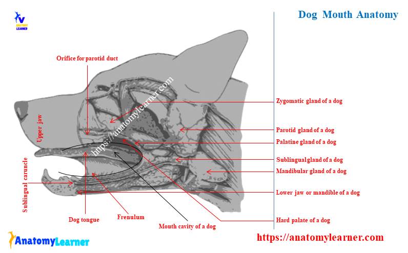
Dog mouth cavity labeled diagram
Now, I will show you almost all structures from the dog mouth anatomy with a labeled diagram. In this labeled diagram, I tried to show you the lips, vestibule, gums, palates, and oral cavity of a dog.
Some structures from the dog mouth may be missed, as I showed in a single labeled diagram. But, you may get a more updated labeled diagram on the dog mouth on social media of anatomy learners.
It will be better to join and search the dog mouth labeled diagram on social media of anatomy learners.
Frequently asked questions on dog mouth anatomy.
In this part, I will try to solve the common inquiries on the dog mouth anatomy. But, it is suggested to read the full article from starting to end to get all the basic knowledge on dog mouth.
What is the side of a dog’s mouth called?
The side of a dog’s mouth is known as the commussiors. This is where the upper (inferior) and lower lips join together on both sides.
What are the parts of a dog’s mouth?
The parts of a dog’s mouth are lips, cheek, gums, and oral cavity. Again, a dog’s oral cavity divides into the vestibule space and oral cavity proper.
You will find a different structure like the soft palate, hard palate, glands, teeth, tongue, vessels, and nerves in the oral cavity proper.
What are the bumps or spikes on a dog’s lips?
What is the oral cavity of a dog?
The oral cavity includes the vestibule and the oral cavity proper. In this article, I have already described the details of the dog’s oral cavity (both vestibule and oral cavity proper).
Conclusion
I am sure that you got the basic idea of the dog mouth anatomy. Now, you may easily identify the different parts of a dog’s mouth. Of course, you should take help from the dog mouth anatomy labeled diagram.
The outer part of the dog’s mouth is the lip. You got the basic idea of the different parts of the dog lip anatomy. Again, it is very important to know and identify the vestibular space and oral cavity proper from the dog’s mouth. The most important parts of the dog mouth anatomy are the hard and soft palate. I hope you learn the basic point of a dog’s hard and soft palate.