The dog pelvis anatomy includes the hip bones, sacrum, muscles, organs, and other associated structures. It is so difficult to explain the detailed anatomical facts of every single part of the dog pelvis in a single article. But, I will try to summarize the basic structures from the dog pelvis with a labeled diagram.
I will show you the anatomical features of the pelvic bones, muscles, and different important organs from the pelvis region. This article will also help understand the basic difference between the male and female dog pelvis anatomy.
Again, I will enlist some of the important arteries, nerves, and ligaments from the dog pelvis at the end of the article. So, if you are interested to know the anatomical facts of a dog pelvis, please continue this article till the end.
Dog pelvis anatomy
To know the dog pelvis anatomy, you might have a good piece of knowledge on the boundary of the pelvic cavity. Again, you should know how the bony pelvis is formed. The muscles of the dog pelvis are also important as the arteries and nerves pass through them.
You will find different important organs and structures in the pelvic region (varies with the male and female). Fine, let’s get a basic idea of the structures and organs from the dog’s pelvic region.
- The boundary of pelvic inlet and outlet of a dog pelvic cavity
- Formation of the bony pelvis in a dog
- The list of pelvic muscles of a dog
- Most important arteries and nerves of the dog pelvis
- The list of important pelvic organs of a dog
You will get very little information on these structures of the dog pelvis in this section. So, make sure you read the full article to learn the dog pelvis’s details and anatomical facts.
The boundary of the dog pelvic cavity
Fine, let’s try to identify the dog pelvic cavity first with the help of the below-mentioned labeled diagram. I am sure that you have a good piece of knowledge on the bones from the dog skeleton anatomy. These might help you to identify the pelvic region easily.
The pelvic cavity of the dog includes the pelvic inlet and an outlet. It has considerable obstetric importance; it must be large enough to allow for the passage of the young during parturition.
First, let us know how the pelvic inlet is formed. So simple, you will find the base of the sacrum and sacral promontory at the dorsal aspect of the dog pelvic inlet.
Laterally, the dog’s pelvic inlet is bounded by the arcuate lines of the sacrum. The arcuate line is the ventromedial border of the body of the ilium bone. It extends from the articular surface to the iliopubic eminence.
There is also a terminal line which is a circular line that outlines the cranial pelvic aperture. Again, the ventral part of the dog pelvis inlet is bounded by the anterior part of the pubic symphysis.
On the other hand, the dog pelvic outlet is the inferior and lesser aperture of the pelvic cavity. Dorsally, you will find the first coccygeal vertebrae (caudal vertebrae) in the pelvic outlet. The ischiatic tuberosity and sacrotuberous ligaments bound the lateral border of the dog pelvic outlet.
Again, in the ventral aspect, you will find the ischiatic arch and posterior part of the ischial symphysis.
The bony pelvis of a dog
The bony pelvis is the most important part of the dog pelvis anatomy. You will find the ossa coxarum and sacrum bones in the formation of a bony pelvis in a dog. Again, you know the bony pelvis (hip bone) consists of three distinct bones – ilium, ischium, and pubis.
The anatomical features of the dog ossa coxarum are somewhat different compared to that of a ruminant. In contrast, you will also find some variation in the sacrum bone of a dog compared to the ruminant. In the next section of this article, let’s find all the important osteological features of the ossa coxarum and sacrum bone.
The sacral part of the dog’s pelvic canal is approximately as long as its floor. Again, the lateral osseous wall of the pelvic canal is formed by the body of the ilium (most part) and ischium bone. In addition, the floor of the dog pelvis is formed by the sacropelvic surfaces of the rami of the pubis and ischium bones.
Important muscles of dog pelvis
Here I will enlist some of the important pelvic muscles from the dog. But, you will learn the detailed anatomical facts of these pelvis muscles later.
The lateral pelvis muscles of a dog include the followings –
- Tensor fasciae latae muscle of dog pelvis
- The gluteus superficial, medius, and profundus muscles of the dog pelvis, and
- Piriformis muscle of the dog pelvis
Again, the medial pelvic muscles of the dog include obturator internus, obturator externus, Gemelli, and quadratus femoris muscle. In addition, I will describe some other muscles from the dog’s pelvis and pelvic limb.
Arteries and nerves of the dog pelvis
The most important arteries of the dog pelvis include – the internal iliac artery, external iliac artery, external and internal pudendal artery, and deep femoral artery. But, you will find other different arteries and veins in the pelvis region of a dog. If possible, I will show you all these vessels with the labeled diagram in another article.
Again, the most important nerves of the dog pelvis anatomy include the obturator nerve, pudendal nerve, and deep perineal nerve. In addition, there are other different nerves present in the dog pelvis. The formation of the ischiatic nerve of a dog also occurs in the pelvic region. You will find a full guide on the ischiatic nerve formation and courses of these nerves here in anatomy learner.
Organs of the dog pelvis
Can you tell me what are important organs do dog’s pelvis have? Fine, here I will enlist the most clinically important organs or part of the organs from the dog pelvis.
So, the most important organs of the dog pelvis are –
- The urinary bladder of a dog – locates on the floor of the pelvis cavity
- The parts (body) of the uterus (in female dog)
- Other organs and structures related to the male and female dogs (will enlist in the figure)
Now, this is time to learn details of these organs and structures from the dog pelvis.
Dog pelvic bone anatomy
The dog’s pelvic bones include the pelvic girdles, thigh, leg, and hind paw. In the pelvic dog girdle, you will find the ilium, ischium, pubis bones. Again, a dog’s thigh is represented by the femur and associated sesamoid bones of the stifle joint.
The leg of a dog’s pelvic limb consists of the tibia and fibula bones. In contrast, the hind paw includes the tarsal bones, metatarsals, and digits that include the three phalanges in each and sesamoid bones.
But, how the dog pelvis anatomy is formed? The dog bony pelvis is formed by the ossa coxarum (both sides) and the sacrum bone. The ossa coxarum of the dog includes the ilium, ischium, and pubis bones.
You already know that the cranial opening of the bony pelvis is considered a pelvic inlet. It is formed by the cranial end of the sacrum, shaft of the ilium and anterior border of the pubic bone. Again, the pelvic outlet is smaller and formed by the caudal vertebrae above the ischial arch and ischial tuberosity below.
In addition, the posterior borders of the sacrosciatic ligament form the lateral walls of the pelvic outlet. Do you know the name of this enclosure? Fine, this enclosure is known as the perineum.
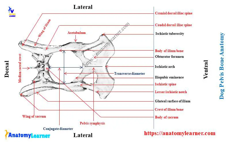
You will find two terms named conjugate and transverse diameter. At the pelvic inlet, the length between the body of the sacrum and the cranial end of the pubic symphysis is known as the conjugate diameter. Again, the distance between the two psoas tubercles is the transverse diameter.
Variation in the conjugate and transverse diameter found in the male and female dog. You will learn these variations of a male and female dog in the next part of the article. First, let’s know some exceptional osteological features of dog’s pelvis bone.
Special features of dog pelvis bones
You will find the following special osteological features in the pelvis bone anatomy of a dog. These osteological features are not enough to learn the dog’s pelvic bones. You might learn the details of each bone of the dog pelvis with a labeled diagram.
- The ilium of the left side and the right side of a dog’s hip bone (bony pelvis) are almost parallel.
- You will find a concave gluteal surface in the dog ilium bone.
- There is no distinct gluteal line in the dog ilium compared to the ruminant.
- The crest of the dog ilium bones is strongly convex.
- There is a twisted ischium bone present in the dog’s bony pelvis.
- The ischial tuberosity is flat, whereas you will find a pointed ischial tuberosity in a small ruminant.
- The superior ischiatic spine is blunt, and the greater and lesser ischiatic notches are shallow compared to the ruminant.
- You will find a wide acetabular notch and deep acetabulum in the dog’s hip bone.
Fine, now you should know the details anatomical facts of ilium, ischium, and pubis bones of the dog pelvis.
But, before going to the description of each bone of the dog pelvis, please try to identify the important osteological features from the below-mentioned labeled diagram.
Okay, now let’s move to the specific description of each bone from the dog pelvis.
The ilium bone anatomy from dog pelvis
The ilium is the most cranial bone in the dog pelvis anatomy. You will find two important parts in the dog ilium bone: a laterally concave wing and an irregular body. The body of the ilium bone expands at the caudal ends and forms the two-fifth of the acetabular cavity.
You will find the iliac crest in the ilium bone that comprises the tuber sacrale and tuber coxae. The iliac crest forms a cranially protruding arc which is thin in its ventral aspect.
“It will be better if you learn the osteological facts of ruminant hip bone first. For that, you may simply go to the osteology section and search for hip bone. This might help you to compare the osteological features of the dog hip with the ruminant.”
Special features of dog ilium bone
The dorsal border of the iliac crest is thicker in the dog. Fine, let’s try to identify the following features from the dog ilium bone labeled diagram –
A cranial and caudal dorsal iliace spine of dog ilium bone (tuber sacrale)
A cranial ventral iliac spine of dog ilium
The tuber coxae and alar spine
The dog ilium’s cranial and caudal dorsal iliac spines are the tuber sacrale. Again, caudal to the tuber sacrale the dorsal border of the ilium body gently concave and forming the greater ischiatic notch. In addition, the dorsal border of the ilium continues with the dorsal border of the ischium as the ischiatic spine.
The gluteal surface of the iliac wing faces laterally and dorsally. Again, the sacropelvic is the medial surface of the iliac wing that articulates with the wing of the sacrum.
You will find an iliac surface in the ilium bone, a square and smooth flat part of the sacropelvic surface. There is an arcuate line present in the ventromedial border of the ilium. This arcuate line extends from the articular surface to the iliopubic eminence.
The ischium bone of the dog pelvis
The ischium bone of the dog pelvis forms the caudal third of the os coxae. It also helps form the acetabulum, obturator foramen, and pelvis symphysis. In a dog ischium bone structure, you will find a body, ramus, and tuberosity.
The body of the ischium of a dog pelvis is the cranial part of the bone that lies lateral to the obturator foramen. You will find a thick dorsal border (faces laterally) and continue with the dorsal border of the ilium bone as an ischiatic spine.
The dorsal border is flattened at the caudal to the ischiatic spine. You will find some shallow groove at the flattened dorsal border of the ischium bone.
The ramus of the ischium bone is medial to the obturator foramen and continuous caudally. Again, the medial border of the ramus of ischium bone forms the ischiatic symphysis. The ischium bone is curved and forms the caudal part of the lateral boundary of the pelvic cavity.
Again, the caudomedial border of the ischium bone forms the deep ischial arch. The angle of the ischial arch may vary in male and female dogs.
The ischiatic tuberosity is the caudolateral part of the ischium bone and lateral to the ischial arch. It is wide and become gradually thicken from the medial to lateral slide.
The ventral aspect of the ischiatic tuberosity gives rise to the largest muscles of the thigh (biceps femoris, semitendinosus, and semimembranosus muscles).
The pubis bone of a dog
The pubis bone of the dog pelvis extends from the ilium and ischium laterally to the symphysis pubis medially. This bone of the dog pelvis is dorsoventrally compressed and curved. The caudal border of the pubis is thick and forms the cranial part of the obturator foramen.
You will find a body and two rami in the anatomy of a dog pubis bone. The body of the dog pubis bone is the central flat triangular part of the bone. This body of the pubis forms the craniomedial border of the obturator foramen. It fuses with the ilium bone and contributes to forming the acetabulum.
You will find two rami in the dog pubis bone – cranial and caudal. The cranial ramus of the pubis bone fuses with the ilium and helps form the acetabulum. An iliopubic eminence is present at the cranial border of the cranial ramus of the dog pubis bone.
The caudal ramus of the dog pubis bone fuses with the opposite side and forms the pubis symphysis. It also helps to form the obturator foramen in the dog’s hip.
The ventral surface of the pubis bone gives origin to the gracilis, abductor, and obturator externus muscles. Again, the dorsal surface gives origin to levator and obturator internus muscles.
You will find the ventral pubic tubercle on the cranioventral surface of the pubic bone (adjacent to the pubic symphysis). A pecten extends from the iliopubic eminence to the pubic symphysis. The ventral tubercle and pecten serve as the attachment for the prepubic tendon.
The sacrum of the dog pelvis
The bodies and processes of the three sacral vertebrae fuse to form the single sacrum in a dog. You will find a large body in the first segment of the sacrum bone. The sacrum of a dog presents two surfaces (dorsal and ventral), a wing, a base, and an apex. This sacrum help to form the roof of the dog pelvis anatomy.
“If you want to learn the details anatomical facts of animal sacral vertebrae, please go to the osteology section and search for sacrum or sacral vertebrae.”
In the dorsal surface of the dog sacrum, you will find some important structures where the median sacral crest is most important. But, how this median sacral crest is formed in dog’s sacrum? It represents the fusion of three spinous processes of the three sacral vertebrae.
You will also find two pairs of dorsal sacral foramina in the dorsal surface of the dog sacrum bone. These dorsal sacral foramina transmit the dorsal division of the sacral spinal nerves and spinal vessels.
There is also an intermediate sacral crest present in the dorsal surface of the sacrum bone. The cranial articular processes of the dog sacrum are large, faces dorsomedially, and articulate with the seventh lumbar vertebrae. Again, the caudal articular processes are small and articulate with the first caudal vertebra.
The pelvic surface of the dog sacrum
The ventral or pelvic surface of the dog sacrum is concave and presents some important structures. Here on the pelvic surface, you will find two transverse lines. Again, there are two pairs of pelvic sacral foramina present in the ventral surface of the sacrum.
You will find a thin lateral sacral crest in the pelvic surface of the sacrum that terminates caudally as a flattened and pointed process. Let’s know a little about the wing and base of the dog sacrum bone.
The wing of the dog sacrum is the enlarged lateral part that possesses a large and rough articular surface. Again, the base of the sacrum faces cranially. The apex of the dog sacrum is the caudal extremity that articulate with the first caudal vertebra.
Male and female dog pelvis anatomy
In the male and female dog pelvis anatomy, you will find the same structures (bones, muscles, vessels, and nerves). Here, I will provide some of the exceptional osteological features of the hip bones that might help you to differentiate the male from the female.
- You will find the more conjugate and transverse diameter in the female os coxae.
- In the female, the bone is more inclined forward.
- The pelvic outlet and the ischial arch are larger in the female pelvis than in males.
- Again, the ischia of both sides join in more wide-angle in females and make the pelvic cavity roomier.
I hope you will identify the male and female pelvis bone practically with the help of the above-mention points. Please, compare every single feature of the male pelvis with a female and note down it for future study.
Dog pelvic limb anatomy
I have a detailed guide on pelvic limb anatomy here in anatomy learner. If you are interested to learn the details anatomical facts of the dog’s pelvic limb, you may read that article. Again, I will summarize the dog’s pelvic limb anatomy with the labeled diagram.
If you have a good piece of knowledge on the dog pelvic limb, you may skip this part. There are pelvic girdles, thing region, leg region, and hind paw or pes present in the pelvic limb. What should you know from these regions? You should know only the bones and their special osteological features.
I have already described the bones of the pelvic girdle (ilium, ischium, and pubis), so I am going to skip that now. Let’s enlist some of the important osteological features from the other bones of the pelvic limb of a dog.
- You will not find any third trochanter and supracondyloid fossa in the dog femur.
- The greater trochanter of the dog femur is at the lower level to that of the head.
- Again, the lesser trochanter of the dog femur is tuberculous.
- The dog’s patella is comparatively longer, and the anterior surface is more convex than the ruminant.
- The tibial crest is very prominent in dogs compared to the goat.
- Again, the fibula is a long and thin bone that extends the full length of the tibia bone.
- There are seven tarsal bones in a dog arranged in three rows.
- You will find five metatarsal bones in a dog where the first one is ill-developed.
So, there is very little information on the dog’s pelvic limb. Again, I request you to learn the detailed anatomical facts of the pelvic limb of a dog from other articles.
Dog pelvis muscles anatomy
The dog pelvis muscles extend between the pelvis and the thigh. In a dog, you will find the two groups of pelvis muscles (lateral and media). Here, I will only show you the dog’s most important pelvis muscles anatomy.
First, let’s know the muscles you will learn from the dog pelvis? Well, the list of the dog pelvis muscles are below –
- The lateral pelvic muscles – include tensor fascia lata, gluteus superficialis, gluteus profundus, gluteus medius, and piriformis muscles.
- The medial or small pelvis muscles – include obturator extrenus, obturator internus, Gemelli, and quadratus femoris muscle.
Okay, let’s try to know some of the important anatomical facts of these pelvis muscles of a dog.
Tensor fascia lata muscle of a dog
The tensor fascia lata muscle of a dog is a triangular shape that attaches to the ilium from the tuber coxae to the alar spine. You will find a good relationship with the Sartorius, medial gluteus, and quadriceps femoris muscles. The gluteus cranial nerve innervates this muscle.
You will find two parts in the tensor fascia latae muscle – cranial and caudal. The cranial part of the tensor fascia latae is more superficial and inserted on the lateral femoral fascia. Again, the caudal part is deep and inserted into the layer of lateral femoral fascia. It also runs deep to the biceps femoris muscle of the dog hip.
What are the main functions of the tensor fascia latae muscle in a dog? This muscle helps to flex the hip, abduct the limb, and extend the stifle joint.
The gluteus superficialis muscle of a dog
You will find three gluteus muscles in the dog pelvis area – superficial, profundus, and medius. The superficial gluteus is the most superficial, small, flat, and rectangular. This muscle extends between the sacrum and first caudal vertebrae proximally.
The gluteus superficialis muscle arises from the gluteal fascia, deep fascia, and tuber sacralae. Again, the thick caudal part of the muscle comes from the lateral part of the sacrum bone and first caudal vertebra.
The gluteus superficial muscle of a dog covers the gluteus medius, piriformis, and sacrotuberous ligaments. Again, the tendon of the superficial gluteus muscle runs over the major trochanter and fuses with the aponeurosis of the tensor fascia latae muscle.
The gluteus caudal nerve innervates the superficial gluteus muscle of the dog. This muscle helps in the extension of the hip joint.
Gluteus profunds of a dog
This is the deepest, broad, and fan-shaped muscle in the gluteus group of a dog. The gluteus medius and piriformis muscles completely cover this muscle.
The gluteus profunds muscle arises from the lateral surface of the body of the ilium bone near the ischiatic spine. It will form a thick tendon that inserts cranially on the trochanteric major. This gluteus profunds muscle is innervated by the gluteus cranalis nerve.
This muscle helps extend the hip joint of a dog. It also has an abduction function for the pelvic limb.
The gluteus medius muscle of a dog
The superficial gluteus muscle covers the gluteus medius muscle. This muscle lies on the gluteal surface of the ilium bone of a dog. The gluteal cranial nerve also innervates it.
The gluteus medius muscle arises from the iliac crest and tuber sacralae. Again, you will find some fibers that come from the sacroiliac ligament, gluteal fascia, and deep fascia. A large part of the muscle lies deep to the gluteal fascia and skin.
Again, the deep part of the gluteus medius arises from the transverse processes of the last sacral and first caudal vertebra. And it is inserted into the trochanter major by a narrow tendon.
The gluteus medius helps extend the hip joint in a dog. Again, this muscle has a medial rotatory function and prevents lateral rotation during weight-bearing.
The piriformis muscle of the dog pelvis
The piriformis muscle of a dog pelvis is completely covered by superficial gluteus muscle. This piriformis lies caudal and medial to the gluteus medius muscle. It arises from the lateral surface of the third sacral and first caudal vertebra.
The tendon of the piriformis muscle inserts the aponeurosis part of the gluteus medius muscle. This muscle also helps extend the hip joint of the dog.
That’s fine; let’s know the medial pelvic muscle of the dog pelvis. The medial pelvic muscle lies caudal to the gluteus profunds muscle and the hip joint. They extend from the inner and outer surface of the ischium to the femur bone.
The obturator internus muscle of a dog
The obturator internus is a large, fan-shaped muscle of the dog pelvis that covers the obturator foramen internally. This muscle arises from the pelvic surface of the rami of ischium and pubis. It also arises from the ischiatic tuberosity and ischiatic arch.
The fibers of the obturator internus muscle pass over the smooth surface of the greater ischiatic notch directly. This muscle lies deep between the edges of the broader Gemelli muscle.
Again, the obturator internus muscle help in lateral rotation of the hip joint and prevent medial rotation while bearing the weight.
The obturator externus of a dog pelvis
The obturator externus is also a fan-shaped muscle in the dog pelvis. It arises from the ventral surface of the pubis and ischium adjacent to the pelvic symphysis. This muscle is separated from the ischial symphysis by the adductor muscle.
The obturator externus muscle covers the obturator foramen externally. Again, the caudal border of this muscle is covered by the quadratus femoris, and the adductor muscle hides the cranial border.
The tendon of the obturator externus muscle insert to the trochanteric fossa. This muscle also helps in the lateral rotation of the hip joint. It also prevents the medial rotation on weight-bearing.
The Gemelli muscle of the dog
The Gemelli is the fusion of two muscle parts in a dog. It lies between the terminal portion of the obturator internus and external muscles.
This muscle arises from the lateral surface of the body of the ischium in the arch ventrally to the lesser ischiatic notch. The tendon of the Gemelli’s muscle inserts in the trochanteric fossa.
The ischiadicus nerve innervates the Gemelli muscle of a dog. This muscle also has the same function as the obturator externus muscle. It helps in lateral rotation of the hip and prevents medial rotation on weight-bearing.
Quadratus femoris muscle of the dog pelvis
The quadratus femoris is a short, fleshy muscle in the dog pelvis anatomy. This muscle arises from the ventral surface of the ischium medial to the lateral angle of the ischiatic tuberosity. The quadratus femoris is surrounded by the adductor, biceps femoris, semitendinosus, semimembranosus, and obturator externus muscles.
The muscle extends in almost sagittal direction cranially, just medial to the biceps femoris muscle. It bends slightly laterally and runs distally to reach the distal part of the trochanteric fossa. The tendon of the quadratus femoris is inserted in the trochanteric crest.
What is the action of the quadratus femoris exhibited in the dog pelvis? Well, it helps extend the hip and perform the lateral rotation of the hip joint.
If you wish to learn the other different muscles from the pelvic region, you may read other articles from anatomy learners.
Arteries of the dog pelvic limb
It is so difficult to explain all the courses of arteries from the pelvic region and pelvic limb of a dog. Please learn the detailed anatomical facts of the pelvic dog region from other articles of anatomy learners. Here, I will show you the most important arteries from the dog pelvic limb with a labeled diagram.
“Please learn all the arteries from the dog pelvis region.”
Okay, let’s know some of the important arteries from the dog pelvis.
External iliac artery of a dog
The external iliac artery is the largest in the pelvic limb. It arises from the lateral surface of the abdominal aorta at the level of the sixth and seventh lumbar vertebrae. This artery runs caudoventraly and relates with the psoas minor and iliopsoas muscles.
You will find different branches in the external iliac artery of the dog. I will enlist the branches of the external iliac artery later.
Each of the external iliac arteries passes downward and backward up to the cranial border of the pubis and continues as the femoral artery. This femoral artery passes through the femoral canal with a saphenous nerve in front and the femoral vein behind.
The same artery continues as the popliteal artery below the stifle joint. Beyond the proximal third of the tibia is continued as the cranial tibial artery. Again, below the tarsus, the same artery continues as the dorsal pedal artery in the dog. Then the same artery will form the metatarsal arteries and common digital arteries.
Now, let’s enlist the branches of the external iliac artery of the dog –
- The deep femoral artery of a dog
- The pudentoepigastric trunk (caudal epigastric, external pudental, and caudal superficial epigastric artery)
- Medial circumflex femoral artery of a dog
- Caudal abdominal artery of a dog
- The femoral artery (superficial circumflex iliac, proximal caudal femoral, middle caudal femoral, distal caudal femoral, descending genicular, and saphenous artery)
- A popliteal artery (caudal tibial and cranial tibial artery)
- Arteries of the hind paw of a dog (dorsal pedal, dorsal digital common arteries, medial and lateral plantar arteries)
Now, I will discuss the most important arteries from the external iliac artery of the dog pelvic limb.
Branches of the dog external iliac artery
The deep femoral artery of the dog pelvis arises from the caudomedial surface of the external iliac artery at an angle of approximately 45 degrees. It runs obliquely distocaudally over the medial surface of the external iliac vein.
The pudendoepigastric trunk is short and extends to the deep abdominal inguinal ring. Again, the caudal epigastric artery arises from the deep femoral artery. The external pudendal artery arises as to the ventral terminal branch of the pudendoepigastric trunk.
You will find a small caudal superficial epigastric artery in males compared to females. Again, the caudal abdominal artery is a small artery that arises from the cranial surface of the external iliac artery just after the deep femoral artery arises.
Femoral artery of the dog pelvis anatomy
This is one of the most important arteries in the dog pelvis anatomy. The femoral artery continues the external iliac artery at the thigh region. You will find many branches of the dog femoral artery.
Here, I will enlist some of the major branches of the dog femoral artery –
- The superficial circumflex femoral artery of the dog
- A lateral circumflex femoral artery of a dog
- The proximal caudal femoral artery
- A saphenous artery of the dog
- The descending genicular artery of a dog
- A middle caudal femoral artery
- The distal caudal femoral artery
Okay, let’s know a little about these branches of the dog femoral arteries.
The superficial circumflex femoral artery is a small branch of the femoral artery. It arises from the lateral surface of the femoral artery close to the lateral circumflex femoral artery. In a dog, the lateral femoral artery is also known as the cranial femoral artery.
You will find the proximal caudal femoral artery at the caudal side of the femoral artery. It crosses the pectineus and adductor muscles. The saphenous artery is very short and arises from the medial surface of the femoral artery. This artery disappears deep into the semimembranosus muscle.
The descending genicular artery arises from the femoral artery distal to the origin of a saphenous artery. Again, the middle caudal femoral artery is lateral to the semimembranosus muscle.
The distal caudal femoral artery arises from the caudolateral surface of the femoral artery.
The popliteal artery continues the femoral artery through the popliteal fossa. You will find two main branches of the popliteal artery – the cranial tibial and the caudal tibial. If you want to know more about the popliteal and hind paw arteries, please read the details guide from the cardiovascular section.
Internal iliac artery of the dog
The dog’s internal iliac artery originates at the level of the seventh lumbar vertebra from the abdominal aorta. You will also find different branches of the internal iliac artery of a dog. The internal iliac artery terminates as the caudal gluteal artery and internal pudendal artery.
I will not show you every single branch of the internal iliac artery of the dog. I will only show you the important major arteries from the internal iliac artery of the dog.
The umbilical artery is one of the major branches of the internal iliac. It supplies the urinary bladder male and female organs.
The internal pudendal artery is another important branch of the internal iliac artery of a dog. This is a small ventral terminal branch of the internal iliac artery. It lies in contact ventrolaterally with its satellite vein. Again, it runs caudally on the terminal tendon of the psoas minor muscle.
Nerves in the dog pelvis
Here, I will discuss the major nerves from the dog pelvis. But, you might read the details on all pelvic nerves (like lumbosacral plexus).
“You will find the details guide on the lumbosacral plexus nerves here in anatomy learner with the labeled diagram.”
Here, you will learn the below-mentioned nerves from the dog pelvis region –
- The pelvic nerve of the dog
- A perennial nerve of a dog
- The pudendal nerve of a dog and
- A caudal cutaneous femoral nerve
The pelvic nerve of the dog is formally known as the pelvic splanchnic nerve. This nerve arises from the ventral branch of the first and second sacral nerves. They may also arise from the pudendal nerve.
You may find the pelvic nerve as a single or two separate nerves that independently run to the pelvic plexus. They supply to the colon, colic flexure, viscera of the pelvic cavity, and urinary bladder.
The pudendal nerve of the dog pelvis arises from the ventral branches of all three sacral nerves through the sacral plexus. This nerve is obliquely caudoventrad to the pelvic outlet. It lies lateral to the coccygeus muscle and appears superficially medial to the superficial gluteus muscle.
You will find the internal pudendal vessel with the internal pudendal nerve.
The caudal cutaneous femoral is nearly large as the pudendal nerve. This caudal cutaneous femoral nerve arises from the first and second sacral nerves. Sometimes, it may also arise from the third sacral nerve.
You will find two branches in the perennial nerve – superficial and deep perineal. The superficial perineal nerve sends several branches to the skin and perineum. Again, the deep perineal nerve has several branches that leave the pudendal nerve at the pelvic outlet.
Dog pelvis labeled diagram
Now, I will provide a dog pelvis diagram to practice what you have learned from this article. You may also get help from the dog pelvis labeled diagram. I tried to show almost every structure and organ from the dog’s pelvis in the diagram.
If you want more labeled diagrams on dog pelvis, you may join with anatomy learners on social media.
Some of the suggested reading from anatomy learners –
Anatomical features of dog abdominal region with labeled diagram
Dog neck anatomy with labeled diagram
Frequently asked questions on dog pelvis anatomy
This is the section where I will discuss the different inquiries of the dog pelvis anatomy learner. I hope this may help you know some information on the pelvic dog region.
Where is the pelvic area of a dog?
The pelvic area is located between the abdominal cavity and the pelvic limb. You will find the pelvic bones, muscles, nerves, and vessels in the dog’s pelvic area structure. I have already described everything about the dog pelvic in this article. If you want, you may read the full article to know the detailed structure of the dog’s pelvic area.
What is a dog’s pelvis called?
The dog pelvis is also called the ossa coxarum or pelvic area. The ossa coxarum is formed by the ilium, ischium, pubis, and sacrum bones. You will find a detailed description of these bones in the first part of this article. So, please move to the first part and read this article to know the features of the dog pelvis bones.
Can a dog recover from a fractured pelvis?
Yeah, a dog can recover from a fractured pelvis if you provide proper management.
Does a dog have pelvises?
Yes, the dog gave two pelvises (both sides) articulated by the pelvic symphysis. In the pelvic symphysis of the dog pelvis, you will find the pubic symphysis and ischiatic symphysis.
Conclusion
The information mentioned earlier might help you get the basic knowledge of the dog pelvis anatomy. As a veterinary student, you might know the basic structure (bones, muscles, nerves, vessels) of the dog pelvis properly. I have planned to provide more labeled diagrams on the dog pelvis in the coming day.
The dog’s pelvic anatomy also includes the anatomical facts of the pelvic limb. So, it is your responsibility to learn the details anatomical facts of the pelvic limb bones. This is also important to differentiate the male and female pelvis practically.