Before studying the animal appendicular skeleton, you might have good knowledge about the parts and structure of a long bone. You will find three major parts (body and extremities) and almost seven major structures in a long bone of an animal. This article will provide detailed information on a long bone’s gross and microscopic features with a labeled diagram.
Again, I will provide the examples and functions of the long bones from different animals like dogs, cats, and ruminants. You will come to know the modified long bones and miniature long bones in different animals.
Structure of a long bone
The long bones of an animal skeleton are typical of elongated cylindrical form with enlarged extremities. They occur in both the thoracic and pelvic limbs, acting as supporting columns and as levers. For studying the structure of a long bone, you need both the longitudinal and transverse section samples.
First, let’s see the humerus of the dog; it is an elongated cylindrical form that possesses two large extremities. This bone occurs in the thoracic limb of the dog skeleton and supports the column and whole body. So, the humerus is the long bone of the dog skeleton.
Thus, a long bone of any animal possesses two (three) important parts –
- First, cylindrical elongated body or shaft – a tubular part that encloses the medullary cavity that contains the bone marrow, and
- Second, two proximal and distal extremities possess articular surfaces, articular cartilages, and others.
Again, the longitudinal section of a long bone shows different structures. First, from the labeled diagram, let’s see the structures found in an animal’s long bone.
Now, I will enlist the structures that you have been seen in the longitudinal section of a long bone –
- Articular cartilage or hyaline cartilage,
- Epiphysis head (proximal and distal),
- The diaphysis of the bone (shaft or body),
- An epiphyseal plate of the long bone,
- Metaphysis (optional),
- Spongy or cancellous part of the long bone,
- Compact part of the long bone,
- Periosteum and endosteum (covering) of the long bone, and
- The medullary cavity of the long bone.
Let’s discuss the features of these structures; as mentioned above, the long bone.
Identifying the long bone structures
I hope the following little information might help you identify the structures from the longitudinal section of a long bone.
Articular cartilage or hyaline cartilage: covers the bones’ extremities, prevents them from rubbing together and absorbs shock.
Epiphysis: the long bone’s head or end parts or extremities that grow separately from the shaft
The diaphysis is the shaft, body, or central part of the animal’s long bone.
The metaphysis is the growing part of the diaphysis adjacent to the epiphyseal cartilage.
Epiphyseal plate: these are the thin layers of cartilage present in between the metaphysis and epiphysis. In this area, the longitudinal growth of the long bone occurs.
The spongy substance is delicate bony plates and spicules that run in various directions and intercross. The interval between the delicates plates occupies by marrow and is known as the marrow spaces.
Compact substance: consists of dense, hard calcified interstitial substance (bone matrix or lamella). The thickness of the compact substance varies greatly in various situations.
Periosteum: the outer surface of the long bone is covered by a membrane except at the articular cartilage. This is the periosteum of the long bone.
Endosteum: a thin fibrous membrane that invests the medullary cavity of the long bone.
Bone marrow and cavity: central cavity of the bone shaft covers with compact substances and contains red or yellow marrow.
Again, you should learn the microscopic features of a long bone (transverse section). The compact and spongy bones show two different microscopic figures under the light compound microscope. Let’s see the special microscopic structures in compact and spongy parts of a long bone.
Special features of compact and spongy substances
So, the compact part of the long bone is a dense and hard substance located at the external aspect of the bone. This compact part also surrounds the spongy substances of the long bone. You will see a Haversian system (bony tissue) arranged in a definite pattern.
This Haversina system of the compact part of the long bone consists of –
- Several concentric lamellae,
- Minute lacunae that provide accommodation to osteocytes,
- Canaliculi – very minute canals radiate from lacunae,
- The interstitial lamellae – irregular bony deposits,
- Haversian canals (numerous narrow canals),
- Transverse Volkmann’s canals, and
- Periosteum and endosteum of the bone.
The spongy part of the long bone structure is also known as the cancellous bone. You will find a large number of small bony plates or trabeculae. They unite irregularly, enclose the small marrow spaces between them, and form a rigid structure of the spongy substances.
The bony trabeculae of the spongy substances arrange along the line of maximal internal stress so that the bone can resist any stress. You will not find any Haversian system in the structure of the spongy substances.
Generally, the spongy substances of the long bone remain at their extremities (proximally and distally). You will also find this spongy substance in the small and irregular bones of the animal skeleton.
Example of long bones
Let’s see the example of the long bones from the dog’s appendicular skeleton. You know the appendicular skeleton of a dog consists of the bones of both thoracic and pelvic limbs. Fine, if you see the skeleton of a dog, you will find the following long bones from the thoracic and pelvic limbs –
- The humerus of the dog,
- Radius and ulna bones of the dog, and
- Tibia and fibula bones of the dog.
Again, you will find some miniature long bones in the thoracic and pelvic limbs of the dog skeleton. If you don’t know the term miniature long bones, then this information is for you – these are the small long bones in the appendicular skeleton of some species like dogs, cats, rabbits, and others.
These miniature long bones also possess the same features as the long bone. You will find the cylindrical shaft or body and two expanded extremities (proximal and distal). The same structural features in the miniature long bone are also present in the normal long bones.
You will see the following miniature long bones in the skeleton of the dog –
- Metacarpal bones of the dog’s thoracic limb, and
- Metatarsal bones of the dog’s pelvic limb.
Again, you will find some modified long bones in the skeleton of a dog. These modified long bones possess an elongated structure, but they do not possess any medullary cavity.
The best example of the modified long bone is the clavicle of the dog, rabbit, and bird.
The modified long bone diagram shows the two clavicles joining ventrally and forming the furculum.
Are ribs long or flat bone?
What about the ribs? Are they long or flat bones in the animal? In most vertebrates, ribs are the long curved bone as they possess almost all the features of the long bone. The dog ribs are considered to the long curved bone. But, in some species, like a monkey or goats, the ribs are flat bones.
If you see a dog’s rib, you will find the cylindrical and elongated body or shaft. Again, there are two extremities present in the rib of a dog. The proximal extremity of the dog rib is the vertebral end that possesses the head and tubercle.
Again, the distal end of the dog ribs is the sternal end that possesses the articular surface for the cartilage. You may know details about the dog ribs from another article by an anatomy learner.
Dog rib cage anatomy with a labeled diagram
Long bones of ruminant
You will find a little difference in the example of the long bone in the ruminant compared to the dog. Here the metacarpal and metatarsal of the ruminant skeleton (cow, goat, and others) are under the main long bones.
So, you will find the following list of the long bones from the ruminant appendicular skeleton –
- The humerus of ruminant,
- Radius and ulna bones of the ruminant,
- Metacarpal bones of the ruminant,
- Femur bone of the ruminant,
- Tibial and fibula bones of the ruminant, and
- Metatarsal bones of the ruminant.
Again, the ribs of the ruminant are the modified curved long bones. You will not find any miniature long bones in the appendicular or axial skeleton of the ruminant.
In addition, you will see some ill-developed modified long bones in the appendicular skeleton of the ruminant. You will find ill-developed small metacarpal and small metatarsal bones in the appendicular skeleton of the ruminant and horse.
Parts of a long bone description
You already got the basic idea of the different parts and structure of a long bone of an animal. But, in this section, I will try to describe all the parts (two or three) from a long bone with examples and labeled diagrams.
The dog humerus shows a slightly sigmoid-shaped long body and two expanded extremities (proximal and distal). You will find the head, neck, greater tubercle, lesser tubercle, intertubercular groove, teres major tuberosity, and teres minor tuberosity in the proximal expanded part of the dog humerus.
Again, the distal proximity shows important features like trochlea, capitulum, radial fossa, epicondyles, and supratrochlear foramen. In addition, the distal extremity of the dog humerus shows lateral supracondylar fossa and crest.
So, in this long bone from the dog appendicular skeleton, you find the long shaft and two extremities.
Again, the ulna is a separated, ill-developed long bone (compared to the ruminant) with a long shaft and two extremities. At the proximal extremity of the dog ulna, you will se the olecranon process, olecranon tuber, anconeal process, coronoid process, trochlear notch, and ulnar tuberosity.
The distal extremity of the dog ulna comprises a styloid process.
The dog femur is the heaviest long bone in the appendicular skeleton that possesses a long cylindrical body and two expanded extremities. You see, the body of the dog femur is straight proximally and arched cranially at the distal part. There are four different surfaces in the dog femur bone – cranial, caudal, lateral, and medial.
It is very hard to demark the cranial, lateral, and medial surface of the shaft of the dog femur. But, the caudal surface is flatter and comprises rough lines and medial and lateral lips.
A miniature long bone of the dog
You already know that the metacarpals and metatarsals are the miniature long bones in the appendicular skeleton of a dog. Because these metacarpals and metatarsals possess the ideal features of a long bone, the size is small.
There are five metacarpal bones in the dog – metacarpal I to V. In all metacarpal bones, you will see the cylindrical-shaped body and two extremities. The proximal extremity of these metacarpals is the base, and the distal extremity of these bones is the apex.
Metacarpals II to V is the main metacarpal bones in the dog’s thoracic limb. The metacarpal II and V are shorter, and III and IV are four-sided structures.
Again, you will find the five metatarsal bones in the pelvic limb of the dog skeleton. Metatarsal I is atypical and possesses a small cylindrical body. You will find a similar structure in the metatarsal bones II, III, IV, and V. Both metatarsal bones of the dog’s pelvic limb comprise a base, a body, and an apex.
Parts of the modified long bone of an animal
The clavicle is not articulated with the skeleton of the dog. This bone locates at the tendinous part of the brachiocephalic muscle. Again, the medial part of the clavicle attaches to the sternal fascia by the distinct ligamentous bands.
This clavicle of the dog is thin and slightly concave both in longitudinally and transversely. The clavicle is more closely joined with the clavicular tendon between the cleidocephalicus and cleidobrachialis muscles.
The best example of the modified long bone is the bird clavicles. These are the thin, rod-shaped bones in the skeleton of a bird. They attach ventrally and form special structures (furculum).
Gross structure of a long bone description
In the structure of a long bone, you might know the details of epiphysis, diaphysis, articular cartilage, compact substance, spongy substance, medullary cavity, and others. Now, I will show you the details features of these structures from the long bone of an animal.
There are two main important substances in the long bone – compact and spongy substances. The distribution of these compact and spongy substances varies from bone to bone. In the long bone, you will find a large elongated cavity (known as the marrow cavity).
Both the compact and spongy bone encloses this elongated cavity of the long bone. There is bone marrow (red or yellow) present in this medullary cavity.
You will see the bone trabeculae and plenty of small spaces in the spongy bone feature. These small spaces of the spongy bone also remain occupied by the bone marrow.
Articular cartilage of a long bone
The two expanded ends of a long bone possess articular cartilage. Generally, most of the articular cartilage is hyaline. So, they cover the articular surface of the long bones and provide a smooth surface for the synovial joint.
You may learn the structure of a synovial joint from the below-mentioned article –
- The synovial joint structure of an animal with the labeled diagram
This hyaline cartilage on the articular surface of a long bone normally does not ossify. And this cartilage receives nutrition from synovial fluid and surrounding vessels.
Do you know the main function of a long bone’s articular cartilage? The hyaline articular cartilage provides a frictionless environment for the two adjacent bones during the movement.
If you see the histology slide of this hyaline cartilage, you will find around or elongated cells that arrange singly or in a group. Again, the ground substance shows transparent or granular appearances.
The shaft of a long bone
You know the diaphysis is the shaft of a long bone. The length and diameter of the shaft may vary from bone to bone and even the species to species.
Grossly, the shaft structure of a long bone shows a periosteum, dense, compact bone, little spongy bone, endosteum, medullary cavity with bone marrow, and different vessels. I will describe all these osteological features of the shaft separately.
The main component of a bone shaft is hard and dense compact bone. You will find a great variation in the thickness of the compact substance in different long bones. This compact bone of the shaft encloses the medullary cavity, filled with the bone marrow (red or yellow).
Generally, four different surfaces are present in the shaft of a long bone – cranial, caudal, lateral, and medial. These four surfaces of the shaft possess various osteological features in different bones.
Again, you will find four borders (which may vary in different long bones) in the shaft of a long bone. But, there is no angle formed in the long bone of an animal.
Now, I will point out some important features from the shaft of various long bones with the diagrams.
A shaft of the humerus bone
The shaft of the animal humerus is irregular cylindrical, and twisted. You will find a smooth and spirally curved lateral surface in the shaft of a humerus bone that forms the musculospiral groove. There is a deltoid tuberosity at the lateral surface of the humerus where the deltoid muscle inserts.
Again, the medial surface of the animal humerus is nearly straight in its length and rounded from side to side. You will see a teres major tuberosity at the proximal end of the middle surface of the humerus. Again, you may also see some nutrients foramina on the medial surface of the humerus bone.
The cranial surface of the shaft is irregular, wide and smooth proximally, narrow and roughened distally. Here, the cranial surface and lateral surface form the cranial border, known as the cranial border. This cranial border of the humerus is also the crest of the humerus.
In the crest of the goat, dog, and horse humerus, you will see two important structures – deltoid tuberosity and teres minor tuberosity.
In addition, the caudal surface of the shaft of the humerus is smooth and rounded from side to side.
The shaft of the radius bone
Do the radius of a dog, horse, and goat possess the same surfaces and borders? No, the radius of a dog, horse, and goat possesses two defined surfaces and two borders.
The shaft of the radius is curved in its length and flattened craniocaudally; thus, it forms cranial and caudal surfaces. You will find a slightly convex, smooth cranial surface rounded from side to side.
Again, the caudal surface of the shaft of a radius bone is concave in length and flattened in the transverse direction. You will see two interosseous spaces (proximal and distal interosseous spaces) in the whole length of the radius shaft.
In addition, the shaft of the radius bone possesses two borders – medial and lateral. The medial border of the shaft is slightly concave, whereas the lateral border is more strongly curved.
The shaft of the metacarpal
The shaft of the metacarpal bone shows two surfaces and two borders. Here, you will see the dorsal and palmar surfaces and lateral and medial borders.
The dorsal border of the metacarpal shaft is convex and smooth. It shows a verticle or longitudinal groove and two foramina (proximal and distal). Again, the palmar surface of the metacarpal shaft also shows a longitudinal or verticle groove with two foramina (proximal and distal).
The lateral border of the shaft forms when the dorsal and palmar surfaces meet at the lateral aspect. Again, the medial border of the metacarpal shaft form when the dorsal and palmar surfaces meet at the medial aspect. Both the lateral and medial borders of the metacarpal shaft are smooth.
A shaft of a femur bone
The shaft of a dog, horse and goat is cylindrical but flattened caudally and in two extremities. You will see four different surfaces (cranial, caudal, medial, and lateral) in the shaft of the femur bone. So, you will also find the four borders in the shaft of an animal femur bone.
There is a lesser trochanter at the proximal part of the medial border of the shaft of the femur bone. Again, the lateral border of the shaft possesses two important features – supracondyloid fossa and lateral supracondyloid tuberosity.
A shaft of the tibia bone
The shaft of the tibia bone is large, three-sided proximally, and small and flattened distally. So, you will see three defined surfaces (medial, lateral, and caudal) and three borders in the shaft of the tibia bone.
You will find the identifying features at the lateral surface of the tibial shaft. It is spiral and smooth in appearance. But, the medial surface of the tibia shaft is rough, whereas the caudal surface is flattened and possesses popliteal lines. You will also find nutrient foramina at the caudal surface of the shaft of the tibia bone.
The three surfaces of the shaft form the three borders in the tibia – cranial, medial, and lateral. You will see a tibia crest at the cranial border of the shaft with some tibial tuberosities. The lateral border of the shaft shows a concave feature.
Structure of periosteum of the long bone
The periosteum is a membrane that invests the outer surface of the bone, except where it covers the articular cartilage. In the structure of the periosteum of a long bone, you will find specialized connective tissue. Again, the periosteum comprises two distinct layers – the outer protective fibrous layer and the inner cellular osteogenic layer.
You will see the well-developed osteogenic layer during the active growth. This layer comprises osteoblast cells that help in ossification. The thickness of the outer fibrous layer varies with different locations.
Again, the adhesion of the periosteum to the bone also differ greatly in various location. Generally, you will find the thin periosteum in the area where it covers the muscular tissue. The degree of vascularity shows the more or fewer activities of the periosteum.
Where will you not get the periosteum? You will not find periosteum on the following surfaces –
- Articular cartilage that covers the bone, and
- The outer surface of the sesamoid bones.
You will find three major functions of the periosteum of a long bone –
- As a cover, it protects the long bones,
- It helps the growth and repair of the long bone, and
- Periosteum helps in the nutrient supply to the outer part of the long bone.
I hope you can understand the features of the periosteum of a long bone. Now, you should know the features of the endosteum of the long bone.
Endosteum of a long bone
The structure of the endosteum is similar to the periosteum of the long bone but is thinner. This endosteum lines largely the medullary cavity of the long bone. Again, you will find the lining of the endosteum in the various small marrow spaces, which also contain numerous blood vessels.
As the structure of the endosteum is similar to the periosteum, you will also find two layers – fibrous and cellular layer. Again, the cellular layer of the endosteum comprises the osteoblast cells that help in the ossification process.
If an emergency-like fracture of the bone occurs, the osteogenic layers of both the endosteum and periosteum provide osteoblasts that help repair the bone. But, sometimes, you may find the over-repairing in the injured area with a very poor quality bone.
Thus, there may develop a bony bulge at the site of injury. This osseous bulge of the bone is known as the exostoses.
Structure of medullary cavity of a long bone
The shaft of a long bone consists of a medullary or marrow cavity along its length, which is enclosed by compact substances. In the structure of a medullary cavity of a long bone, you will see the soft and pulpy tissues. These soft and pulpy tissues of the medullary cavity are the bone marrow.
Again, you will also find different small marrow spaces containing the bone marrow. There are two types of bone marrow in the medullary cavity and the small marrow spaces of the long bone –
- Yellow bone marrow (medullar ossium Flava), and
- Redbone marrow (medulla ossium rubra).
The composition of the bone marrow of a long bone may vary with age and species. In the young animal, you will find only the red marrow, but later this red marrow replaces in the medullary cavities by the yellow bone marrow.
The red marrow of the medullary cavities comprises several cells and blood-forming substances. In contrast, you will find the originally fatty tissues in the yellow marrow of the medullary cavity and small marrow spaces of the long bone.
The yellow marrow of the bone is formed by the regressive changes in the red marrow. It causes fatty infiltration and degeneration of the various types of cells. Within the age, bone marrow undergoes gelatinous degeneration and forms the gelatinous marrows.
Red marrow of the bone
What are the structural components in the red marrow of animal bone? You will find the reticular connective tissue, various blood cells at different stages of development, lymphoid cells, adipose cells, and blood vessels.
Again, you will also find the megakaryocytes or giant cells in the structure of the red marrow of the animal. Redbone marrow persists in the sternum throughout the life of an animal.
Again, you will also find the red bone marrow in the ribs, skull, vertebrae, cancellous part of some short bones, and ends of the long bones. The marrow of the cranial bone may degenerate in older animals.
Yellow bone marrow
So, where will you find the yellow bone marrow in an animal? The medullary cavity of the long bone diaphysis and the hollow interior part of the bone comprise the yellow marrow.
In the yellow bone marrow composition, you will see the connective tissue, fat cells, blood vessels, and some mature and immature red blood cells. But, the most important component of the yellow marrow is the fat and stem cells. These steam cells of the yellow marrow become fat, cartilage, and cells of the bone.
Compact substances of the long bone
The compact substance is the hard and dense substance that remains at the external aspect of the long bone and surrounds the spongy substances. You will find a variation in the thickness of the compact substances in the different portions of a long bone.
Generally, this compact substance of the long bone is thicker in the shaft and thin in the extremities (proximal and distal). Again, you will find the greatest uniform thickness in the circumference of the bone.
The maximum thickness of the compact substances may find in the shaft of the femur and humerus bones of the animal. But, sometimes, the thickness may increase due to an increase in the tension of muscles and ligaments.
In the structure of a compact substance of a long bone, you will find a bony tissue that arranges in a defined pattern and form the osteons or Haversian system. Again, the Haversian system of the long bone consists of Haversian canals, lamellae, lacunae, canaliculi, interstitial lamellae, and Volkman’s canal.
In the next part, I will describe all these structures from the compact substance (microscopic features of a long bone). So, you may jump to that portion and know the details features of the compact substance.
The spongy substance of the bone
In the structure of a spongy substance of a bone, you will see delicate bony plates and spicules. These bony plates and spicules run in various directions and intercrosses. This arrangement of the spongy substance is due to the mechanical requirements of the bone.
You will find the interval between the bony plate filling with the bone marrow. This interval of these bony plates is the marrow spaces.
You know the spongy substance makes the bulk of the short, irregular bone and the extremities of the long bones. But, you may find a little extension of the spongy substances into the shaft of a long bone.
Some of the bone contains air spaces within the compact substances instead of spongy substances and marrow. These are the pneumatic bone of the animals’ skeleton; the best example of the pneumatic bone is the femur of a bird.
So, all the air spaces or cavities of the compact substances are the sinuses and lines with the mucous membrane. They communicate with the external air and nasal cavity directly or indirectly.
Sometimes you may not find any spongy substance in between the compact substance of the long or other bones. In that case, the compact bone may be thin and transparent.
In the microscopic structure of the spongy substance, you will not find any Haversian system in the matrix. Only bony trabeculae and small bony spicules with bone marrow are visible under the light compound microscope.
I have already described the histological features of the spongy and compact bones here in anatomy learner. You may know the details features of the spongy and compact substances from these articles.
Histological features of the spongy substances of the animal bone with the labeled diagrams
Blood vessels and nerves of the long bone
The long and other bones of the animal skeleton are supplied with rich blood vessels and nerves. You will find two sets of arteries in the bone – periosteal and medullary. The periosteal artery of the bone gives small branches that enter the minute opening of Volkman’s canals on the surface.
Again, these small branches of periosteal arteries reach the Haversian canal of the compact substances of the long bone. You will also find other different small branches of the arteries that enter into the extremities of a long bone. These small branches of the arteries supply the spongy substances and the marrow cavities.
In addition, some medullary or nutrient arteries enter into the nutrient foramen and pass into the canals through the compact substances. Do you know where these nutrient arteries supply? These nutrient arteries give branches into the medullary cavities and supply the marrow.
Some other small branches of the periosteal artery also supply to the shaft of the long bone. You will also find metaphyseal and epiphyseal arteries in the long bone that arises from the articular arteries of the joint. These arteries supply the spongy substance and the marrow at the ends of the long bone.
Some lymph vessels exist as a perivascular channel in the periosteum of the long bone and the Haversian canal of the compact substance. They also form the fine subperitoneal network from which the larger vessels produce.
Nerves of the bone
The nerve fibers accompany the blood vessels of the long bone. You will find the vasomotor nerve fibers in the long bone. But, most of the nerve fibers of the long bones are sensory. The sensory nerve fibers have a crucial role in remodeling the bone and maintaining bone mass.
Both the central and sympathetic fibers from the peripheral nervous system are involved in regulating the bone remodeling and maintenance of the bone mass.
Sensory nerves of the long bone carry impulses that may result in pain. Again, the sympathetic nerve fibers play an important role in reflex vasomotor response in the marrow cavity of the long bone.
In addition, some different peripheral nerves and vessels run over the different long bones of the animal skeleton. For example, the femoral artery of an animal passes along the medial aspect of the femur bone. Again, the superficial branch of the cranial tibial artery runs over the cranial surface of the tibia bone.
The axillary artery and its branches run along the medial aspect of the femur (long bone of animals). You may learn about these arteries, veins, and nerves from the specific articles of anatomy learners.
The microscopic structure of a long bone
In the microscopic structure of a long bone, you need to know the features of compact and spongy substances. I have described all the microscopic features of the compact and spongy substances from the bone in anatomy learner. Again, I will focus on the main important microscopic features of the compact substances from the long bone of an animal.
So, what is the compact substance in a long bone? I hope you already know this term; this is a hard and dense substance that covers the external surface of the long bone. The thickness may vary, and you will find a dense and smooth appearance on the joint surface.
First, let’s know what the structures that I will describe here under the microscopic features of the compact substance are –
In the transverse section of the compact substances, you will find different Haversian systems or osteon, circumferential or interstitial lamellae, endosteum, and periosteum.
Again, the Haversian system of the compact substance includes –
- Numerous concentric lamellae – possess minutes spaces (lacunae) that accommodate the osteocytes in their canaliculi,
- Haversian canals in the osteon – longitudinal passages through the compact substance, and
- The Volkman’s canals – transverse canals among the concentric lamellae or osteon.
Now, let’s discuss these structures from the compact substances of the bone.
Lamellae and Haversian canals
Under the microscope, you will see the compact bone comprised largely of the calcified interstitial substance of the bone matrix that arranges in different layers. These calcified bone matrices are the lamellae of the compact bone.
You will find some small cavities throughout the interstitial substance of the lamellae. These small cavities are the lacunae of the compact substance. Again, during these minutes, cavities of the lamellae fill with the osteocytes.
The microscopic features of the compact bone also show that some small, slender tubular passages radiate in all directions from the lacunae. They penetrate the interstitial substances of the lamellae of the compact substance. These are the canaliculi of the compact bone structure.
So, the branching from the canaliculi anastomoses with the adjacent lacunae and forms a continuous system of cavities or passages. These small cavities or passages are essential for passing the nutrition to the bone cells.
In addition, the cartilage cells receive their nutrition from the gel-like hyaline matrix. The calcium salt deposits on the bone’s interstitial substance and reduces the permeability. Thus, the bone (compact substances) needs the vascular system or channel for nutrition.
There are numerous longitudinal channels in the center of the concentric lamellae of the compact substances. These channels are the Haversian canals of the compact substance.
The other name of the Haversian canals is the nutrient canals. They possess one or two blood vessels and connect. You will also find the communication of the nutrient canals with the marrow cavity and with the surface through the transverse channel (Volkman’s canals).
So, the blood vessels from the endosteum and periosteum communicate to the Haversian system through the Volkmann canals.
Volkman canals of the compact substance
So, I hope you can understand what Volkman’s canals are. These transverse passages communicate the two or more osteon or Haversian systems.
You have already seen that the nutrient canals (Haversian canals) communicate with the marrow cavities through these transverse passages (Volkman’s canals).
How is the Haversian system formed in the bone?
The numerous lamellae of the compact substance arrange concentrically around the longitudinal vascular channels. These cylindrical units of the structure around the longitudinal vascular channels are the Haversian systems.
So, in the structure of a Haversian system of a long bone, you will find the lamellae, lacunae, canaliculi, osteocytes, Haversian canal, and Volkman’s canal.
The size and the numbers of the Haversian systems of a compact substance may vary in the different long bones of the skeleton.
Now, let’s see how the Haversian system is formed? The bony lamellae deposit at the outer surface of the growing long bone of an animal. Therefore, you will find several longitudinal grooves at the external surface under the periosteum of a long bone.
Again, in this phase, you will find different blood vessels along the floor of the long bone. In addition, the grooves become converted to tunnels (channels) by further deposition of the bone at the ridge.
The channel also contains the osteoblast cells in the blood vessels. Again, the osteoblast of this structure continues to deposit the calcium salts in a concentric manner around the channels.
Thus, more lamellae are formed around the tunnels or channels. Subsequently, the osteoblast proliferates and differentiates into the osteocytes that form more osteoid or matrix. The channel develops and forms the Haversian or nutrient canals. Thus, the Haversian system of the long bone is formed.
Circumferential lamellae of the compact substance
These are also the bony lamellae that enclose the inner and outer surface of the long bone. They are formed by the deposition of layers of new bone under an osteogenic layer of the periosteum.
The circumferential lamellae of the compact substance are not arranged like the lamellae of the Haversian system. But, these circumferential lamellae are held together by perforating fibers.
Again, the circumferential lamellae of the compact substance help to grow the bone shaft in width (transversely). This process of bone growth is appositional growth.
There are two types of ossification in animal bone – intramembranous or mesenchymal ossification and intracartilaginous or endochondral ossification. If you don’t know the term ossification, then this little information is for you – this is the process of bone formation in the skeleton of an animal.
You may learn more about the ossification process (intramembranous and Intra cartilaginous) from another article by an anatomy learner. Again, you will find a short description of different types of bone cells (osteocytes, osteoblasts, and osteoclasts) in that specific article.
How is the long bone formed?
Except for the bones of the cranial cavity and face ossify in the intracartilaginous process. So, you may tell the long bone to ossify by the intracartilaginous or endochondral ossification process.
You will find different stages of intracartilaginous ossification –
- Condensation of the mesenchymal tissue,
- Formation of a cartilage model,
- Primary areolae formation (primary ossification center),
- Secondary areolae formation with marrow cavity,
- True bone formation (compact substance),
- Another part (epiphysis) formation, and
- Epiphysis and diaphysis fusion (complete bone formation)
You will find the details process of endochondral ossification here in another article by an anatomy learner. So, you may visit that article and learn details of every single stage of endochondral ossification. Let’s share the summary of the endochondral ossification with you.
Process of endochondral ossification
The formation of a long bone will also provide a clear structure concept. First, the cartilaginous model is formed by the condensation of mesenchymal tissue. You will also see a perichondrium that surrounds the cartilaginous model.
During this time, cartilage cells (chondrocytes) proliferate at the midsection and arrange in rows towards the ends. They become hypertrophied, release alkaline phosphate, and precipitate calcium salt in the matrix.
Thus, the primary areolae are formed around the calcified materials, the primary ossification center. Here, the subperiosteal collar bone is formed around the primary ossification center, and the perichondrium turns into the periosteum.
The collar bone erodes by the increased activities of the subperiosteal osteoclasts. Here, you will find periosteal buds that contain the osteoblast, osteoclasts, and blood vessels.
The osteoclasts absorb the irregular calcified material and form the secondary areolae. Again, the secondary areolae help to form the medullary cavity in the bony matrix.
The osteoblasts form lamellated bony plates, and thus numerous longitudinal grooves appear on the outer surface of the bone. Small blood vessels appear, and the groove turns into the tunnels.
In this process, the long bone developed longitudinally. Again, the appositional growth also increases the width of the bone.
This is a very short process for long bone ossification. I again request you to read the full guide from the specific article published because you will find the details of every single stage.
Composition of the long bone
The bone is a mineralized connective tissue that consists of inorganic mineral slats and organic matters. You will find the ratio between these two substances is 1:2. The composition of the bone may vary greatly with the age and location of the bone.
You will find the membrane, cells, vessels, nerves, cartilages, fluid, and marrow under the organic matters of the bone. Again, the inorganic mineral salts of the bone include calcium phosphate (86%), calcium carbonate (6%), magnesium phosphate (3%), sodium carbonates, and sodium fluoride (5%).
The function of long bone
The long bone of an animal skeleton provides different functions. Here, I will provide the most important functions of the long bone of an animal skeleton.
- The long bone of the animal skeleton provide support and protect the framework of the body,
- Again, the long bone of the skeleton works as a lever that bears the weight of the body and helps in different types of movement,
- Long bone serves as a storehouse for the calcium and phosphorus, and many other elements in small amounts,
- The long bone of the skeleton serves as a factory for red blood cells and several kinds of white blood cells. Again, in a normal older animal, the long bone store the fats.
So, these are very short descriptions of the functions of the long bone from the animal skeleton.
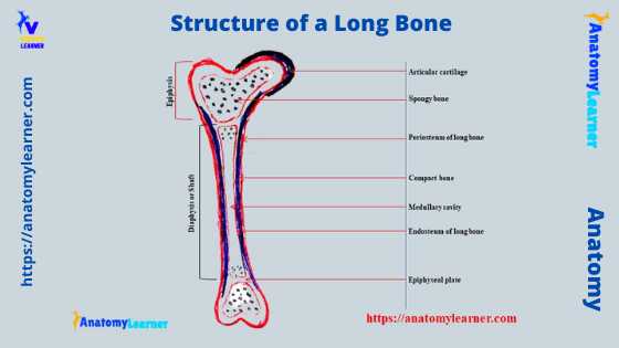
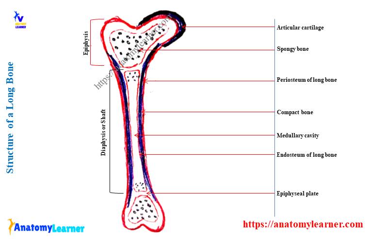
Long bone diagram labeled
I hope you already got the different labeled diagrams on the structure of a long bone throughout the article. Now, I will again provide some important long bone diagram labeled pictures that might help you achieve the best knowledge.
So, what I will complete here in the diagrams –
- Longitudinal section of a long bone (gross features),
- Transverse section of a long bone (gross and histological),
- Longitudinal section of compact substance (histology),
Again, in another diagram, I will try to show you the different types of bone cells and the formation of bone (the process of ossification). Or you may find these labeled diagrams (on bone cells and ossification) here on social media for anatomy learners.
Longitudinal section of the long bone (gross)
The longitudinal section of the long bone shows different features in the below-mentioned diagram. I tried to show you the epiphysis, diaphysis, epiphyseal plate, and metaphysis from the long bone labeled diagram.
Again, the diagram shows the endosteum and periosteum from the compact substance of the long bone. Compact and spongy substances are marked in the labeled diagram from the long bone.
The long bone labeled diagram also shows the medullary cavity that contains the yellow bone marrow. Few small blood vessels are identified in the long bone labeled diagram (shaft).
Transverse section of the long bone – gross
The transverse section of the long bone shows some of the important features. Here, the long bone transverse section labeled diagram shows the periosteum, compact bone, and endosteum.
Again, a central medullary cavity is present in the middle of the long bone labeled diagram. You will see some blood vessels at the outer surface of the compact substance of the long bone labeled diagram.
The transverse section of the long labeled diagram did not show any spongy substances.
Transverse section of compact substance
The transverse section of the compact substance also shows the different histological features of the long bone. Here, I tried to show you the different Haversian systems or osteon in the labeled diagram. The labeled diagram also shows the Haversian canal, several concentric lamellae, lacunae, and canaliculi.
Again, the long bone labeled diagram shows the circumferential lamellae directed in various directions. Volkman’s canals (transverse canals) are present in the labeled diagram.
Now, I will show you the different types of bone cells hosted in the lacunae of the Haversian system of a long bone. I tried to show you all the bone cells in this single labeled diagram – osteocytes, osteoblasts, and osteoclasts.
Other bones of the skeleton
You will also find other different types of bone in the skeleton of an animal. However, I have already described these different types of bone in another article by an anatomy learner. Again, I will provide you with a summary of the other different types of bones from the skeleton of an animal.
So, in the skeleton of an animal, you will find the following other types of bones –
- A short bone of the skeleton,
- The irregular bone of the skeleton,
- Sesamoid bones of the skeleton,
- Flat bones of the skeleton.
Let’s know a little information about these bones from the skeleton of an animal.
The short bone of an animal
The shape of the short bones varies from typical cuboidal to the irregularly compressed rod. You will find the short bone in the carpus and tarsus region of the limbs. They possess six surfaces where one surface is non-articular.
The non-articular surface of the short bone provides an area for ligaments and blood vessels. Example of the short bones – seven carpal of the thoracic limb of an animal.
The short bone comprises the compact substances, periosteum, endosteum, and other structures.
Flat bones of the animal
The flat bones are found in the pelvic girdle, shoulder girdle, and skull. In the structure of a flat bone, you will find two layers of compact substances and intermediate uniting spongy substances. The spongy substance of the flat bone structure is the dipole.
The flat bones of the skull surround and protect the internal organs of the skull. Some of the flat bones of the skull possess air spaces that form the pneumatic bone.
Irregular bones of the animal
These are the irregular shape, small, and unpaired bones in the skeleton of an animal. They remain in the midline of the body in the vertebral column. That means all the vertebrae bones are under the irregular bones.
In the structure of irregular bone, you will also find almost similar structures to a long bone. The periosteum of the irregular bone is thin. Again, the marrow spaces of the vertebral bone possess red bone marrow.
Sesamoid bones of the animal
These are the small bones present near the moving joint of an animal’s body. They are usually formed in the tendons, but they may be developed in the ligamentous tissue over which the tendon passes.
You will find only one articular surface that glides over the surface of the other articular surface of the long bone. The main function of this sesamoid bone is to reduce the friction between the two adjacent bones.
Frequently asked questions in long bone features
So, now I will try to solve the common questions asked by the anatomy learners. But, if you read the previous information in this article, you may skip this part.
What is the structure of a long bone?
Grossly, the long bone comprises the epiphysis, diaphysis or shaft, metaphysis, and epiphyseal plate. Again, you will find two important components in the features of a long bone – one is a compact substance, and another is a spongy substance.
There is a periosteum that surrounds the outer part of the compact substance. The thickness of the periosteum may vary with the location and types of bone. Again, the endosteum lines the inner surface of the compact bone or medullary cavity.
Another most important feature of the long is the medullary cavity which remains enclosed by the compact bone in the center of the long bone. This medullary cavity of the long bone comprises the bone marrow (yellow marrow).
There are also some periosteal and medullary arteries present in the long bone.
What is the structure and function of a long bone?
I have already described every feature of a long bone with the diagram. So, from the diagram, you will find the compact and spongy substance in the component of a long bone. Again, there are endosteum, periosteum, epiphysis, shaft, epiphyseal plate, and blood vessels in the features of a long bone.
The main function of the animal’s long bone is the support the body and protect the framework. Again, they act as a body lever that bears weight and helps in movement. They also store calcium and phosphorous and serve as a red blood cell factory.
What are the three structures of long bones?
Do you want to know the three major structures from the long bone of an animal? Well, there are three major structures in a long bone: the shaft or body or diaphysis of the long bone, and another two are, the extremities.
You will see two different extremities – proximal and distal (an expanded portion of the bone) in a long bone of an animal. You may learn the details facts about the proximal and distal extremities from the different long bones of the animal skeleton from the first section of this article.
What are the 5 major parts of a long bone?
You have already found different structures in a long bone. From these structures, I will enlist the major 5 structures of a long bone –
- Epiphysis or extremity of a long bone,
- Diaphysis or body or shaft of a long bone,
- Endosteum and periosteum of a long bone,
- Compact substances of the long bone, and
- The spongy substance of a long bone.
You will get a details description of the structures mentioned above of the long bone in the first part of this article.
Conclusion
So, all the information on the structure of a long bone might help you learn it perfectly. The compact substance and spongy substance are the two important components in the structure of a long bone of an animal. Again, there are other important structures in a long bone like epiphysis, diaphysis, epiphyseal plate, periosteum, and endosteum.
The compact substance of a long bone shows the most important features – the Haversian system, circumferential lamella, and Volkman’s canals. Now, let’s practice and identify all the gross and microscopic features from the long bone structure labeled diagram.