Hair under a microscope shows a hair follicle and a cylindrical hair shaft. In a hair shaft, you will find columns of keratinized cells organized into three layers – medulla, cortex, and cuticle. Again, the hair follicle is the epidermis structure that develops as down growth of the epidermis into the dermis and possesses different parts.
This article might help you to know the different features of a hair (shaft and follicle) under a light microscope with their concise description. Again, I will try to show you the hairs of different animals like rabbits, cats, and dogs with their specific features.
So, if you want to know the microscopic features of the animal hair shaft and hair follicles, let’s continue this article till the end.
Hair under microscope
So, hair is an epidermal down growth embedded into the dermis or hypodermis of the animal’s skin. You will find two main parts in hair – a cylindrical shaft and a terminal hair follicle.
The cylindrical shaft of the hair under a microscope shows three layers (medulla, cortex, and cuticle) of keratinized cells. Again, the terminal end of the hair follicle shows an expanded hair bulb composed of connective tissue papilla and hair root.
First, let’s see the microscopic image of an animal’s hair and try to identify its following features. All the following microscopic features are well identified in the hair-labeled diagram.
- Epidermis and dermis layer of the skin with hair,
- Different parts of a hair shaft – medulla, cortex, and cuticle,
- Hair root in the epidermis or hypodermis of the animal’s skin,
- Connective tissue layer that surrounds the hair shaft or follicles,
- The inner and outer root layers of the hair,
- Hair bulb with the papilla,
- A sebaceous gland that attaches to the hair shaft,
- Opening of the sebaceous gland that opens into the hair shaft,
- Adipose tissue near the hair bulb.
Again, let’s see the cross-section of the hair follicles and try to identify the following features from the labeled diagram.
The cross-section of the hair follicle shows four main features –
- Hair root – contains medulla, cortex, and cuticle,
- Inner root layer – contains outer cuticle, Huxley’s layer, and Henle’s layer,
- The outer root layer of the hair,
- A glassy membrane, and
- The connective tissue layer surrounds the hair follicle.
I hope you got the basic idea of the different microscopic features of the hair of animals. Now, you may learn the details of microscopic features of every single structure of the animal’s hair.
Hair identifying characteristics in a microscope
So, let’s know the specific identifying features of a hair under a light microscope. Here, I will enlist some important identifying features of hair and hair follicles for transverse and cross-sectioned samples.
- The provided sample tissue shows a cylindrical shaft embedded into the dermis and hypodermis layer of the skin.
- There is an expanded hair bulb (consists of papilla and hair root) at the terminal end of the hair follicle of the provided sample.
- The sample also shows the external root layer and adipose tissue near the hair bulb area.
- Again, the cross-section of the provided sample tissue shows four different structures – hair root, inner and outer root, and a glassy membrane.
- The root of the hair clearly shows the medulla, cortex, and cuticle.
I think these identifying features of the hair shaft and follicle might help you identify the hair at your laboratory with the help of a light microscope.
Summary of microscopic hair
So, hair is made of a hair follicle and hair shaft. There are columns of keratinized cells that organize into three layers in the shaft of a hair. These three layers are – a central medulla, a keratinized cortex, and a thin hard outer cuticle. The outer cuticle is a highly keratinized structure in the hair shaft.
- Medulla – is a thin core of the hair whose cells contain soft keratin,
- Cortex – is the bulk of the hair, composed of highly keratinized cells,
- Cuticles – are composed of highly keratinized cells that overlap each other.
The hair follicle remains in the dermis and contains the following different features –
- The inner and outer root layers of the hair follicles,
- A glassy membrane of the hair follicle,
- Hair bulb and matrix,
- The dermal papilla of the hair.
The outer root layer of the hair follicle is continuous with the epidermis. This layer does not take part in hair formation. You will find a glassy basement membrane that separates this outer root layer from the surrounding connective tissue.
There is an inner root layer that remains inside the outer root layer. This layer contains the softer keratinized cells derived from the cells in the hair matrix. You will also find three layers in the composition of the inner root layer – Huxley’s layer, Henle’s layer, and the outer cuticle layer.
There is no inner root layer at the neck of the hair follicle where the ducts of the sebaceous gland open into the hair follicle. Again, it forms a lumen into which the sebum is delivered.
Hair bulb and dermal papilla
The hair bulb represents the hair matrix, and hair follicles stem cells. These hair matrices and stem cells are responsible for forming the hair. The stem cells proliferate, move upwards, and gradually become keratinized to produce the hair.
Again, the hair matrix stem cells help form the inner and outer root layer of the hair follicle. There is a dermal papilla at the base of the hair bulb and remains in the skin’s dermis.
This dermal papilla provides nutrition (blood supply) to the hair of an animal. A basement membrane separates the dermal papilla from the hair matrix.
There is arrector pili muscle, a small bundle of smooth muscle cells associated with the hair follicles. The contraction of these arrector pili muscle help to raise the hair. Again, it helps to release the sebum from the gland into the duct.
Sebaceous gland features of a hair
Sebaceous are the branched acinar holocrine glands that attach to the hair follicle. You will find different secretory cells in the structure of the sebaceous gland of an animal’s skin.
The cells rupture to secrete an oily secretion into the lumen of the hair follicle. Again, the ruptured cells are continuously replaced by stem cells located at the edges of the glands.
You will learn more about the microscopic features of the sebaceous glands of the hair follicle in the next section of this article.
Microscope features of hair
Hair is present on the skin, covering most of the whole body. But, the length and texture of the hair are different in the different regions of the animal or human body.
The animal with a thick coat of hair helps them warm their body. But, in humans, this function is performed by the subcutaneous fat.
You will quickly identify and differentiate the animal hair from human hair under a light microscope with the help of their cuticle and medulla features. Generally, the animal hair contains a continuous or stacked medulla in its hair shaft.
On the other hand, human hair contains fragmented, interrupted, or continuous medulla in its shaft. Again, there is some significant difference in the scale patterns of the cuticles of human and animal hairs.
You will also find the difference in the hair roots of a human and animals. In a dog, you will often find a spade-shaped hair root, whereas a cat shows frayed at the base of their hair.
But, you need to know the basic structure of the hair first, including different parts, structures of the hair shaft, and follicles.
Parts of a hair
Each hair of an animal consists of two parts – one visible on the body surface and another part anchored in the thickness of the skin. The visible part of the hair is known as the shaft, and the embedded part is the hair root.
There is an expanded lower end known as the bulb in the root. Again, the bulb enters the dermis and forms the papilla hair. A network of blood vessels presents at the hair papilla supplies nutrients to the hair and helps them grow.
The root of each hair is surrounded by a tubular layer called the hair follicle. You will find different layers of cells in the hair follicles that derives from the different layer of skin.
The hair root is always attached to the dermis obliquely; thus, the existing hair of the skin is also oblique and easily lies flat on the skin surface.
Again, the hair shaft comprises the keratin protein that makes hair both strong and flexible. You will find a similar structural pattern in the keratine protein like all other body proteins. So, there is a chain of amino acids in the helical or spiral arrangement in the keratin protein.
There are strong bonds between the amino acids of these helices; thus, these bonds make strong hair.
Structure of hair shaft under a microscope
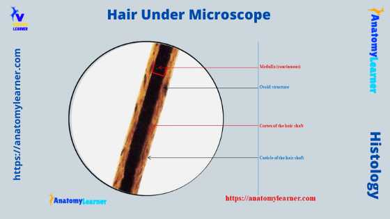
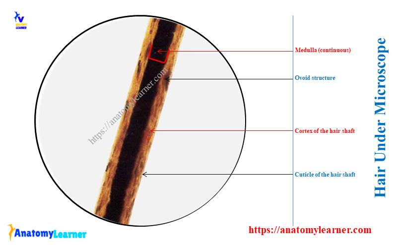
The hair shaft under a microscope shows three different distinct layers – an inner medulla, a cortex, and an outer cuticle. The structure of a pencil may be a good analogy for the structure of a hair shaft.
The outer covering of the pencil is similar to the cuticle of the hair shaft. Again, the wood of the pencil is analogous to the hair shaft’s cortex. In addition, the pencil’s graphite shows a similar structure to the medulla of the hair shaft.
So, the cuticle is a thin external membrane that covers the surface of the hair shaft. You will find flattened cornified cells in the cuticle structure under the microscope. Each of the cells possesses a free edge that overlaps with the next cells.
The cortex of the hair shaft lies next to the cuticle. You know, the cortex is acellular and made up of keratin. Again, the medulla of the hair shaft is the inner layer that runs down the center of the cortex.
The thick hair of animals shows the irregular cornified cells in its medulla. But, the thin hair of animals does not contain the medulla.
The medulla’s cornified element contains melanin responsible for the color of hair. Microscopically, you will find minute air bubbles both in the medulla and the cortex of the hair shaft.
The amount of air bubbles in the cortex or medulla increases with the age of the animal. Again, the loss of pigmentation from the medulla and cortex is responsible for greying of the hair.
Types of the cuticle of the hair shaft
So, the cuticle of the hair shaft is the outermost layer. It is made of scales that overlap and protect the inner layer of hair. These scales point from the proximal end to the distal end of the hair shaft.
If you don’t know the proximal and distal end of the hair shaft, then this is for you –
The proximal end of the hair shaft closes to the scalp, whereas the distal end is farthest from the scalp.
You will find different types of scales in different animal species. But, the basic pattern of the scales are three types –
- Coronal scales pattern,
- Spinous scales pattern, and
- Imbricates scales pattern.
Let’s know a little about these scales’ patterns with diagrams.
Coronal or crown-like scales
You will find a very fine diameter in these crown-like scales compared to the others. These scales resemble a stack of paper cups.
These coronal scales are mainly found in the hair shaft of rodents and bats. But, you may rarely find this type of scale in the hair shaft of humans.
In the diagram, you will see the longitudinal view of the coronal scales of goat hair.
Spinous or petal-like scales
Spinous or petal-like scales of the cuticles are more or less triangular-shaped. These scales frequently protrude from the hair shaft of the animals.
You will find these spinous scales in the hairs of animals like dogs, cats, and money. But, these spinous or petal-like scales are not found in the human hair shaft.
Again, in the labeled diagram, you will see the longitudinal section of the spinous or petal-like scales of the cuticle from animal’s hair.
Imbricate or flatten scales of the cuticle
You will find the overlapping scales with a narrow margin in the imbricate or flattened scales of animals and humans. The diagram shows the imbricate pattern of the cuticle scales from human hair.
You will find these imbricate or flattened scales in humans and animals.
The medulla of a hair under a microscope
The center of hair under a light microscope shows a medulla. It may be a hollow tube (air-filled) or filled with cells. The appearance of the medulla of different types of hair is different. You may find the fragmented, segmented, or continuous medulla in the hair.
Even you may find double medulla in the hairs of some animal species. The medulla contains the pigment granules, though some species have no pigment granules in their hairs.
Under a light microscope, a medulla of an animal’s hair appears as a black or opaque structure. Again, it may appear white or translucent in transmitting lights.
So, if you find the medulla in the hair of an animal or human, you will find mainly three basic types –
- The fragmented medulla of hair,
- A discontinuous medulla of a hair, and
- The continuous medulla of a hair.
But, you may also see the stacked medulla in some of the hairs. Another name for the discontinuous medulla is broken or interrupted medulla.
All these types of medulla of hair are shown on the labeled diagrams.
The medulla of a human hair may be continuous, fragmented, or absent. What about the medulla of the animals? They possess a very regular and well-developed medulla in their hair.
You will find the uniserial, multiserial ladder, vacuolated, and lattice medulla in different animal species. The uniserial and multi serial ladder medulla find in the rabbit hair. Again, the deer possess the lattice type of medulla in their hair.
The cellular or vacuolated medulla are most common in many animal species. All these uniserial, multiserial, cellular or vacuolated, and lattice medulla are shown in the hair labeled diagrams.
What is a medullary index?
The medullary index of a hair is determined by measuring the diameter of the medulla and dividing it by the diameter of the hair. The medullary index for the animal hair is usually greater than 0.5.
Again, the medullary index of humans is generally less than 0.33. So, the medulla of an animal is much larger than it is in humans.
So, you may decide on the base of the medullary index of any hair –
If the medullary index is 0.5 or more, the hair comes from the animals. Again, if a hair’s medullary index is about 0.33 or less, this hair comes from a human.
How to visualize the scales and medulla of hair?
It is very easy to visualize the scales and medulla of a hair shaft. Simply, you should follow a simple procedure and need some instruments.
So, what resources do you need to visualize the scales and medulla of animal hair? Well, you need the following materials –
- A compound microscope for examination purposes,
- The glass slide and coverslip,
- Adhesive material or simple fingernail polish,
- Hair samples from different animals (goat, cattle, dog, and cat) should be collected.
Now, you may follow this simple procedure to visualize the animal’s hairs under the light microscope –
- Collect the hair samples from animals like dogs, cats, sheep, goats, and cattle. You may collect this hair with the help of a comb. Or you may collect the hair from dissected animals with the scalp.
- The hair sample with skin or scalp will be good for visualizing the full structure.
- You should take one glass slide and add some adhesive, or you may paint the center part of the glass slide with nail polish.
- After drying the nail polish, you may place your hair sample on it.
- Now, it’s time to observe the hair sample under the light microscope. Observe the microscope features of cattle, sheep, goat, dog, and cat hairs separately.
So, what have you seen under the light microscope? Write the microscope features of these hairs sample so that you may compare them to each other.
I will show all these microscope figures of different animals’ hair in the labeled diagrams section.
The cortex of a hair
The cortex gives the hair its shape and has two major characteristics – melanin and cortical fusi. You know melanin is the pigment granules that give hair its color. Again, the cortical fusi are air spaces, usually found near the root but may be found throughout the hair shaft.
So, the cortex of a hair shaft is the main body of the hair that consists of elongated and fusiform cells. Again, the cortex contains air spaces which are the cortical fusi, pigment granules, and large oval-shaped structures.
This cortical fusi, pigment granules, and large oval-shaped structures are called ovoid bodies. The cortical fusi is the irregular-shaped air space of varying shape and size.
Observing the hair shaft under a light microscope of an animal or human hair will find this cortical fusi near the root of mature hair. You will also find this cortical fusi throughout the hair shaft of different animals and humans.
The pigment granules are the small, dark, and solid structures in the hair shaft. These pigment granules are more granular and considerably smaller structures than this cortical fusi.
The color, size, and distribution of the pigment granules may vary within a single hair and among the different species of animals. Again, the oval structures are the solid bodies that are spherical to the oval.
You will also find more oval structures in the shaft of cattle or goat hair than in humans.
Structure of hair follicle under a microscope
Under the light microscope, hair follicles may be seen as the epidermis part that enters into the dermis around the hair root. The innermost layer that immediately surrounds the hair root is therefore continuous with the surface of the skin. Again, the outermost layer of the follicle is continuous with the dermis.
The microscopic figure of an animal’s hair follicle shows three distinct layers. From the inner to the outer layer of a hair follicle, you will see the followings –
- An inner root layer is present only in the lower portion of the hair follicles,
- The outer root layer is continuous with the stratum spinosum layer of the epidermis, and
- A connective tissue layer is derived from the dermis layer of the skin.
So, let’s discuss these three different layers of the hair follicle with their microscopic features.
Inner root layer of a hair follicle
It is so difficult to identify the inner and outer root layer of the follicle practically under a light microscope. So, it will be better if you learn these microscopic features with the help of a labeled diagram.
The inner root layer of the hair follicle contains the cuticle, Huxley’s layer, and Henley’s layer. The innermost layer of the hair follicle is the outer cuticle. It lies against the cuticle of the hair and consists of flattened cornified cells.
Again, the microscopic figure of the inner root layer shows one to three layers of flattened nucleated cells. These flattened nucleated cells from the Huxley’s layer or the stratum epitheliale granuloferum. You will find the large eosinophilic granules in the cytoplasm of these flattened nucleated cells of the Huxley’s layer.
Another name for the eosinophilic granules of flattened cells is trichohyaline granules.
Again, the outer layer of the inner root shows a single layer of cuboidal cells with flattened nuclei. This layer is the Henle’s layer or the stratum epitheliale pallidum.
Outer root layer of a hair follicle
The outer layer of the hair follicle starts from the cuboidal cells of the outer layer and continues with the stratum spinosum of the skin. In this outer layer of the hair follicle, you will find the round and nucleated cells.
At the lower end of the hair root, these rounded nucleated cells become continuous with the hair bulb. The cells found in the hair bulb also continue with the stratum spinosum of the skin. So, the cells of the lower end of the hair root and hair bulbs possess the germinative matrix.
Sometimes, a microscopic view shows the great mitotic activities of the hair root and bulb cells. After producing the new cells, they pass superficially and undergo keratinization. Thus, it helps to form the various layer of the hair shaft.
The outer cell layers also give rise to the inner root layer cells. Again, the outermost cell layers of the outer layer and the lowest layer of cells of the hair bulb represent the basal cell layers of the epidermis.
The microscopic figure of the hair follicle shows the basal lamina that separates the outer root layer from the connective tissue layer. This basal lamina is the glassy membrane of the hair follicle structure. The glassy membrane of the hair follicle structure is strongly eosinophilic.
Connective tissue layer of a hair follicle
The microscopic features of the connective tissue layer of the hair follicle are similar to the normal connective tissue. This connective tissue of the hair follicle is made up of tissue continuous with the dermis of the skin.
You will find numerous blood vessels and numerous connective tissue fibers in the connective tissue of the hair follicle. These numerous vessels and fibers form the basket-like network around the lower end of the hair follicle.
Other features of hair follicles under a microscope
Hair under a light microscope also shows the sebaceous gland and arrector pili muscles closely associated with the hair follicles. The sebaceous glands normally open into each hair follicle near its upper end. Again, the arrector pili muscle passes obliquely from the lower part of the hair follicle towards the junction of the epidermis and dermis layers.
- Microscopic features of sebaceous glands, and
- Microscopic features of arrector pili muscle of the hair follicle.
Fine, let’s know some of the important microscopic features of these two structures (sebaceous gland and arrector pili muscle) from the skin histology slide.
Sebaceous gland features
So, the sebaceous glands located in the dermis are closely associated with the hair follicles. Each sebaceous gland consists of numerous alveoli connected to a broad duct. Again, this sebaceous gland duct opens into the hair follicle near its upper end.
The alveolus of a sebaceous gland is pear-shaped and consists of a solid mass of polyhedral cells. You will find the small cells in the sebaceous alveoli at its outermost part. Again, the inner cells of the sebaceous alveolus are larger, rounded, and filled up with lipid.
The lipid of these inner cells is discharged by the disintegration of the innermost cells that are replaced by the proliferation of the outer cells. You will find the oily secretion from the sebaceous gland of any animal that helps to keep the skin and hair soft.
Do you know what the contents of sebum are? Well, sebum consists of various lipids, including triglycerides, cholesterol, cholesterol ester, and fatty acid. The secretion of the sebaceous gland also prevents the dryness of the skin and makes it resistant to moisture.
Sometimes sebaceous gland occurs independently in the hair follicle and directly opens on the skin surface.
Features of arrector pili muscle
The arrector pili muscle is the band of smooth muscle fibers that attach to the dermis and connective tissue layer of the hair follicle. It passes obliquely from the lower part of the hair follicle to the junction of the dermis and epidermis.
Again, the arrector pili muscle lies on the side of the hair follicle and forms an angle with the skin surface. The hair follicle becomes almost verticle relative to the skin surface of any animal. Again, the skin surface overlying the attachment of the arrector pili muscle becomes depressed while the surrounding area becomes raised.
The contraction of the arrector pili muscle pressed upon the sebaceous gland helps them secret within the hair follicle.
Hairs from different body areas
The hair may be straight, curly, and kinly in different animal species. So, in a cross-section of hair, you will see round (straight hair), oval (curly), and crescent-shaped (kinly).
You may determine the hair from the different parts of the animal’s body. The length, shape, size, color, stiffness, curliness, and microscopic features might help you to identify the hairs from the different body areas.
So, hairs from the different body areas are –
- Head hair of any animal,
- Pubic hair of animals,
- Limb hairs (arm or leg hairs),
- Beard of any animals,
- Chest and axillary hairs of any animal.
Head hairs of any animals
These are the long with medium-shaped diameter hair in animals. But, the diameter of this head hair may vary in different animal species. The medulla of the head hair may be absent or continuous and relatively narrow.
But, you will find a relatively wider and continuous medulla in cattle head hair. You may also find the head hair with the cut or split tips. Again, the texture of these head hair is soft or pliable. Sometimes you may see some mechanically damaged hair on the head of some animal.
Features of other hairs of animal body
Let’s see some of the common and important features of the hairs from other different parts of an animal’s body.
Pubic hairs: the diameter of the pubic hair is generally coarse and wiry in appearance. They show a considerable variation in the diameter. You will find a relatively broad medulla that is continuous or sometimes discontinuous.
The root of the pubic hair possesses follicular tags. Again, the tip of the pubic hair shows rounded or abraded features.
Limb hairs: you will find a little variation in the diameter of the limb hairs. The limb hair of different animals is arclike in shape in gross appearance. Again, the medulla of the limb hair is birad, discontinuous with a granular appearance.
Beard: the diameter of the beard is very coarse with an irregular or triangular cross-sectional shape. The medulla is very broad and continuous in the bread of animals.
Chest hair: the shaft of the diameter is moderate but may vary in different species. The tip of the chest hair is long, fine, and arclike.
Hair under microscope labeled
Let’s see some of the hair under microscope labeled diagrams that might help you understand every single feature. Here, I will show you the different labeled diagrams of the hair shaft and follicles.
The first diagram of the hair shows the full structure of the skin. Here, it shows the different epidermis layers and dermis of animal skin with a hair shaft and follicle.
Let’s see the hair shaft; externally, a cuticle covers the hair with a thin membrane. You will easily understand the cuticle layer of hair in the next diagram.
Again, the diagram shows the cortex layer just deep into the cuticle layer. The medulla of the hair is the core, which is also shown in this diagram. In addition, you will understand the medulla feature clearly from the next labeled diagram.
Here, the outer root, inner root, connective tissue layer, hair bulb, and papilla are also identified in the labeled diagram.
Now, let’s see the second diagram (schematic presentation), where the different layers of the hair follicle are seen. This diagram shows the hair root that contains the cuticle, cortex, and medulla. It also shows the Huxley’s layer and Henle’s layer that form the inner root layer of the hair follicle.
Again, this diagram shows different layers of rounded nucleated cells that form the outer root layer of the hair follicle. You will see a glassy membrane in the diagram that separates the inner root layer from the outer root layer.
In addition, the hair labeled diagram shows a highly vascularized connective tissue layer that surrounds the outer root layer of the hair follicle.
Get more microscope hair-labeled diagrams on social media of anatomy learners.
Cattle hair microscopic view
You will find a great variation in the length and color of most of the hair specimens in different domestic animals. The diameter of the cattle hair is more coarse than those of human hair.
You will find a great variation in the medulla of cattle hair. The medulla may be absent or continuous, amorphous or vacuolated with a narrow or broad width.
Again, the cuticle scales are imbricated and with no protrusion from the hair shaft. You will find numerous ovoid structures with pigment granules in the hair shaft of cattle.
The root of the hair follicle is elongated in cattle, and the medullary structure is continuous into the root area. Again, you will find some follicular tissue in the root of the cattle’s hair.
I tried to show you the different parts of the cattle’s hair shaft in the diagram.
Cat hair under a microscope
So, how will you see a cat hair under a light microscope? The diameter of the cat hair is fine, but you may find a little variation in different species of cats. You will find a uniserial ladder continuous medulla in the cat’s hair that occasionally vacuolated.
The scales of the cat’s hair cuticles are spinous and very prominent. But the root of the cat hair is elongated and does not possess any distinct shape.
The base of the root of a cat’s hair shows a frayed fibrils appearance. In the labeled diagram of the cat’s hair, I tried to show some of the important features from the hair shaft and follicle.
Microscope feature of horse hair
The diameter of the horse hair shaft is very coarse. There is a great variation in the medulla of a hose hair shaft. The medulla may be absent in some hair; it may be continuous, cellular, or amorphous.
You will find the imbricate scales in the cuticle of the horse hair without protrusion from the hair shaft. There are fine pigment granules distributed evenly in the medulla of the horse hair. But, you will not find any oval structure in the medulla of the horsehair.
There is a bulb-shaped root found in a horse’s hair. The diagram of horses’ hair shows different important features from the shaft and follicle.
Dog hair under a microscope
I have also examined the dog hair under a light microscope. How will you identify the dog hair from other species? Fine, I will provide some important identifying features of a dog’s hair with its microscopic figure.
The diameter of the hair shaft of a dog varies from fine to coarse. But, it is coarser than those of the cat hair. You will find a great variation in the diameter of the short hairs from different regions of a dog’s body.
The medulla of the dog hair is generally continuous, but it may be vacuolated to amorphous and occasionally very broad. Scales of the dog hair are not generally prominent. The pigment granules are occasionally coarse and distributed into the root of the hair.
I tried to show you all the important features from the shaft and the follicles in the dog hair labeled diagram.
Rabbit hair under a microscope
The rabbit hair is extremely used in felted fabrics, gloves linings, fur trim, coats, and others. You will find the fine diameter in the hair shaft of a rabbit.
The medulla of the rabbit hair shaft is a multiserial ladder and shows a ribbon-like appearance. You will find numerous pigment granules and oval structures in the hair of a rabbit.
Hairs of other different animals
The medulla of the bear hair is continuous and amorphous. Numerous pigments are coarse granular and fairly distributed in bear hair. The scales of the bear hair cuticles are imbricated.
The diameter of the goat hair shaft is coarse. You will find the unbroken lattice medulla in the goat hair. Again, the scales of the goat hair are imbricating.
The fur animal shows very fine to medium diameter, whereas the domestic animals show medium diameter in their hair shaft. Again, the medulla of the fur animal is generally serial or vacuolated, whereas the domestic animal shows the amorphous medulla.
Frequently asked questions on microscope hair.
Let’s discuss some of the frequently asked questions on microscope hair. If you learn the previous section of the hair perfectly, you may skip this part.
How can you tell if the hair is under a microscope?
The hair shows a shaft and a follicle under a microscope. Again, the hair shaft shows three important features – cuticle, cortex, and medulla- that I have already described with the labeled diagrams.
The hair follicle also shows distinct layers like – inner, outer root layers, a glassy membrane, and a connective tissue layer.
So, when you find these characteristic features under a light microscope, you may easily tell them hairs. Again, you may identify the hair based on specific identifying features in different animals.
What does healthy hair look like under a microscope?
The healthy hair of any animal consists of a hair shaft and hair follicle. Again, the shaft of healthy hair comprises of cuticle, cortex, and medulla. The microscopic figure of the healthy hair shows overlaying scales.
Again, the cortex of the healthy hair consists of keratin and pigment granules. There are also air sacs (cortical fusi) in the cortex of healthy hair.
There are three scales in different types of healthy hair of animals – coronal, spinous, and imbricate. The healthy hair of animals also shows the continuous, discontinuous, fragmented, and stacked medulla.
What microscope can see hair?
The compound microscope can see the hair easily. I have already described how to visualize the hairs under a light microscope easily.
Conclusion
So, the hair under a light microscope shows two important features – hair shaft and hair follicle. The important features of the hair shaft are the cuticle, cortex, and medulla, which you might identify with the help of a light microscope. Again, the hair follicle shows four different important layers – inner root, outer root, a glassy membrane, and a well-vascularized connective tissue layer.
Hairs from different animals show specific characteristic features under the light microscope. I hope you already got these specific characteristics of hairs from different animals and can identify them under a light microscope.