The dog uterus anatomy consists of three main parts – horns, body, and cervix. You will find a significant variation in both external and internal features of a dog’s uterus compared to the cows.
As a veterinarian, I have studied the anatomical features of a dog uterus with actual live samples. Thus, I may help you get the proper dog uterus knowledge with a labeled diagram.
Quick overview of dog uterus: Female dogs have a V-shaped uterus with a cervix, a short body, and two long horns. Each of the horns cranially continues with the uterine tube and is internally devoided of cotyledons.
I will identify and describe the unique features of 3 parts of the dog uterus. You will also know the details of ligaments, vessels, and nerves from the dog uterus with a diagram.
Finally, I will help you to differentiate the gravid dog uterus from the normal female dog uterus. Again, you will find enough information to differentiate various animals’ uterus from dogs.
So, let’s get into the article to know the detailed anatomical facts of the dog uterus with a diagram.
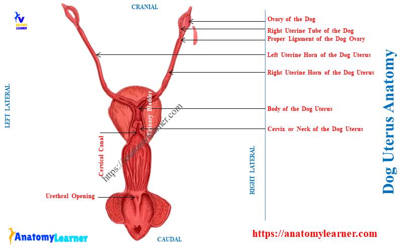
Dog uterus anatomy
The dog uterus is the thick-walled, hollow muscular organ. It serves to conduct spermatozoa to the uterine tube for the fertilization of the oocytes.
The dog uterus extends from the sublumbar region to the pelvic region. First, I would like to show the essential anatomical features of the dog uterus.
Let’s see and try to identify the below-mentioned features from the dog uterus labeled diagram –
- Three parts of the uterus – neck, body, and horns,
- Broad ligament of the dog’s uterus,
- Round ligament and mesometrium,
- Uterine tubes and ovaries attached to the dog uterus,
- Proper and suspensory ligaments of the ovary,
- Internal uterine velum,
- Cervical canal in the canine uterus,
- Internal and external orifices of the cervical canal and
- Ovarian and uterine arteries,
The labeled diagram identifies all the above-mentioned anatomical features of the dog uterus.
Now, you may easily point out the unique features of dog uterus structure. Here, in Table 1, I tried to show the key features of the dog uterus anatomy –
| Dog uterus anatomy | Key features |
| Shape of dog uterus | Hollow muscular Y-shaped organ |
| Location of the uterus | Sublumbar region to pelvic cavity |
| Parts of dog uterus | 3 parts – cervix, body, horns |
| Horns of the uterus | Longer and V-shaped in a dog |
| Body of the uterus | Short and have uterine velum internally |
| Cervix of the dog uterus | Possess cervical canal Have external and internal orifices |
| Ligaments of the uterus | Broad ligament Round ligament of the dog uterus |
| Vessels of the dog uterus | Uterine arteries and veins |
| Innervation of the canine uterus | Right and left hypogastric nerves Pelvic nerves |
Where is a dogs uterus?
The dogs uterus is located almost in the abdominal cavity and slightly in the pelvic cavity. It lies between the urinary bladder below and parts of the large intestine above.
This organ is also attached to the sublumbar region and to the lateral wall of the pelvic and abdominal cavity. Here, the parts of dog uterus are connected with these cavities by the broad ligament.
You might have deep knowledge of the boundary of the body cavities (including pelvic and abdominal) and their content. The below-mentioned article might help you to learn the details about the dog’s body cavities –
- Body cavities and organs with the labeled diagram – the major and minor body cavities of the animals,
The broad ligament of the dog uterus is formed by the double folds of the peritoneum. Structurally, this ligament consists of connective tissue and muscle fibers.
There are a few modifications of this broad ligament of the uterus. I will provide a detailed guide of the broad ligament in the dog uterus ligament section.
Summary of the dog’s uterus location: Most of the part of the uterus remains in the abdominal cavity, and less part in the pelvic cavity. The parts of the large intestine will be found above, whereas the urinary bladder remains below the dog uterus.
Suggested article for you to understand the location of the uterus between the urinary bladder and parts of the intestine –
- Dog urinary bladder anatomy – location and structure with diagram and
- How long is a dogs small intestine?
What does a dog uterus look like?
The whole structure of a dog uterus looks like Y –shaped. It possesses a short body and two very long horns. These two horns of the dog’s uterus look like V-shaped structures.
From the body to the uterine tube, each horn of the dog’s uterus shows the same diameter. Thus, the horn will not tapper like the horn of the cow’s uterus.
You will also find almost similarly shaped horns in the cat’s uterus. But you may also see long horns in other different female animals.
Let’s see the appearance of the animal’s uterus with their long or short horns from Table 2 –
| Animal’s uterus | Shape of uterus | Shape of horns |
| Dog | Y –shaped | Long horns V –shaped |
| Cat | Roughly Y-shaped | Long horns V or U – shaped |
| Cow | Hollow muscular organ | Longer and curved |
| Horse | Hollow | Short and blunt |
| Pig | Y-shaped Long tubular flexuous tube | Long flexuous horns Have U-shaped colis |
| Rabbit | Cylindrical tube | Longer, slender Slightly flexuous tube |
“The horns of the dog uterus look like typical V-shaped; but, the cat have roughly U-shaped uterine horns.”
What are the parts of dog uterus?
The dog uterus has 3 distinct parts –
- The cervix or neck of the dog uterus – is the caudal part of the uterus that has a thick wall,
- Body of the uterus – it is the very small part of the dog uterus that connects with the right and left uterine horns and
- Two uterine horns – consist of right and left longer and straight horns that cranially continue with a uterine tube,
These 3 parts of the uterus are not only found in dogs but also found in all other female animals. You will only find the differences in the size and appearance of these 3 parts of the uterus.
I will discuss all the variations of these 3 parts of the uterus in different animals. But first, let’s know the detailed anatomical facts of these three parts from the dog uterus.
The cervix of the dog uterus anatomy
The cervix of the dog uterus anatomy is about 3 – 4 centimeters long. It is the caudal part of the dog’s uterus that cranially attaches to the body part.
However, there is no clear demarcation between the neck (cervix) and the body of the dog’s uterus. The wall of the cervix is thick in the dog’s uterus.
The cavity of the cervix is narrow and known as the cervical canal. I would like to show you some unique features from this cervical canal.
Here, I tried to show some spiral mucosal folds and plugs of mucus from the cervical canal. These folds and plugs of mucus form the cervical rings in the dog uterus.
This cervical canal represents two orifices – external and internal uterine orifices. The internal uterine orifice of the cervical canal faces almost directly dorsally. Again, the external uterine orifice faces ventrally toward the caudal part after the uterus (shown in the diagram).
You will see numerous muscular folds on that surface where the external uterine orifice projects.
“Another name of the dog’s cervical canal is birth canal.”
The length of the cervical canal of the dogs varies. The uterus of a medium-sized dog possesses an average 0.5 – 1 centimeter-long cervical canal.
Again, the number of mucosal plugs is unique for the various animals. The small ruminants possess more mucosal plugs compared to the dogs.
How do you differentiate the cervix from the body of a dog’s uterus?
Both external and internal appearance help you to differentiate the cervix from the body of a dog uterus. Externally, the cervix is hard in conformity due to the presence of internal mucosal plugs.
But, the small body part is not so hard as the cervix. Again, you will see the constricted external orifice (caudally) in the cervix. The opening of the body towards the horns is larger and has no constriction.
The internal appearance will confirm the parts of the body and cervix of the dog uterus. Internally, the cervix of the dog uterus shows the narrow cavity or cervical canal.
You may easily confirm it through the presence of mucosal folds and plugs within the canal. However, there are no folds or plugs in the internal structure of the body part of the dog’s uterus.
Body of the canine uterus anatomy
The body of the dog’s uterus is located both in the abdominal and pelvic cavities. Typically, the largest part of the body remains in the abdominal cavity.
In multiparous dogs, the entire body of the uterus may remain cranial to the brim of the pelvic cavity.
The body of the dog uterus extends from the area of convergence of the uterine horns to the cervix. Thus, you may say the following about the body of a dog’s uterus –
- Cranially, the body of the uterus bifurcates to form two uterine horns and
- Caudally, this body continues with the cervix,
External features and attachment of the body of the dog uterus
The body of the dog uterus is cylindrical and short compared to the cervix and horns. But, the body appears to be longer in the fresh condition.
This is due to common peritoneal covering on the caudal part of the horns before their union. The body is dorsoventrally flattened and attached laterally with the broad ligament.
Again, the wall of the body of a dog’s uterus is thicker than the horns. You will find various organs that are directly related to the body of a dog’s uterus.
Let’s see the relationship of the various organs with the body of the canine uterus –
- The dorsal part of the uterus body – is related to the colon and other parts of the large intestine and
- The ventral part of the body – is related to the bladder and a few parts of the small intestine,
How many uterine horns does a dog have?
A dog has two uterine horns (right and left) in its uterus. Both the right and left uterine horns of the dog uterus anatomy are usually of the same size.
Each horn of the dog uterus is a cylindrical and spiral muscular tube and possesses two extremities –
- Cranial extremity, and
- The caudal extremity of the uterine horn,
The cranial extremity of the horns receives the uterine tube at the tubouterine junction. Again, the uterine tube connects with the respective ovaries by the proper ovarian ligaments.
Let’s know the details of anatomical features of the dog ovaries (right and left) from the below-mentioned article –
The caudal extremity of these two horns shows a slightly larger diameter than the cranial extremity. Both caudal extremities of the right and left uterine horns join together to form the body of the dog uterus.
The average length of the uterine horns of the dog uterus varies from 10 – 15 centimeters. Both horns are located in the abdominal cavity, starting below the sublumbar region.
The dorsal surface of the uterine horn is slightly concave and related to the sublumbar muscles. Again, the ventral surface of the dog’s uterine horn is convex and free.
Why does a dog have a longer uterine horn?
A dog has longer uterine horns compared to others due to adaptation for litter bearing. These longer uterine horns of the dog uterus carry several developing young.
But, the horse has relatively short horns and a longer body due to the adaption of litter within this body. Let’s see the animals that possess longer and shorter uterine horns from Table 3 –
| Uterine horns | Species | Unique features |
| Long uterine horns | Dog Pig Cow Goat Sheep | V –shaped U –shaped colis A curved, have caruncles Curved and colis Curved and colis |
| Short uterine horns | Horse | Possess blunt ends |
Dog uterus anatomy ligaments
I tried to show you the broad ligament that attaches the uterus and ovaries to the body walls. But, this broad ligament of the dog uterus primarily divides into three portions –
- The mesometrium part of the ligament,
- Mesovarium part, and
- The mesosalpinx part of the broad ligament,
Here, the mesometrium is the larger part of the broad ligament in the dog uterus. It attaches the uterus to the dorsolateral body wall.
The mesovarium is that part of the broad ligament that connects with the respective ovaries of the dogs. Finally, the mesosalpinx is the part of the broad ligament that attaches to the two uterine tubes.
The mesometerium of the broad ligament begins on the transverse plane through the cranial end of the uterine horn. This part of the ligament extends caudally up to the caudal part of the cervix.
The mesometrium attaches peripherally to the lateral wall of the pelvic cavity. You will find a triangular-shaped double-layer peritoneum in the medial surface of uterine tubes.
Two uterine horns are connected with each other with this triangular double-layer peritoneum. This triangular structure is known as the genital folds.
The mesometrium and lateral ligament of the urinary bladder are fused at their attachment. And you know their attachment has occurred in the lateral wall of the pelvic cavity.
What is a round ligament of the dog’s uterus?
The broad ligament gives a fold to the uterus on either side. These folds extend up to the internal inguinal ring and are known as the round ligament of the dog’s uterus.
So, the round ligament of the dog uterus attaches to the cranial tip of the ipsilateral uterine horns. These are actually the caudal continuation from the proper ligament of the ovaries.
These two round ligaments of the dog uterus are the remnant of embryonic gubernaculum. They consist of large smooth muscles and connective tissue. The large amount of smooth muscles allows them to stretch during pregnancy.
The round ligaments run in the free edge of the peritoneal fold. It has given off from the lateral surface of the mesometrium.
This structure extends caudally, ventrally, and medially towards the deep inguinal rings. Thus, it coursed through the inguinal canal and is innervated by the genitofemoral nerves.
You will also find the external pudendal artery and veins supplying the round ligaments.
Structure of the dog’s uterus
The wall of the dog’s uterus consists of three main layers –
- Outer serous coat – perimetrium,
- Middle muscular coat – myometrium, and
- Inner mucous coat – endometrium,
These three layers of the dog uterine wall are well-visible under the light microscope. Let’s learn the unique features of 3 layers of dog uterine wall from the below-mentioned article –
- Uterus histology – histological features of the endometrium, myometrium, and perimetrium with the diagram,
Let’s see how to grossly identify these three layers from the dog’s uterus.
The perimeterium is the outermost coat of the dog’s uterus. It derives from the peritoneum and covers the whole organ except the lateral borders. Microscopically, you will find a layer of simple squamous epithelium on the perimetrium of the dog uterus.
Unique features of myometrium of the dog uterus
The myometrium is the middle and thickest coat of the dog uterine wall. It consists of thin outer longitudinal and thick inner circular muscle layers.
You will also find numerous vessels, nerves, and circular and oblique muscles within the circular layer. The circular muscle layer is thick at the cervix region of the uterus.
The thick circular muscles at the cervix act as the sphincter. Again, these layers of muscles of the uterus help in different conditions.
A strong circular contraction and longitudinal shortening advance the fetus. Again, the transverse circular contraction helps to move the fetus from the uterus.
Finally, the contraction of the abdominal and other associated muscles caudal the final expulsion of the fetus. Most of the dog uterine vessels remify in the muscular layer of the uterus.
What are the unique features of the endometrium of a dog uterus?
The endometrium of the dog’s uterus is thick compared to the other animals. This layer of the dog uterus consists of surface epithelium and lamina propria.
The luminal surface of the endometrium is lined with the low cuboidal epithelium cells. But, you will find a periodic change in the lining epithelium of the endometrium.
There are simple branched tubular glands in the lamina propria of the endometrium. But, you will not see these endometrial glands grossly from the dog uterus.
Grossly, you will see the reddish color in the mucosal surface of the dog uterus. You will also find smooth or low longitudinal ridges on the endometrial surface.
The endometrium of the dog’s cervical canal does not contain relatively high mucosal folds. You will not find any caruncle or cotyledons on the endometrium of the dog’s uterus.
Vessels of the dog uterus
The dog’s uterus is supplied with the ovarian and uterine arteries. These two arteries arise from the aorta of the dogs.
The uterine branch of the ovarian artery is anastomosed with the uterine artery. Finally, they enter the mesometrium of the dog’s uterus at the level of the cervix.
The artery lies relatively close to the body of the dog’s uterus while entering the broad ligament. It diverges from the uterine horn until it approaches the cranial extremity of the horns.
The dog uterine artery remains in the wall of the dog uterus and in the mesometrium. These branches supply both sides of the dog’s uterine horns.
You will also find the ovarian and uterine veins in the dog uterus anatomy. These veins of the dog uterus also follow a similar course to the arteries, except for their termination.
The right ovarian vein empties into the caudal vena cava at the level of the right ovary. Again, the left ovarian vein enters into the left renal vein.
Both the dog’s ovarian veins are tortuous in their course between the peritoneal layer of the broad ligament. The pressure in the dog’s uterine veins is higher than in the femoral veins.
Innervation to the dog’s uterus
The dog’s uterus receives both sympathetic and parasympathetic innervation through the pelvic plexus. Here, the sympathetic innervation reaches the pelvic plexus through the right and left hypogastric nerves.
The parasympathetic innervation reaches the pelvic plexus through various pelvic nerves. Again, the dog’s uterus receives visceral afferent fibers through the pelvic nerves and plexus.
The internal pudendal and genitofemoral nerves also innervate the various parts of the dog’s uterus.
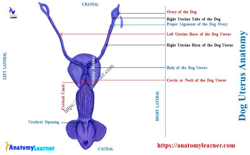
Dog uterus anatomy diagram
First, I would like to show the actual location of the dog’s uterus within the abdominal and pelvic cavities. The labeled diagram identifies the extension of the dog’s uterus from the sublumbar to the pelvic regions.
I have also identified the main three parts of the canine uterus from the labeled diagram. The diagram shows the longer muscular tube-like uterine horns.
The uterine horns continue with the uterine tube and ovaries. The right and left ovaries from the dog uterus are also identified in the diagram.
The diagram also shows the extension of the broad ligament with their modifications. Again, the specific portion of the mesometrium, mesosalpinx, and mesovarium is also identified from the broad ligament.
The surface and topographic location of the dog’s uterus are also shown in the diagram. Here, you will find the organs that are dorsally and ventrally related to the body and cervix of the uterus.
Now, let’s see the internal structure of the dog’s uterus with the labeled diagrams. A unique feature of the dog uterus is found in the cervix region.
Here, the diagram shows the cervical canal, mucosal plugs, and orifices from the cervix of the dog’s uterus. The internal surface of the dog’s uterus also shows the mucosal folds from the body and horns.
Let’s find more labeled diagrams on dog uterus structure here.
Differentiate dog uterus from cow, horse, and goat
By their external appearance, you may easily differentiate the dog uterus from the cow, horse, goat, pig, and rabbit. But you might know the basic structure of a dog’s uterus with its location.
Let’s see the differentiating features of the dog uterus from the cow, horse, pig, and goat from Table 4 –
| Uterus of animals | Neck of the uterus | Body of the uterus | Horns of the uterus |
| Dog | Short Narrow cervical canal Few mucosal plugs | Short body | V –shaped Longer horns No caruncles |
| Cow | Long cervix Spiral cervical canal More mucosal plugs | Short body | Curved caudally Tapered at ends Have caruncles |
| Horse | Straight cervical canal Less mucosal folds | Longer and wider | Short horns Devoid of cotyledons |
| Pig | Long elongated Have round prominence | Smaller body | Long tubular flexuous horns Have numerous U-shaped colis |
| Rabbit | Double cervical opening | Very short | Curved, narrow horns Slightly flexuous and thick walled Have longitudinal folds with nodules |
You may easily differentiate the dog’s uterus from others from the information mentioned above.
Dog uterus vs cow uterus
The cow uterus is the thick-walled, hollow muscular organ of 3 parts. You will find similarities in the basic structure of the cow’s uterus, like the dog’s.
The cow possesses long horns and a short body like the dog and pig. However, the appearance of the horns of the cow uterus is different.
The end extremity of the cow uterine horn is coiled up like the ram’s horns. Again, these horns of the cow’s uterus are parallel to each other.
The right and left uterine horns of the cow uterus bind together by intercornual ligament. This ligament provides the false impression of a long uterine body in a cow.
The most unique feature of the cow uterus is the presence of caruncles. These are the small fleshy masses present on the internal surface of the uterine horns.
The caruncles are regularly spaced, circular to ovoid, internal, specialized thickening of the cow’s endometrium. It makes up the maternal component of the placenta.
Again, the fetal component is the cotyledon. Together, these caruncles and cotyledons make up a placentome in the cows.
The cow has a very long cervix compared to the dog cervix. It is characterized by the transverse folds that interdigitate with each other to occlude effectively.
These transverse folds make the cervical plugs and make the canal torturous.
Unique features of cow uterus compared to the dog uterus
- The neck of the cow uterus is longer than the dog uterus,
- You will find a small body in the cow uterus (but grossly, it seems to be larger),
- The two horns of the cow’s uterus are longer like the dog’s, but the appearance is different,
- The cervical canal is more torturous and longer compared to the dog’s cervical canal,
- You will find more transverse folds in the cow uterus than in the dog uterus,
- The cow uterus has the caruncles and cotyledons in its horns,
So, these might help you to differentiate the dog uterus from the cow uterus.
Dog uterus anatomy vs horse uterus
I will help you to differentiate the dog uterus anatomy from the horse uterus. The horse has short horns and a longer body, so you will quickly determine their uterus.
Two horns of the horse’s uterus are short of equal length to its uterine body. You will not find any cotyledon or caruncles in the internal structure of the horse’s horns.
Again, the appearance of the horse’s uterine horns is not as V-shaped as the dogs. This might be a good differentiating point between the dog and horse uterus.
The end part of the horse’s uterine horns is blunt but not blunt in the dogs. Again, the diameter of the horse’s uterine horns is more than the dog’s.
The horse has a simple cervix compared to the cows and dogs. You will find the straight cervical canal in the horse uterus than the dogs.
The body of the horse’s uterus is longer and wider than those of the cows and dogs. This is another important differentiating feature of the dog uterus from the horse uterus.
Frequently asked questions on the dog uterus anatomy
Here, I will enlist the frequently asked questions on the dog uterus structure. Here, you will get the concise answer to these questions related to the canine uterus anatomy.
But you might go through the whole article to know the details of the dog’s uterus. Let’s see the commonly asked questions on dogs and other uteruses.
Do dogs have two uteruses?
Dogs have only one uterus with a single neck, body, and two uterine horns. But, different animals like giraffes and rabbits have double cervical openings.
Most domestic mammals have only a single external opening of the cervical canal. You will find two external cervical orifices in the rabbit uterus.
The horns of the rabbit uterus are curved, narrow, and slightly flexus tube-like. You will find 3 – 4 longitudinal folds of the mucosa in the uterine horns of the rabbit uterus.
Again, these longitudinal folds of the rabbit uterine horns present nodular elevations along its length. The caudal part of the uterine horns remains in the close apposition without any union between them.
Thus, you will not find any real body area in the rabbit uterus anatomy. The end part of the rabbit uterine horns opens separately and is hence termed the double cervix uterus.
How big is a dog uterus?
The size of the dog’s uterus varies with different factors. A normal dog has 10 – 14 centimeters long uterine horns with 0.5 – 1 centimeter diameter.
The body of the dog’s uterus is 1.5 – 3.5 centimeters long and up to 1 centimeter in diameter. Again, the average length of the dog’s uterine cervix is about 2 – 4 centimeters.
Conclusion
The dog uterus anatomy comprises two V-shaped uterine horns, a body, and a neck. All these three parts of the dog’s uterus appear as the larger Y shape.
The longer horns of the dog uterus anatomy are devoid of caruncles. Again, the shorter neck of the dog uterus possesses a torturous cervical canal with longitudinal folds.