The dog ovary anatomy shows distinct surfaces, borders, extremities, and ligaments. You will also see the defined medulla and cortex in the ovary of a dog which possesses different structures and features.
But, the location and structure of the dog’s ovary may vary with cows. The location of the canine ovary is significant as you need it for practice.
Summary of dog’s ovary: ovary is the paired gonad in a female dog that lies caudal to respective kidneys. You will externally find 2 extremities (tubal, uterine), 2 surfaces (lateral, medial), and 2 borders (free, attached) in the dog ovary. The main contents of the medulla are vessels, nerves, and lymphatic, whereas the cortex of the dog ovary consists of numerous follicles.
But, in this guide, you will know the anatomical facts of the dog’s ovary with its specific location. In the last section of this article, I will also compare canine, feline, bovine, and equine ovaries.
So, if you are interested in learning the anatomical features of dog ovaries with the labeled diagram, let’s continue this article till the end.
Dog ovary anatomy
You will find 2 – right and left ovaries in the female dog. Both the right and left ovaries of female dogs locate near the caudal poles of respective kidneys.
Here, the right ovary of a female dog lies cranial to the left ovary and is dorsal to the descending duodenum. In contrast, the left ovary is between the descending colon and the abdominal wall.
You will see a thin-walled peritoneum sac (ovarian bursa) that encloses each ovary of the dog. This ovarian bursa is made of mesovarium and mesosalpinx and opens to the peritoneal cavity through a slitlike orifice on its medial surface.
Now, let’s see some of the unique features of dog ovary anatomy in a very short compared to other animals like cows and goats –
- These are the 2 oval elongated structures that locate below the third to fourth lumbar vertebrae (caudal to kidneys),
- Each of these ovaries completely encloses by a peritoneal pouch (commonly known as the ovarian bursa),
- These 2 ovaries of the dogs represent 2 extremities, 2 surfaces, and 2 borders,
- You will find 2 ligaments in the dog ovary – suspensory and proper ligaments of the ovary,
- The longitudinal section of the female dog ovary clearly shows 2 distinct areas – cortex and medulla,
- The cortex of the dog ovary represents different types of follicles – primordial, primary, secondary, and tertiary follicles,
- Main vessels of dog ovary that supply or drain blood – an ovarian artery and vein,
After ovulation, you will find a remarkable change in the structure of the female dog’s ovary. This article will discuss these changes in the dog’s ovaries in detail.
Now, let’s get into the details of the anatomical facts of both the right and left ovaries of the dog.
Dog ovary location – where are dogs ovaries
As I told you before, the dog ovaries lie just the caudal poles of the respective kidney. But, it is hard to practically identify the dog ovaries’ location from the external approach.
Thus, you need to know the specific location to identify the right and left ovaries of the dog from an external approach. Let’s see the exact location of the right and left ovaries of the dog below –
- Left ovary of the dog – located a few centimeters (10 – 15 cm) caudal to the middle of the thirteenth rib and a few centimeters (1 – 4 cm) caudal to the left kidney. Generally, this left ovary of the dog lies between the descending colon and abdominal wall.
- Right ovary of the dog – locates a few centimeters (8 – 10 cm) caudal to the last rib of the right side of the dog and caudal to the right kidney.
So, you can understand that the right ovary lies slightly cranial to the left. If you considered the vertebrae, then you will find the below–mentioned information –
The right ovary of the dog lies under the second to third lumbar vertebrae, whereas the left kidney lies under the third to fourth lumbar vertebrae.
I hope this information might help you to identify the right and left ovaries of the dogs by an external approach. But, you might also have a clear concept of the anatomical facts of a dog uterus to understand the exact location of dog ovaries.
For that, you may read the below-mentioned article to get a clear concept on dog uterus anatomy –
- Dog uterus anatomy – horn, body, and cervix with the labeled diagram,
Variation in a location of the dog ovaries
Do the location of these ovaries are same in young female dogs and pregnant dogs? The answer is no; you may find a slight variation in the location of the ovaries in the young female dog and pregnant dog.
In the young female dog, the right ovary lies ventral to the adipose capsule of the right kidney. This ovary also lies dorsal to the descending duodenum of the dog.
What about the pregnant dog? Well, female dogs that have undergone numerous pregnancies may have a slight variation in the location of their ovaries.
Both the right and left ovaries in pregnant dogs shift caudally and ventrally. You will find a significant amount of fats in the older dog, especially on the mesosalpinx. Thus, this amount of fats can partially obscure both the right and left ovaries in dogs.
The ventral border and medial surface of both the right and left ovaries of the dog have a connection with the mesovarium.
Dog ovary shape and size
While learning dog ovary anatomy, you might know the exact shape and size of it. The typical shape of the dog’s ovary is an elongated oval structure. But, the size of the dog’s ovary may vary in different breeds of dogs and within the same breed.
Let’s see the typical size of the dog ovary from Table 1 –
| Dog Ovary Anatomy | Measurement |
| Length of dog ovary | 1 – 1.5 centimeter |
| Width of dog ovary | 0.5 – 0.8 centimeter |
| Thickness of dog ovary | 0.3 – 0.5 centimeter |
| Weight of dog ovary | 0.3 – 0.5 gram |
So, the average length of the dog ovary is about 1.25 centimeters, whereas the width is about 0.6 centimeters (average). Again, the thickness may vary within the same dog species, but the average thickness is about 0.4 centimeters.
In a multiparous (having several litters) dog, you will find the left ovary larger than the right ovary.
The average weight of the dog’s ovary is about 0.4 grams. All these measurements are applicable for average-sized (medium) dogs.
You will find a significant variation in the shape and size of the cow ovary compared to the dog. In a cow, you will see the oval shape of the right and left ovaries that attach to the cranial end of the uterine tube.
Again, the length of the cow’s ovary is about 3 – 4 centimeters, and the weight is more than the dog’s ovary. The average weight of a medium-sized cow is about 15 – 21 grams which is 40X higher than the dog’s ovary.
External features of dog ovary (right and left)
Externally the dog ovary attaches by the mesovarium to the body wall and the mesosalpinx. You know, the mesovarium is the modification of the broad ligament of the dog uterus that specifically attaches to the ovary (only).
Again, the mesosalpinx is the part of the dog’s broad ligament that only attach to both uterine tubes. You will see 2 parts of the mesovarium that attach the dog ovary –
- Mesovarium proximal – extends from the body wall to the origin of mesosalpinx, and
- Mesovarium distal – extends from the origin of mesosalpinx to the ovary,
This mesovarium distal segment forms part of the wall of the ovarium bursa in the dog. In the normal position, both the right and left ovaries of a dog represent –
- Tubal (cranial) and uterine (caudal) extremities,
- Attached (mesovarium) and free borders, and
- Smooth and rounded lateral and medial surfaces,
At the age between 6 – 9 months of age, dogs possess smooth ovaries. But, in the multiparous dog, you will find the rough surface in its ovaries.
The lateral surface of the dog ovary represents follicles of various sizes (at different stages of development). Mostly, you will find the graffian follicles (rounded bodies) that remain scattered at the periphery of the dog ovaries.
Sometimes, you may also find the remnants of the ruptured follicles that form the corpus luteum (yellowish) structure in the dog ovary. This structure is well-developed in pregnant dogs.
Extremities and borders of dog ovaries
You may easily identify the 2 different extremities and 2 borders of the dog ovaries. Here, the tubal or cranial extremity of the dog ovary is rounded and related to the fimbriated end of the uterine tube.
Again, the uterine or caudal extremity of the dog ovary is also rounded and connected to the horn of the uterus. But how does the caudal extremity of the dog ovary attaches to the horn of the uterus?
The caudal end of the dog ovary attaches to the horn of the uterus by the proper ligament of the ovary. This is the true ligament (proper) of the dog ovary, also known as the ovarian ligament.
Let’s see the borders from the dog’s right and left ovaries. Here, the free borders from both the right and left ovaries are convex.
Again, the attached or mesovarium borders of both right and left ovaries are also convex and enclosed by the broad ligament. This part of the broad ligament is known as the mesovarium.
Dog ovary anatomy ligaments – which structures support ovaries?
If you notice the dog ovary anatomy, you will find 3 distinct ligaments. These 3 ligaments (mesovarium, proper, and suspensory) and surrounding numerous amounts of fats support the dog ovaries.
These 3 major ligaments and surrounding fats help the dog ovaries keep their position. So, you might understand and identify these 3 major ligaments from the dog ovaries –
- Mesovarium of the dog ovary,
- Suspensory ligament of the dog ovary, and
- Proper ligament of the dog ovary,
Let’s know the anatomical facts of these ligaments (3 major ligaments) in little. As I already describe the mesovarium ligament from the dog ovary in the last section of this article; so, let’s get into the anatomical facts of suspensory and proper ligaments of the dog ovary.
Suspensory (broad) ligament of the dog ovary
The suspensory ligament of the dog ovary is the cranial part of the broad ligament. This ligament lies between the two layers of the peritoneum in the free border of the mesovarium.
Again, this suspensory ligament of the dog’s ovary continues caudally with the proper ligament. Now, let’s see how this suspensory ligament of the dog ovary attaches.
The suspensory ligament of the dog ovary attaches cranially to the middle and ventral third of the last two ribs.
Again, it attaches caudally to the ventral aspect of the ovary and mesosalpinx. Finally, this suspensory ligament of the dog ovary lies between the opening of the ovarium bursa and the ascending uterine tube.
The anatomical name of the suspensory ligament of the dog ovary is ligament suspensorium ovarii.
Proper ligament of the dog ovary
You know the suspensory ligament of the dog ovary caudally continues as the proper ligament. The anatomical name of the proper ligament of the dog ovary is ligament ovarii proprium.
This proper ligament attaches to the uterine extremity of the dog ovary to the cranial end of the uterine horn. Now, this ligament of the ovary continues with the round ligament of the dog uterus.
And you know, the round ligament extends caudally towards the inguinal canal and passes through it.
I hope you can understand the segment of the suspensory and proper ligaments from the dog’s ovaries. Both these suspensory and proper ligaments of the dog’s ovaries comprise connective tissue and smooth muscle fibers.
Structure of dog ovary
The internal structure of the dog ovary shows 2 distinct areas – an outer cortex and an inner medulla. Here, the medulla of the dog ovary consists of blood vessels, nerves, lymphatics, smooth muscle fibers, and connective tissue fibers.
Again, the cortex of the dog ovary composes of connective tissue stroma. But, the important structures of the dog ovary’s cortex are the presence of numerous ovarian follicles.
The internal structure of the dog’s ovary may be easily understood under a light microscope. For that, you may read the below–mentioned article to get a full concept of the internal structures (cortex and medulla) of the dog ovary –
- Ovary histology slide – histological features of cortex and medulla of the animal’s ovary with labeled diagram (with real microscope figures),
The outer surface of the dog’s ovary is covered by connective tissue. This is the tunica albuginea of the dog ovary (condensed connective tissue).
Again, this tunica albuginea is also covered by the visceral layer of the peritoneum. Thus, histologically, you will find the superficial epithelium on the outer surface of the dog ovary. You may also be called this epithelium, the germinal epithelium of the dog ovary.
The different types of ovarian follicles present deep in the tunica albuginea of the dog ovary. You will see the followings types of ovarian follicles on the cortex of a dog ovary –
- Primordial follicles of the dog ovary,
- Primary follicles of the dog ovary, and
- Tertiary follicles of the dog ovary,
You will find the histological definition of these different types of ovarian follicles in the above–mentioned suggested article.
Let’s see how you may differentiate the different stages of follicles from the dog’s ovary.
Follicles in dog ovary
Except for the graffian follicles of the dog ovary, it is very hard to identify other different follicles from the external surface grossly. So, if you want to identify all types of ovarian follicles, you might follow their histological features of them.
In the primordial follicle of the dog ovary anatomy, you will find the oocyte that surrounds by a single layer of granulosa cells. These granulosa cells enclose a basement membrane that separates the follicle from the ovarian stroma.
In each estrus cycle of the female dog, several ovarian follicles mature. A single layer of cuboidal cells surrounds the single oocyte and forms the primary follicle.
Again, several layers of granulosa cells appear around the oocyte in further maturation. A small cavity is formed among the granulosa cells of the dog ovary. Thus, the secondary follicles of the dog ovary are formed.
Now, you will find multiple layers of granulosa cells and a fluid-filled cavity within these cells. These are the tertiary or vesicular follicles of the dog ovary. Again, these follicles of the dog ovaries are also known as the graffian follicles.
These are the only follicles of the dog ovary that can easily be visible and identifiable from the external surface. In this graffian follicle of the dog ovary, you will find cumulus oophorus, zona pelucida, and corona radiata.
Follicular fluid increase in the cavity, and follicles migrate to the periphery of the dog ovary. Excessive follicles and other factors help to rupture the graffian or mature follicle.
Thus, the oocyte is released from the graffian follicle of the dog ovary into the ovarian bursa. Now, the oocyte is swept by the ciliary action into the infundibulum of the dog’s uterine tube.
What is the corpus luteum?
There are different follicles in the dog ovary, but not all these follicles become ovulated. Most of these ovarian follicles degenerate at different stages of development throughout a dog’s life.
After successful ovulation, a relatively slight hemorrhage occurs in the dog’s ovary. Thus, the follicular cavity of the dog ovary is tinged by blood and termed the corpus hemorrhagicum.
When this structure (corpus hemorrhagicum) is absorbed, the cells from the theca interna and granulosa form the corpus luteum.
Sometimes, fertilization does not occur in the dog; then, the corpus luteum gradually degenerates into a connective tissue scar. You know, this degenerative connective tissue scar of the dog ovary is the corpus Albicans.
But, if fertilization occurs in the dog, the corpus luteum remains fully developed throughout the pregnancy. It produces progesterone and regresses after parturition.
Now, the evolution of the corpus luteum from the dog ovary again allows other vesicular follicles to mature.
Vessels of dog ovary anatomy
Ovarian arteries (right and left) are the main vessel that supplies blood to the dog’s ovary. Again, the right and left ovarian veins to drain blood from the right and left ovaries of the dog.
The ovarian arteries of the dog ovaries arise from the aorta directly. It locates approximately one-third to half of the distance from the renal arteries to the deep circumflex iliac arteries.
Here, the right ovarian artery of the dog’s right ovary arises slightly cranial to the left. In the nulliparous female dog, the artery extends laterally almost at the right angles from the aorta.
But, in late pregnancy, the ovarian artery is drawn cranioventrally along with the ovary. You will find small branches in the ovarian arteries of the dog that supply the adipose and fibrous capsules of the kidney.
Again, the small tortuous branches of the ovarian artery also supply to the uterine tube and uterus. Now, the uterine branch of the dog ovarian artery anastomosis with the uterine artery.
So, the uterine artery of the dog uterus also supplies blood into the ovary. All these ovarian arteries and uterine arteries supply both the cortex and medulla of the dog ovary. They also supply the theca externa and interna cells of the ovarian follicles.
Veins of dog ovary
In the dog ovary anatomy, you will find right and left ovarian veins, which have different termination. Here, the right ovarian vein drains into the caudal vena cava of the dog, whereas the left ovarian vein of the dog ovary enters the left renal vein.
The ovarian and uterine veins anastomose between the peritoneal layers of the broad ligament of the dog uterus. Here, the ovarian veins of the dog ovary receive small branches from the medial edges of the suspensory ligament and lateral surface of the kidney.
Sometimes, you may see the anastomoses of the ovarian vein with the deep circumflex iliac vein. Again, the lymphatics from the dog ovary drain into the lumbar lymph nodes.
I hope you got the basic idea of the arteries and veins that supply and drain blood from the dog ovary.
Nerves of the dog ovary structure
The nerve supply to the dog’s ovaries is from the sympathetic division of the autonomic nervous system. Axons from the fourth, fifth, and sixth lumbar sympathetic trunk ganglia join to form the renal and aortic plexus.
The nerve reaches the right and left ovaries from this renal and aortic plexus of the dog nervous system. They (nerves) pass along with the ovarian arteries of the dogs.
The ovary of the dog also receives the sensory nerve fibers from the spinal ganglia of the thoracic and lumbar segments. All these nerves of the dog ovary help mature the ovarian follicles and have a significant role in successful ovulation.
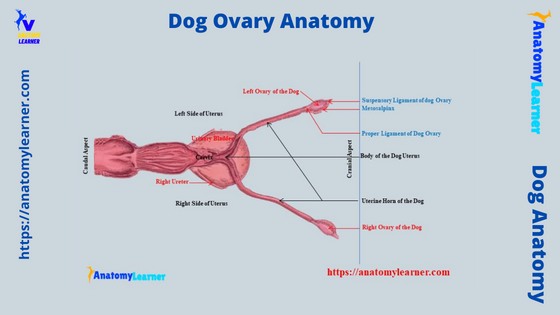
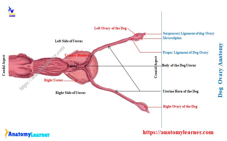
Dog ovary anatomy labeled diagram
In this section, I will show you the different diagrams of the dog ovary structure. First, let’s see the location of the dog ovary with the horn and uterine tubes of the dog from the cavity.
Here, the diagram of the dog’s abdomen shows the exact position of both the right and left ovaries of the dog. It shows the right ovary of the dog is cranial in position compared to the left.
Now, let’s see the external anatomical facts of each ovary from the dogs. Here, both the right and left ovaries of the dog show surfaces, extremities, and borders.
The diagram shows the 2 extremities (tubal and uterine), 2 borders (attached and free), and 2 surfaces (lateral and medial) of the dog’s right ovary. Again, the same features are found in the left ovary of the dog.
Now, let’s see the attachments of both the right and left ovaries of the dog with the abdominal wall and uterus. The diagram shows the mesovarium that attaches to the ovary to the uterus wall.
Again, the labeled diagram also shows the suspensory and proper ligaments of the dog ovary. This video might help you to clear the concept of the dog ovary structure (especially their location and external and internal features).
Now, the labeled diagram of the dog ovary shows the internal cortex and medulla with their different features. Here, the most important feature is the presence of ovarian follicles in the cortex area of the dog ovary.
The external surface (lateral surface) of the dog ovary also shows the mature ovarian follicle. Here, the diagram (find more) shows these mature or graffian follicles from the lateral surface of the dog ovaries.
Can you feel a dog’s ovaries?
No, you can not feel the dog’s ovaries from the external approaches. But, if you open the abdominal cavity and explore the ovary, you can identify them with its physical properties.
Why can you not feel a dog’s ovaries through external approaches or palpation? Well, to understand it, you might know the exact location of both the right and left ovaries of the dogs.
In very short, dog ovaries (both right and left) lie just caudal to the caudal pole of the corresponding kidneys. Right? But where are the kidneys located in a dog?
You know both right and left kidneys are located below the bodies of lumbar vertebrae (first 3 lumbar vertebrae). If you want, you may know the details anatomical facts of the dog kidney anatomy with their topographic and surface location from the below-mentioned article –
- Dog kidney anatomy – right and left canine kidneys location with labeled diagram,
As the location of the kidneys of a dog is under the bodies of lumbar vertebrae, they are hard to palpate from a surface (except for any disease condition). Again, the dog ovaries are located caudal to the corresponding kidneys (under the bodies of the respective lumbar vertebrae – 2 – 4).
So, you can not feel the dog’s ovaries from external palpation.
But, when you dissect the abdominal cavity and explore the ovary, you may quickly find these ovaries in a young female dog. But, in an older dog, these ovaries may be hard to find as there is a lot of fact enclosing them.
So, you might remove the surrounding fats first; then, you will find the dog’s right and left ovaries.
Frequently asked questions on dog ovary anatomy
Now, let’s see the commonly asked questions on dog ovary anatomy by canine anatomy learners. Here, I will enlist the related questions on the canine ovary structure along with their concise answer.
But, it is recommended to read the whole article to get the full concept of the dog ovary structure. Okay, let’s see the commonly asked questions by the canine anatomy learner on their ovaries.
Do dog dogs have 2 ovaries?
Yes, dogs have 2 ovaries – right and left. The shape and size of both the right and left ovaries of the dog are similar in the young female.
But, the size may vary in the older (multiparous female dog) dog. Usually, the multiparous female dog possesses a larger left ovary than the right.
How many ovaries does a bird have?
If you compare the bird with the female dog, you will find only one ovary and one oviduct in the bird. Here, the right set of the oviduct and ovary undergoes degeneration in early life.
But, the left set of the oviduct and ovary persist throughout the bird’s life. The bird’s ovary usually locates at the upper part of the abdominal cavity, just below the last two ribs.
You will find a great number of follicles on the bird’s ovary.
How many ovaries does a dog have?
Dogs have 2 well-developed ovaries – right and left ovary. Here, the right ovary is further cranial to the left ovary and lies just caudal to the corresponding kidneys of the dog.
You will find similar anatomical facts in both the right and left ovaries of the dog. In an older dog, the surface of the ovaries is slightly rough, but it is smooth in young female dogs.
How will you differentiate the mare ovary from the dog?
You will find a significant variation in the external and internal anatomical facts between the mare and dog ovaries. The ovary of the mare (female horse) is more prominent and bean-shaped compared to the dog.
They (ovaries of a mare) locate in the sublumbar region. You will see a depression on the free border of the mare ovary, which is identical features.
This depression on the free border of the mare ovary is known as the ovulation fossa. In the dog or cow ovaries, you will not find such type of fossa.
What size are dogs ovaries?
The size of the dog’s ovaries may vary according to the dog’s age, body weight, and species. A medium-sized dog has an ovary that shows an average 1.25-centimeter length, 0.6-centimeter width, and 0.4-centimeter thickness.
Conclusion
So, you got the basic idea of the dog ovary anatomy with the labeled diagram. Practically, the dog ovaries’ anatomical location (both topographic and surface) is significant for the learners.
The information mentioned above on the dog’s right and left ovaries anatomy might help you identify them from the samples. Now, you may also compare the anatomical facts (features) of the ovary of different species with the dogs.