The cat tail bones are the smallest caudal vertebrae of the vertebral column. These are the parts of the cat’s axial skeleton. The number of these bones is variable in the cat tails.
Here, I will discuss the unique osteological features of the tail bone from the cats. You will also know the actual numbers of their tail bones with the diagram from this article.
Quick overview: the cat’s tail has the smallest caudal vertebral bones. You will find 19 – 25 bones in the structure of the cat’s tail. The V-shaped hemal arches and hemal process are present at the ventral surface of the first few caudal vertebral bones.
I will also share other associated structures of the cat’s tail, like muscles, nerves, and vessels, with a diagram. Finally, after completing this article, you can differentiate cat tail bone from other animals’ tail bones.
So, let’s learn the details of a cat tail bone structure.
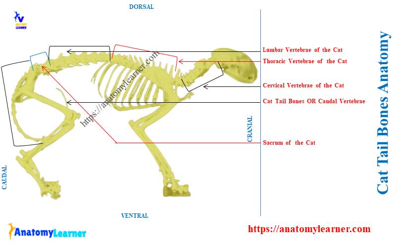
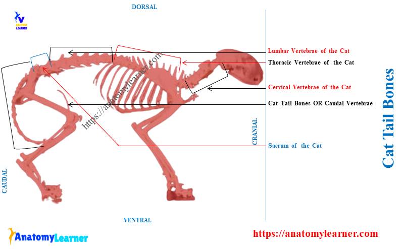
Cat tail bones
The cat tail bones are the caudal part of its vertebral column. They are the smallest bones compared to the other vertebral bones of this series.
All the vertebral bones of a cat are the parts of the axial skeleton. You may know cat’s other bones of both the axial and appendicular skeletons from the below-mentioned article –
The vertebral bones of the cat are single, unpaired, and located in the midline of the body. Here, I will help you to explore the features of the caudal vertebral bones from the cat’s tail.
Let’s see what are the unique features of the bones that formed the cat’s tail –
- The most cranial bones of the tail exhibit a typical vertebral characteristics,
- But the more caudal vertebral bones of the tail rapidly lose their typical features and become simple cylindrical structures,
- Two small hemal processes are found on the ventral surfaces of the body of a cat’s tail bone,
- The V-shaped hemal arches (chevron bone) present on the ventral surface of the cat’s caudal vertebral bone,
- You will also find the gradual decreases of the body, arches, and processes of the tail bones,
- The V-shaped hemal arches of the cat’s caudal vertebral bones house the caudal vessels,
- The cranial articular process of the feline tail bone gradually loses their articular functions,
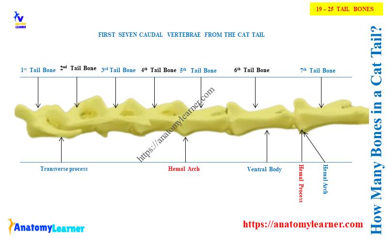
Here, the diagram shows the number and appearance of the tail bones from the cat’s vertebral column.
The hemal arches and processes are rarely identifiable as they have a tendency to lose during skeletal preparation. Thus, you need proper care in cat skeleton preparation to get the hemal arches.
How many bones are in a cat tail?
There are 19 – 25 bones in a cat tail anatomy. Most of the cat’s breed show an average of 21 bones in its tail.
The number of these bones in their tail varies according to the breed, size, and other factors. Let’s see the vertebral formula of a cat –
C7T13L7S3C19- 25
(Here, C = cervical vertebrae; T = thoracic vertebrae; L = lumbar vertebrae; S = sacral vertebrae; and C = caudal or tail bones)
Thus, you will find 49 – 55 vertebral bones in the cat’s vertebral column. This arrangement and number of different vertebral bones of a cat are almost similar to the dogs.
Even the anatomical features of these tail bones are almost similar to the dog’s tail bones. A significant variation (structure and number) may be found in the cows’, horses, and goat’s tails compared to the cats.
Let’s see the number of tail bones in various animals from Table 1 –
| Species | Number of tail bones |
| Cat | 19 – 25 |
| Dog | 6 – 21 |
| Cow | 15 – 25 |
| Horse | 17 – 22 |
| Pig | 18 – 22 |
| Rabbit | 12 – 15 |
However, the length and width of these tail bones greatly vary in various animals. In anatomy learner, you will find a detailed guide on various animal skeletons, including their tail bones.
Are cats tail bones or cartilage?
The cats tails are bones and caudal segments of the vertebrae. These bones form the base of the whole tail.
Again, the cat’s tail is not a cartilaginous structure. But, you will find the intervertebral cartilage disc in the cranial bones of the tail.
The structures that are found in the cat tails are bones due to the following reasons –
- They are the most rigid structure of the cat’s tail,
- They are the irregular bones of the cat skeleton as they have rough and irregular surfaces,
- You will find many projections of these tail structures that help in the attachment of various muscles,
- These hard structures of the cat tail consist of spongy substance with a thin covering of compact substance,
Again, these are the basic structures for tail movement in a cat. Thus, the tails of the cat contain the bones.
Besides these bony structures of the cat tail, you will also find the vessels, nerves, ligaments, and cartilage. Let’s see the structure of the cat tail, where I tried to show every single component with the diagram.
Cat tail bones anatomy
To describe the cat tail bones anatomy, you might have a good idea of the features of a typical vertebra. A typical vertebra of any animal has three distinct parts – a body, two arches, and three processes.
Here is a video that might help you to understand the features of a typical vertebra. The cat’s typical vertebra has the following –
- The body of the vertebra – is a solid cylindrical rod that support the other structures,
- Two arches of the vertebra – both right and left lateral arches have ventral pedicle and dorsal lamina and
- Three main processes – are spinous, transverse, and articular,
These three processes of the cat’s typical vertebrae possess identical features. Here, I tried to show the unique features of the cat’s typical vertebral bone with a diagram.
The cat typical vertebra labeled diagram shows the body, two arches, and three processes. Now, you may easily identify and describe the unique features of the tail vertebral bone compared to this.
Let’s discuss the unique features of the body, arches, and processes from the cat tail bone.
Body of the cat tail bone
The body of the cat tail bone is a solid cylindrical rod. It supports the aches and processes of the tail or caudal vertebral bones.
The cranial end of the tail bone is convex. In contrast, the caudal end of this tail bone is concave. But these cranial convexity and caudal concavity are not prominent like the typical vertebra.
Both the cranial and caudal extremities of the body of a tail bone attach to the corresponding intervertebral disc. And you know, the intervertebral discs are the fibrocartilaginous structures between two corresponding vertebrae.
The body of the cat tail bone possesses 2 surfaces – dorsal and ventral. Here, the dorsal surface of the tail bone’s body is flattened. It forms the floor of the neural canal of the cat’s tail.
The ventral surface of the body of a cat’s bone is constricted in the middle. It possesses ill-developed spines for muscular attachment.
Now, let’s see what are the special features of the body of a cat tail bone.
Unique features of the body of a cat tail bone
- The body of the first tail bone is as wide as it is long,
- In the middle of the tail, you will find a gradual increase in the length of the bones,
- The body of the tail bone becomes progressively shorter at the caudal aspect of this series,
- You will see a gradual decrease in the wide of the body of these tail bones from cranial to the caudal,
- The body of the last tail bone is minute and ends as a tapering process,
So, the features of the bodies of feline tail bones are different than the other vertebral bones. With these unique features, you may easily differentiate these tail bones from the vertebral column.
Arches of the cat tail bone
There are 2 right and left lateral arches in the cat tail bone that remain on the body. Each arch of the cat tail bone consists of two parts – the pedicle and the lamina.
Here, the pedicle is the lower part of the arch and forms the lateral wall of the neural ring. You will also find small notches at the cranial and caudal ends of the tail bones.
The respective cranial and caudal notches of the adjacent tail bones form the intervertebral foramen. But, not all these caudal vertebral bones form the intervertebral foramen in the cat.
Again, the notches are easily identifiable in the first few tail bones. The notches of the arches are not typical for the cat tail bone.
The lamina of a tail bone is the dorsal part of the arches. It is a flat and thin plate-like bone that remains dorsal to the pedicle of the arch.
The lamina of the arches form the roof of the neural ring of the caudal vertebrae. Again, the right and left laminae join the midline of the body and form the spinous process in a cat tail bone.
Unique features of the arches of a cat tail bone
Let’s see what the unique features of the right and left arches of a cat’s tail bone –
- The right and left arches are well-developed in the first few tail bones,
- The neural ring that is formed by the arches becomes gradually smaller from cranial to caudal,
- You will find the neural rings up to the sixth or seventh tail bones,
- As the caudal tail bones don’t possess any neural rings, the arches are hard to identify,
You will find a groove in the last caudal tail bones that cranially continue with the neural canal. Here, you will see the caudal spinal nerves that supply the cat tail.
How is the neural ring formed in a cat’s bone?
The neural ring of a cat tail bone is formed by the dorsal surface of its body and two lateral arches. Let’s see the boundary of the neural ring that formed in the cat tail bone –
- The ventral aspect of the neural ring – is formed by the dorsal flattened surface of the body of the tail bone,
- The dorsal aspect of the neural ring – is formed by the right and left dorsal laminae of the cat tail bone,
- Lateral wall of the neural ring – the right and left pedicles of the cat tail bone form the lateral wall of the neural ring,
Here, the diagram shows the neural ring from the cat tail bone. Not all these caudal vertebral bones form the neural rings in cats.
Processes of the cat tail bones
The cat tail bones also possess three processes – articular, transverse, and spinous process. But, you will find these three identifiable processes in the first few tail bones.
Here, the articular processes are two types in the cat tail bone – cranial and caudal. You will see two cranial and two caudal articular processes in the first few bones of the tail.
First, let’s see the summary of the 3 processes of the tail bones from Table 2 –
| Process of cat tail bone | Location | Unique features |
| Cranial articular process | Cranial border of the arch | Two in number Concave articular surface |
| Caudal articular process | Caudal border of the arch | Two in number Convex articular surface |
| Transverse processes | Junction between body and arch | Right and left processes First 4/5 pairs are typical |
| Spinous process | Junction between right and left dorsal laminae | Single and project upward Small and disappears in last |
Now, let’s discuss the unique features of these three processes from the cat tail bone with diagram.
Articular processes of the cat tail bone
Typically, the articular processes are larger in cat vertebrae. They contain convex or concave articular surfaces that join with the similar processes of the corresponding bones.
But, both articular processes of the cat tail bone are different than the typical. The cat tail bone exhibits the cranial articular process. But, these cranial articular processes of the tail bone lose their articular activities.
You will find the mammillary process along with the cranial articular process of the cat tail bone. They may persist caudally in the series until all trace of the articular process has vanished.
The cat tail bone also shows the caudal articular process in the first segment. They project from the caudal border of the right and lateral arches.
These two caudal articular processes of the cat tail bone are frequently asymmetric. They gradually disappear in the craniocaudal sequence of the cat tail.
The tail bones with the articular processes form the diarthrodial articulation with similar processes. They help to prevent undesirable movement between the adjacent tail bones.
Transverse and spinous processes of the cat tail bone
The transverse process of the cat tail bone is a large and plate-like structure. It arises from the two roots, one from the arch and one from the body of the tail bone.
Thus, the transverse process of the tail bone projects outward from the lateral aspect of the neural ring. The first four or five pairs of the transverse process of the cat tail are well-developed or typical.
Caudal to the sixth cat tail bone, the transverse process reduces in size. Again, they will disappear at about the fifteen to last tail bones.
The spinous process of the cat tail bone is single and projects upward from the junction of the laminae. It is very small and found in the first few tail bones.
This single spinous process may disappear from the seventh or eighth tail bone. Thus, the typical features of the spinous process are rarely identifiable in the cat tails.
What are the hemal arches of the cat tail bones?
The hemal arches are the V or Y-shaped structure that articulates with the ventral surface of the tail bones. These are the separated bones that are found at the caudoventral ends of the bodies of the 4th – 6th tail bones.
The hemal arches slope caudally and are shaped like V or Y. The hemal arches are also known as the chevron bones.
These arches protect the median artery of the cat, which passes through them. Just caudal to the hemal arches, you will find paired hemal processes on succeeding bone.
The remnant of the hemal processes can also be identified from the caudal tail bones. Proper care should be needed during cat tail bone preparation to identify the hemal process.
What are the cat’s tail muscles?
The cat’s tail exhibits several intrinsic muscles. These intrinsic muscles perform the typical tail movement of the cat.
Most of the intrinsic muscles of the cat tail are the continuation of the lumbar muscles. Here, the extensor caudae medialis and multifidus spinae help to raise the cat’s tail.
Let’s find the idea on various muscles of the cat from the below-mentioned article –
The multifidus spinae that helps to raise the cat tail is the extensive muscle. It can be distinguished from the cat’s vertebral column in the lumbar region.
It arises from the transverse processes of the cat’s lumbar vertebrae. Again, this muscle inserts on the neural spine of the more caudal vertebrae.
Frequently asked questions on cat tail bone
Now, I will enlist the frequently asked questions on the cat tail bone anatomy. The cat tail bones lack different typical osteological features compared to the other vertebrae.
Let’s see the commonly asked questions on cat tail bone by the anatomy learners. Here, I will provide a concise answer to these questions related to the cat tails.
Are there bones in the cats tail?
Answer: Yes, there are bones in the cat’s tail. On average, you will find 21 bones in the cat’s tail. However, all these bones do not possess the typical features of a vertebra.
Besides these bones, you will also find some other structures in the feline tail. You will find the muscles, nerves, vessels, ligaments, and intervertebral discs in the cat tail structure.
How many bones does a cat have in its tail?
Answer: the cats have an average of 19 – 25 bones in their tails. The number of the tail bones in the cats varies with their breed and size.
The first few bones of the tail show the features of a vertebra. These
bones form the rigid structure of the tail and control its movement.
What are cat tail bones called?
Answer: the cat’s tail bones are called the caudal vertebral bones. Again, these tail bones are also called the coccygeal vertebral bones.
These coccygeal or caudal vertebrae are the smallest and lack typical features of the cat vertebra.
Conclusion
So, the cat tail bones are the caudal or coccygeal vertebral bones of the spine. The tail bones vary from 19 to 25 in the cats.
Except for the first few tail bones in the cats, you will find atypical osteological features. The hemal arches and processes are the unique features of the few tail bones of the cats.