The lymph is a clear, colorless fluid in the lymph capillaries, vessels, trunk, ducts, and sinus of the lymph nodes. Its composition is similar to the plasma (with slight variation) and returns to normal blood circulation through the cranial vena cava.
But how is lymph formed and transported into normal blood circulation? If you have these specific inquiries about lymph formation, then this article might help you to get a short answer to it.
Quick answer: lymph is formed by oozing plasma through the small pores of blood capillaries into the interstitial tissue. A set of lymphatic vessels collected lymph from the tissue space and drained it into the cranial vena cava through the right and left (thoracic duct) lymphatic ducts.
But, if you want to know the details of the lymph formation in the tissue space and its transportation into the right atrium of the heart, then let’s continue this article till the end. Here, I will try to explain why lymph is formed in the tissue space and how they are transported through the lymphatic vessels.
Okay, let’s get into the main (lymph) part of the article to know the basics of lymph formation and transportation.
How is lymph formed
I hope you have a basic concept of general blood circulation in the animal body. Generally, oxygenated blood is transported through the aorta (with its different branches) into the different body parts of an animal.
In different tissues or organs, it removes oxygen and enters carbon dioxide; thus, deoxygenated blood is formed. Now, the deoxygenated blood is transported into the right atrium of the heart through the cranial and caudal vena cava.
Again, the deoxygenated blood is transported to the lungs through the pulmonary artery, where the purification occurs. Thus, the oxygenated blood comes back into the left atrium of the animal’s heart.
If you want to know the details of this general systemic blood circulation, you may read the below-mentioned suggested article –
What is the difference between pulmonary and general systemic blood circulation,
From the general blood circulation in the animal’s body, you can easily understand that blood circulation pressure is more in the arteries and capillaries than in the veins. Again, in any capillaries’ structure, you will typically find the pore in their wall.
These pores are normal in the structure of a blood vessel (arteries and capillaries). As because, through these pores, the nitrogenous products produced by the interstitial cells pass into blood vessels. Again, cells in the tissue get their nutrients from the blood cells through diffusion.
Factors involving the lymph formation in the tissue space
There are two main factors that involve lymph formation in the tissue space –
- The extra pressure in the blood capillaries, and
- Pores in the structure of the blood capillaries,
Now, let’s see how lymph is formed in the tissue space. And how these 2 factors are involved in the formation of lymph in an animal’s body tissue.
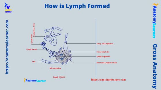
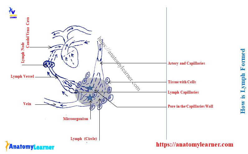
The excess pressure on the blood vessels (capillaries) leads to oozing plasma through the pores of the wall. As the diameter of the red blood cells is more, they are unable to pass through these pores of the blood vessel wall.
So, only the white blood cells from the blood ooze from the pores of the vessel’s wall. As there is no red blood cell, this oozing fluid looks white and is stored in the tissue space.
This clear (white) fluid is the lymph in the tissue space (shown in the diagram). You know that circulation is a continuous process, so the formation of lymph will be continued. Thus, white-colored fluid (lymph) will accumulate in the tissue space.
These clear tissue fluids need to remove from the tissue space. Otherwise, they will lead to the swelling of the specific parts (tissue) of the animal’s body.
But, this condition does not occur normally; the animal’s body has another set of vascular channels that remove this excess fluid (lymph). These extra set of a vascular channels of the animal’s body is the lymphatics (capillaries and vessels).
I hope you got the answer to your question – how is lymph formed in the tissue space? Let’s see how these lymphatic vessels remove lymph (limf) from the tissue spaces.
How is lymph moved through lymphatic vessels?
First, let’s see the structure of the extra set of the vascular channel that removes the lymph from the tissue space. These lymphatic vessels (capillaries) are the blind end structure located in the interstitial tissue.
So, how does this lymph enter these lymphatic vessels? These lymphatic vessels (capillaries) have simple squamous epithelium cells that overlap (shown in the diagram).
The pressure of the excess fluid (lymph) in the interstitial tissue will push these cells of the lymph capillaries. Thus, a pathway will be created, and lymph will enter through these passes.
Here, the lymphatic capillaries possess valves like the veins to have. There is no backflow of the lymph within the lymphatic vessels. Usually, lymph goes in a direction due to pushing excess fluid.
Now, the lymphatic capillaries join with the lymphatic vessels. Again, these lymphatic vessels join to form a different lymphatic trunk. Finally, the different lymphatic trunks of the animal’s body join to form the lymphatic ducts.
But, when the lymph passes from the capillaries to the vessels, you will see different lymph nodes in their passages. Lymph passes at least once through the sinuses of the lymph node before entry into the venous system of the body.
There are 2 lymphatic ducts in the animal’s body – right and left lymphatic ducts. Here, the left lymphatic duct is known as the thoracic duct, which is larger than the right lymphatic duct.
The length of the right lymphatic is so small and opens into the right common jugular vein (sometimes in the right subclavian vein). At the same time, the left lymphatic (thoracic duct) also opens into the right common jugular vein (sometimes the left subclavian).
Finally, lymph right and left common jugular veins to drain into cranial vena cava and enter general circulation.
How do lymph nodes filter the lymph?
As you see in the previous discussion, lymph can easily enter into the blind end capillaries of the lymphatic system. But not only does lymph enter the lymph capillaries, but some tissue debris and other particles may also enter the lymph capillaries as the space is more.
Suppose the specific tissue has an infection or foreign material or bacteria. Then these foreign bodies, bacterias, and tissue debris pass through the capillaries of the lymphatic system.
So, if there is no filtration, all these agents might be transported into the right atrium of the heart. Ultimately, they will go into the general systemic circulation and spread throughout the animal’s body.
But, this condition does not usually occur as the animal’s body has another defense mechanism. There are different lymph nodes in the course of the lymphatic (capillaries and vessels).
You know, in the lymph node, you will find the aggregation of the lymphocytes (nodules). When the foreign bodies enter the sinuses of lymph nodes with the lymph, more white blood cells or lymphocytes accumulate.
They identify these foreign bodies and try to combat them. More foreign bodies or bacteria or other agents will lead to more production or accumulation of lymphocytes or white blood cells in the lymph node.
So, if more infections occur, the animal’s lymph nodes become swollen due to accumulating more lymphocytes. These swollen lymph nodes in the animals indicate the infection in their body.
What are the right and left lymphatic ducts?
These are the main vessels that collect lymph from different regions of the body. Here, the right lymphatic duct of the lymph vascular system collects lymph from the right head, neck, right thorax, and right forelimb of the animals.
The left lymphatic or thoracic duct collects lymph from a wide range. It collects lymph from the left thoracic cage, left arm, left neck, head (minor), left and right hind limb, and abdominal cavity (cisterna chyli).
So, the lymph collection site for the thoracic duct is wide compared to the right lymphatic duct. But how is this left lymphatic or thoracic duct formed? To know the thoracic duct’s formation, you might first know the cisterna chyli.
What is cysterna chyli?
These are the elongated and dilated sacs that work as lymph reservoirs. Typically, this cisterna chyli is formed by the union of lumbar and gastrointestinal lymphatic trunks.
The cisterna chyli locates at the right of the abdominal aorta at the vel of the second or third lumbar vertebrae. You will find a close relationship of the cisterna chyli with the right face of the crus of the diaphragm.
From the cranial end of the cisterna chyli, the thoracic duct (left lymphatic duct) starts.
Where are the lymph vessels absent?
You will not find any lymph vessels in certain regions of the animal’s body. Let’s see which organs and areas have no lymphatic vessels –
- Bone marrow and central nervous system of animal’s body (except meninges),
- Umbilical cord and embryonic membrane,
- Hyaline cartilage of any structure,
- The epithelial layer of the skin,
- Cornea, lens, and vitreous humor of the animal’s eye,
Functions of lymph or lymphatic system
If you notice how lymph is formed and transported into the blood (general circulation) through the lymphatics, you will find the following 2 important functions –
- The lymphatic system or vessels remove or clear the excess fluid (lymph or limf) from the interstitial tissue space,
- It performs a defense mechanism in different ways,
If microorganisms enter through the lymphatic vessels, they will be destroyed by the lymphocytes present in the lymph node. Thus, the lymph node filtrated the microorganism from the lymph and protected the animal’s body.
These are 2 major functions of the lymphatic system or lymph in the animal’s body. In addition, the lymph and lymphatic system also possess the below-mentioned functions –
This system of the animal’s body balances the fluid – drainage tissue fluid (lymph) into the bloodstream through the lymphatic vessels,
Help in fat absorption from the digestive tract and passes through the lymphatic vessels into the venous circulation,
I hope you can understand the primary function of the lymph and lymphatic system in the animal’s body.
Lymph circulation flow chart
You see, how is lymph formed and transported into the general blood circulation? Now, it will be better if I provide a lymph circulation flow chart that represents the summary of all (formation and transportation).
Here, in the flow chart of lymph circulation, I tried to show the below-mentioned topics –
- Normal blood circulation and plasma oozing from the blood capillaries,
- Formation of lymph into the tissue spaces,
- Formation of a new set of lymph capillaries (lymph vascular system),
- Capillaries join from the lymphatic vessels,
- Lymph nodes in the lymphatic channels (filter lymph),
- Lymphatic vessels join to form the lymphatic trunks,
- Formation of the lymphatic ducts – right and left lymphatic ducts, and
- Passes lymph into the cranial vena cava from the right and left lymphatic ducts,
So the lymph circulation flow chart represents the summary of the lymphatic system. It is enough to understand the basics of the lymph, lymphatic vessels, and lymph circulation in the animal’s body.
How is lymph formed and transported labeled diagram
Now, here the labeled diagram will also represent the formation and transportation of the lymph through the lymphatic vessels and finally draining into the right atrium of the heart. If you follow my previous discussion on lymph formation and transportation, you will easily understand the below-mentioned labeled diagram.
In this diagram, I tried to show the different structures of the lymphatic system, like the lymph, lymphatics (capillaries, vessels, trunks, and ducts), and lymph nodes. Here, I have drawn different lymphatic vessels from the different body regions of the animals.
Different lymphatic trunks like the tracheal, hepatic, gastric, lumbar, celiac, intestinal, jejunal, colic, and visceral are also shown in the labeled diagram (few are missed). Again, the labeled diagram show 2 major ducts (right and left lymphatic ducts) from the animal’s lymphatic system.
Finally, you will see the dilated and elongated sac-like cisterna chyli in the labeled diagram. You may find more diagrams here on the lymphatic system of animals.
More inquiries on the lymph formation
Now, let’s see some of the common inquiries on the lymph and lymphatic system of the animal. Here, I will enlist some of these major questions on lymph asked by the anatomy learners. Let’s see these inquiries on the lymphatic system or lymph or lymphatics.
What is the lymphatic system and lymph vascular system?
In the broad sense, the lymphatic system is a description and functional aspect of the lymphatic tissue. The lymphatic tissue includes the lymphatic vessels (lymphatics), organs, and cells.
Here, the lymphatics of the lymphatic system include –
The lymph capillaries and vessels,
Lymph trunks and ducts,
In addition, you will find 2 types of lymphatic organs in the animal’s lymphatic system –
Primary lymphoid organs – include bone marrow and thymus, and
Secondary or peripheral lymphoid organs – include the spleen, lymph nodes, tonsil, and payer’s patches of ileum,
In the lymphoid cells of the lymphatic system, you will find the following cells –
T –lymphocytes, B-lymphocytes, macrophages, and natural killer cells,
But what does the lymphatic vascular system means? The lymph vascular system includes capillaries, vessels, trunks, and ducts. The lymph nodes or other organs will not be included in the lymph vascular system.
How is lymph formed short answer?
Lymph is formed in the tissue spaces when the plasma escapes from the pore of the blood vessel wall. This escaping or oozing of plasma is occurred due to the excess pressure on the blood capillaries and vessels.
So, accumulating oozed plasma into the tissue space is clear as it doesn’t contain red blood cells. Thus it gains the white color and is termed the lymph.
How is lymph different from blood?
You know that lymph is a clear, colorless fluid found in the tissue space near the blood vessels. Whereas the blood is reddish, fluid passes through the blood vessels (veins and arteries).
The lymph is the part of the lymphatic system, whereas the blood is the part of the general circulatory system of the animal’s body. If you think about the compositional difference between the lymph and blood, you will find plasma, red blood cells (RBC), white blood cells, and platelets in the blood.
In addition, the lymph contains plasma and little amount of platelets. But, there are no red blood cells in the composition of lymph.
The amount of oxygen carried through the blood is more than the lymph. You will find a suitable defense mechanism performed by the lymph in the animal’s body. Thus, the lymph is considered part of the immune system of the animal’s body.
The blood possesses a wide range of functions in the animal’s body. They associate with the circulation of oxygen and carbon dioxide into the body tissue. Again, they involve circulating different substances like waste products.
What is a lymphocenter, and where will you find them?
These lymph nodes or groups of lymph nodes constantly occur in the same region of the animal’s body. These lymphocenter receives the afferent vessels from approximately the same region in all animal species.
You will find 7 – 8 lymphocenters in the animal’s body. Here, I will show these lymphocenters from the animal’s body with some of their important superficial lymph nodes –
- Lymphocenter in the head region – includes mandibular, parotid, and retropharyngeal lymph nodes,
- Neck lymphocenter – includes superficial and deep cervical lymph nodes,
- Lymphocenter in the thoracic limb of animal – includes the axillary lymph nodes,
- Thoracic cavity lymphocenter – possesses the dorsal and ventral thoracic lymph nodes, mediastinal lymph nodes, and bronchial lymph nodes,
- Lymphocenter in the abdominal and pelvic wall – posses lumbar, iliosacral, inguinofemoral (superficial inguinal), and ischiatic lymph nodes,
- Pelvic lymphocenter – includes iliofemoral and popliteal lymph nodes, and
- Lymphocenter in the abdominal viscera – possesses the celiac, cranial and caudal mesenteric lymph nodes,
You know these lymphocenters and their different lymph nodes play a great role in filtering the lymph while it passes through the vessels and nodes. An animal’s swollen lymph nodes indicate an infection, so that you might know the location of some of the superficial lymph nodes from the animals.
Thus it will help you to palpate these lymph nodes when they swell due to improper lymph drainage or other problems present in the lymph.
Conclusion
I hope you got the perfect answer to the question – how is lymph formed and transported into the general blood circulation? The lymph formation starts from the oozing of plasma from the blood vessels into the tissue spaces.
The lymphatic capillaries (small) collect the lymph from the interstitial tissue space and transport it through the lymph vessels into the lymphatic ducts. The right and left lymphatic ducts of the lymph vascular system drain the filtered lymph (filtered by lymph nodes) into the heart’s right atrium through the cranial vena cava.