The structural and functional unit of the nervous system is the neuron that may easily observe under a light microscope. Neurons may vary considerably in size, shape, and other features. Here, I will provide details information and identifying points so that you may easily identify the neuron under a microscope.
A neuron consists of a cell body that gives off a variable number of processes – axons and dendrites. You will find different types of neurons (like unipolar, bipolar, multipolar, sensory) in the animal body, but the basic structure is almost similar. The main purpose is to introduce you to the structure of different types of neurons and their associated components under the light microscope.
In addition, I will also show you different types of neuroglial cells and synapses under a light microscope with their proper identifying points. You will also learn how the myelin sheath is formed at the end of this article. So, this article might be a great resource to know details about the neuron structure and its associated components.
Neuron under microscope
If you want to identify the neuron under a microscope, you might have a good piece of knowledge on its structure. I will describe everything about the neuron structure in the next section. But, now, I would like to provide the most appropriate identifying points for the neuron with the slide image.
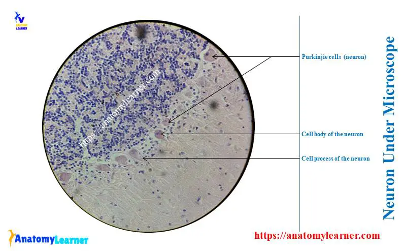
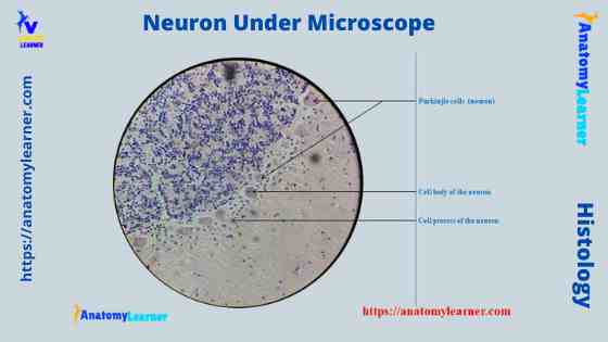
The neuron structure has two main components: the cell body and the neuron processes (axons and dendrite). Let’s see the neuron histology slide labelled diagram and try to find out the below-mentioned characteristics –
- Presence of an identifiable cell body (soma) that locates in the brain’s grey matter (according to the slide image).
- The cell body possesses spherical, euchromatic, and large eccentric nuclei containing a prominent nucleolus.
- Presence of easily visible cytoplasm and plasma membrane (neurilemma) in the body of a neuron.
- There are highly branched short dendritic processes present around the neuron’s cell body.
- Presence of a relatively long, cylindrical axon process that arises from the neuron’s axon Hillock (shown in slide image diagram).
So, the sample tissue represents the neuron (multipolar). Now, let’s know the basic structure of the neuron with slide images and labelled diagrams.
But, before going to the details description of the neuron from the animal body, you might have a good piece of knowledge on the nervous tissue.
Nervous tissue under a light microscope
You know the nervous tissue is a specializedspecialized tissue for receiving a stimulus. These stimuli are analyzedanalyzed and integrated to produce the appropriate and co-ordinate responses. So, you will find three main features in the nervous tissue of an animal –
- SpecializedSpecialized tissue for a reception the stimuli,
- Analyses and integrates the stimuli, and
- Produces the appropriate response and co-ordinate that response to the body.
I hope you can understand what nervous tissue is. Observing the nervous tissue under the light microscope will find neurons (nerve cells) and supportive (glial cells) in its parenchyma. And you already know that the neuron is the structural and functional unit of the nervous system.
Again, the supportive or glial cells of the nervous tissue consist of neuroglial and ependymal cells in the central nervous system (CNS). And the supportive cells of the peripheral nervous system (PNS) include Schwann and Satellite cells.
Structure of neuron under light microscope
So, the neurons are the structural and trophic unit of the nervous system that transform and sustain what they innervate. They are typically incapable of mitosis except for the olfactory receptor cells.
Morphologically, you will find the cell body and neural processes under a microscope. The cell body is also known as the soma or perikaryon and consists of a mass of cytoplasm surrounded by a plasma membrane. Again, the neuron features elongated processes that extend variable distances from the cell body.
Generally, a typical neuron possesses one long axon and multiple dendrites. You will also find the telodendrite at the terminal end of the axon of a neuron.
Metabolically, the neuron is actively involved in maintaining its structural integrity. They also help synthesizesynthesize, package, transport, and release secretory products. You know the neuron are specializedspecialized in excitability and communication with other neurons.
The neuron is involved in ions passing through the protein channels embedded in the neuronal plasma membrane. They also communicate with other neurons by releasing chemical agents like neurotransmitters and neurohormones.
Now, let’s discuss the different parts and components of a typical neuron (multipolar motor) with a labelled diagram. But, first, let’s try to identify the following features from a neuron with the help of a labelled diagram.
- Cell body or perikaryon of a neuron
- Nucleus, cytoplasm, the plasma membrane of a neuron
- Nissl bodies in the cell body of a neuron
- An initial segment of axon and axon hillock
- Dendrites and axons of a neuron
- Axolemma and myelin sheath
- Oligodendrocytes and Schwann cells
- Nodes of Ranvier
- Collateral branch, terminal knob, and motor endplate
I think all these features are easily identifiable from the neuron labelled diagram. Now, let’s discuss the cell body and neuron processes of a neuron.
The cell body of a neuron
The cell body of a neuron is easily identifiable with the routine histological stain. I will provide very concise and important features from the cell body so that you may easily understand and memorizememorize them.
So, the cell body of a neuron consists of a spherical nucleus, cytoplasm, and plasma membrane. You will also find other organelles like the Golgi complex, microtubules, neurofilaments, and others like a typical cell. Again, the cytoplasm of a neuron contains a clump of chromatophilic substance – the Nissle body (will describe later).
There is a conical portion of the cell body of a neuron (axon hillock) from where the axon arises. The cell body of a neuron is mostly located in the grey matter of the brain and spinal cord.
You know the brain’s grey matter remains periphery, whereas, in the spinal cord, it remains on the inner side. If you don’t know what the grey and white matters are – then this is for you –
The grey matter contains all the cell bodies of neurons, their unmyelinated dendrites, and all the cells that support the neuron. That means you will find the neuroglia cells in the grey matter portion.
Again, the white matter contains myelinated neurons and a small number of cell bodies.
The nucleus of a neuron
Generally, you will find a centrally placed nucleus in a neuron. But, sometimes, you may find an eccentric nucleus in some neurons of an animal. The nucleus is the spherical or elliptical structure in the neuron containing euchromatic staining (pale staining).
Again, the shape of the nucleus of a neuron is generally large because of the little cell body cytoplasm. There is a prominent nucleolus evident in the nucleus of a neuron. In the female of some animal species like cats and rodents, you will find the Barr body near the nucleolus. But, in primates, you may find structures in the nuclear membrane.
Cell body cytoplasm of a neuron
The cell body cytoplasm is mainly involved in protein syntheses like cytoskeleton protein, membrane protein, and secretory protein. If you stain the neuron with aniline dyes, then the cytoplasm of a neuron shows a clump of chromatophilic substances.
These chromatopilic substances are the Nissle bodies of a neuron. These are the aggregation of rough endoplasmic reticulum, free ribosomes, and polyribosomes.
The Nissle bodies may also extend into the trunk of the neuron’s dendritic tree. But, you will nerve find these nissl bodies in the axon hillock of a neuron.
If there is any injury in a neuron, the cell body swells. The nucleus shifts to the eccentric position, and the ribosome disappears. So, the nissl bodies also will chromatolize from the cytoplasm of a neuron.
Others organells of neuron
There is a prominent Golgi complex in the cytoplasm of a neuron. You will find the secretory vesicles that transport through the axon to the terminal bulbs. There is also evidence of synaptic vesicles in the cell body of a neuron that primarily form and store in the telodendritic bulbs.
You will find the microtubules, neurofilaments, and mitochondria in the cytoplasm of a neuron. The microtubules are involved in the rapid transport of membrane-bound organelle.
It is also evidence of numerous neurofilaments in the cytoplasm of a neuron.
Neuron processes (axon and dendrite) under microscope
You will find a single axon and numerous highly branched dendrites in the cell body of a neuron under a microscope. Again, the terminal branch of the axon shows teoldendrites. This telodendrite synapses with the dendrites and cell bodies of the other neurons.
Let’s discuss a little of the structural features of an axon and dendrites from a multipolar motor neuron –
- Axon of a neuron and
- Dendrites of the neuron.
Axon of a neuron
Axon is the relatively long and cylindrical process of the cell bodies of a neuron. This axon originates from the axon hillock and ends at the terminal branches. The axonal branch of a neuron is sparse.
The initial segment is just distal to the axon hillock, where the action potential begins. This initial segment is the narrow part of the other part of the plasma membrane of the cell body of a neuron. From this initial segment of a cell body, the myelin sheath begins.
You will also find the plasma membrane around the axon, known as the axolemma. The axon branches at the right angle in the axon course and give the collateral branches.
You will find the microtubules, neurofilaments, mitochondria, and smooth endoplasmic reticulum in the axoplasm. The axon can regenerate conditions and carries impulses away from the cell bodies. They convey the excitation signal that arrives at the end of the axon.
Dendrites of the neuron
These highly branched processes receive numerous synaptic contacts from the other neuron. They also receive signals from the body’s environment or sensory epithelium cells. That means the dendrite conveys impulses from the periphery to the cell body.
The dendrite of a neuron is usually short and divided like branches of a tree. Again, the cytoplasm of a dendrite contains Nissl bodies, mitochondria, and neurofilaments.
Terminal brnaches (telodendrites)
The axon of a neuron terminates in a twing-like branching, which is telodendrites. You will find the expansion at the end of each terminal branch of an axon.
The expanded structure of the terminal end is the synaptic button (terminal bulb). This terminal bulb is the site for neurotransmitters where the molecules are packaged and stored within the synaptic vesicles.
In some neurons, you will find a series of expansions at the proximal part of the terminal bulbs. These expansions are the preterminal bulbs and possess the same functions as terminal bulbs.
Classification of the neurons
The classification of the neurons may be based on their number of cell processes, functions, and others. Here, I will only provide the classification based on the number of cell processes and functions. These might help you identify the different neuron types under a microscope.
According to the cell process number (anatomically), a neuron may divide –
- Unipolar neuron
- Bipolar neuron
- Pseudounipolar neuron and
- Multipolar neuron
Unipolar neuron: When the cell body gives off only a single axon, then the neuron is the unipolar neuron. So, here in a unipolar neuron, you will find the cell body and a single axon.
The photoreceptor cells of the retina (rod and cone cells) contain the unipolar neuron. I hope the diagram might clarify the concepts of what the unipolar neuron looks like.
Bipolar neuron: The bipolar neuron possess only one dendrite and one axon. These axons and dendrite of a bipolar neuron locate at opposite poles of the neuron cell body.
You will find the bipolar neuron in the cochlear and vestibular ganglia, retina olfactory mucosa of the nasal cavity.
Pseudounipolar neuron: The psudounipolar neuron posses a single process (axon) close to the cell body. But, this single process divides into two branches and forms a T-shaped with peripheral and central branches.
The peripheral branch of the pseudounipolar neuron extends peripheral ending, and the central branch extends toward the central nervous system (CNS).
The sensory root ganglia of the most cranial nerve possess the pseudounipolar neuron.
Multipolar neuron: The cell body of this type of neuron give rise to several branches. That means you will find several dendrites and a single axon in the multipolar type of neuron.
The motor neuron with cell bodies in the ventral horn of the spinal cord and pyramidal cells of the cerebral cortex possesses multipolar neurons.
Classification of a neuron based on functions
According to the function, the neuron may be classified into – sensory, motor, and interneuron. The other name of the sensory is afferent, and the motor is an efferent neuron.
Sensory neuron: This neuron receives signals from the receptor and conducts impulses to the central nervous system. You will find the sensory neuron in the sensory ganglia.
Moto neuron: The motor neuron conducts the impulses from the central nervous system (CNS) to the effector organs like muscles. You will find the motor neuron in the ventral horn cells of an animal.
Interneuron: The interneuron connects the sensory and motor neuron and completes the functional circuit.
Please read the other different neurons types from another article on anatomy learners or the recommended histology learning books.
Multipolar motor neuron under microscope
The multipolar motor neuron under a microscope shows the typical features. This neuron possesses the cell body, an axon, and several short branched dendrites. The neurons in the ventral horn of the spinal cord and cerebral cortex are the best examples of multipolar motor neurons.
So, the multipolar motor neuron has one axon and two or more dendrites. The direction of the signals is from dendrite to cell body to axon or from cell body to axon.
The dendrites and cell bodies of multipolar neurons are the receptor portions of the cell. Again, the plasma membrane of the dendrite and cell body is specializedspecialized for impulse generation.
In addition, the axon is the condition part of the cell, and its plasma membrane is specializedspecialized for impulse conduction. The terminal part of the axon possesses the terminal knob or synaptic knob that contains the synaptic vesicles.
These are the microscopic images (mentioned above) of the multipolar motor neuron from the brain’s cerebral cortex. Here, I tried to show you the cell body of the pyramidal cells of the cerebral cortex. Again, the microscopic neuron image shows the axon and dendrites.
Bipolar neuron under a microscope
Bipolar neurons are rare in animals that possess one axon and one dendrite. In the receptor of the organs of the special senses (like taste, smell, hearing, sight, and equilibrium), you will find the most bipolar neurons.
These bipolar neurons are generally found within the retina of the eye, cochlear and vestibular ganglia, and olfactory mucosa of the nasal cavity. But, the amacrine cells of the retina have no axon.
The dendrite and the axon of the bipolar neuron locate at the opposite pole of the cell body. In the bipolar neuron labelled diagram, I tried to show you one axon and one dendrite with its cell body.
The components of the cell body of the bipolar neuron show the same microscopic feature as the multipolar motor neuron. So, the cytoplasm of the cell body of a neuron contains the eccentric nucleus, mitochondria, rough endoplasmic reticulum, ribosome, and other organelles.
Sensory pseudounipolar neuron under microscope
The pseudounipolar sensory neuron possesses one single process: the axon. This axon of pseudounipolar neuron divides close to the cell body into two long axonal branches. So, you will find the peripheral and central branches in the axon of a pseudounipolar neuron.
One branch of an axon (peripheral) extends to the periphery, and another branch (central) extends to the central nervous system (CNS). The receptor portion of the cell locates at the peripheral branch of this axon.
Sometimes the pseudounipolar neuron may develop from a bipolar neuron as its axon and dendrite migrate around the cell body and fuse into a single process.
So, where you will find the pseudounipolar sensory neuron? The majority of the pseudounipolar sensory neuron locates close to the central nervous system. Most cranial nerves’ dorsal root ganglia and ganglia are the ideal examples of pseudounipolar neurons.
I tried to show you the pseudounipolar sensory neuron with its different parts (especially the peripheral and central axon and cell body) in the diagram.
Neuroglia under microscope (neuron cells)
While studying the neuron under a microscope, you might also know the different types of neuroglia cells. There are two types of cells in the parenchyma of the nervous tissue – neurons and supportive or neuroglia cells. The nervous system of animals comprises well more than nineteen per cent of neuroglia cells.
These neuroglia cells are also known as gliocytes which are relatively small. In the routine stain, you will find the perikaryon (body) and the nuclei of the neuroglia cells. They are capable of mitosis that gives rise to tumours of the nervous system.
In the central nervous system of an animal, you will find the following neuroglia cells –
- Astrocytes
- Oligodendrocytes
- Microglial cells, and
- Ependymal cells
The astrocytes, oligodendrocytes, and ependymal cells derive from the embryonic neural tube, whereas the microglia cells derive from the mesoderm.
In the peripheral nervous system, you will find the below-mentioned gliocytes –
- Neurolemmocytes – include the Schwann and Satellite cells.
The neurolemmocytes that ensheathes and myelinate the axon are known as the Schwann cells. Again, the Schwann cell becomes encapsulated neuron cell bodies and forms the Satellite cells.
Now, let’s know-how will you identify these neuroglia under the microscope.
Astrocytes cells of CNS
Astrocytes are the start shape, pale, and possess the ovoid nucleus in the routine stain. These are the largest among the glial cells. These astrocytes exhibit numerous processes that contain the glial fibres.
But, the normal routine stain can not reveal these glial fibres of the astrocytes. You might perform the sliver stain to observe the glial fibres under the microscope.
You will find two types of astrocytes in the central nervous system of an animal – fibrous astrocytes and protoplasmic astrocytes. So, you should know the basic structural difference to identify these astrocytes under the microscope.
The fibrous astrocytes possess long, slender, and moderately branched with a start shaped cell body. The nucleus is oval and placed in the centre of the cell body of the fibrous astrocyte.
Again, the processes of the protoplasmic astrocytes are shorter and highly branched. The branches of the protoplasmic astrocytes are thin and small compared to that of the fibrous astrocytes. In addition, the cell body of the protoplasmic astrocytes is also small compared to that of the fibrous astrocyte.
The Galia filaments of both fibrous and protoplasmic astrocytes compose of glial fibrillar acidic protein. This glial fibrillary acidic protein is unique in astrocytes. The lighter glial fibres are fewer and found in the white matter. In addition, the dense glial filaments are numerous and are found in grey matter.
You will find a gap junction between the adjacent astrocytes of the central nervous system of an animal. There is an endfeet at the terminal part of the astrocyte process. These end feet are the terminal expansions of the processes.
The endfeet of the astrocytes form a limiting glial membrane to which the pia mater attaches at the central nervous system surface.
Functions of astrocytes
The astrocytes are prominent in the subependymal glial layer and form the septa in a spinal cord. You will find the following main functions of the astrocytes –
- It provides structural support (by glial fibres).
- Represent a reserve energy source by storing the glycogen and releasing the glucose.
- It regulates the potassium ion throughout the narrow extracellular space of the central nervous system.
- The astrocyte processes insulate synapses that take up neurotransmitter molecules to stop ongoing synaptic activities.
Again, the astrocytes can induce phagocytosis and proliferate to form the glial scars in the central nervous system (CNS) injuries. It also presents antigen to T-lymphocytes for immune functions. In addition, astrocytes transfer molecules and ions from the blood to neurons and thus form the blood-brain barrier.
Oligodendrocytes of CNS
Sometimes you may also find the oligodendrocyte cells and the neuron under a microscope. These oligodendrocytes possess relatively few branches. They possess small, spherical, and densely stained nuclei in a normal routine stain.
You will find a dense electro cytoplasm containing the microtubules and other organelles like rough endoplasmic reticulum and mitochondria. There is no gap junction found in the adjacent oligodendrocytes.
You will find the oligodendrocytes in both white and grey matter of the central nervous system of an animal. There are three types of oligodendrocytes –
- Perivascular or satellite oligodendrocytes,
- Perineuronal, and
- Interfascicular oligodendrocytes
The perivascular oligodendrocytes lie in the perivascular position around the blood vessels. Again, the perineuronal oligodendrocytes find in the grey matter of the central nervous system. These perineuronal oligodendrocytes are closely associated with the cell body of the neurons.
The interfascicular oligodendrocytes confines in the row beside the axon bundle in the white matter. You will get the following functions of the interfascicular oligodendrocytes –
In the grey matter, the oligodendrocytes serve as the perineuronal satellites.
Again, in the white matter, they form the myelin sheath internodes around the axon of the central nervous system.
Microglial cells of CNS
The microglial are the small, elongated, short irregular type cells of the central nervous system. You will find the short irregular processes in the microglial cell of the CNS.
These short irregular processes may see under the silver impregnation technique.
In routine stain examination, the nucleus of the microglial cell is small, elongated, and chromophilic. The microglial cells may find both in the white and grey matter, usually near the blood vessels.
All the microglial cells are of mesodermal origin. Sometimes they are more sparse and difficult to find in normal tissue.
The function of microglial cells
If the central nervous system is damaged, the microglial cell transform into macrophages with antigen-presenting cells and phagocytic capabilities.
Ependymal cells of the central nervous system
The ependymal cells of the central nervous system line the ventricular cavities within the brain of animals. They also line the central canal within the spinal cord.
The shape of the ependymal cells may vary from low cuboidal to low columnar. They also possess numerous motile cilia. In the luminal border of the ependymal cells, you will find the gap junction and zonulae adherents. The zonulae adherents of the ependymal cells are impermeable.
You will also find the modified ependymal cell in the choroid plexus epithelium. They cover the choroid plexus of the villi that produce cerebrospinal fluid.
There is another modified ependymal cell –tanycytes in the animal’s central nervous system. The tanycytes find in the hypothalamic wall of the brain’s third ventricle.
You will find numerous microvilli at the luminal border of the tanycytes.
There are also long processes at the basal border of the tanycytes. These long processes of the tanycytes contact the capillaries and neurons.
Functions of ependymal cells
The ependymal cells of the central nervous system (CNS) are responsible for secreting cerebrospinal fluid. They facilitate the movement of cerebrospinal fluid (Learn the cerebrospinal fluid pathway).
Neurolemmocytes
These are the gliocytes of the peripheral nervous system that possess mainly two cells – Schwann cell and satellite cells. It is very difficult to identify the Schwann cell or satellite cells from the axon of a neuron under a microscope.
The neurolemmocytes (Schwann cells) ensheathes and myelinated the axon of the peripheral nervous system. Again, another neurolemmocytes (satellite) encapsulates the cell bodies. And thus, they form the ganglionic gliocytes.
The neurolemmocytes of the peripheral nervous system provide a protective environment for the peripheral neurons. They are also enclosed within the basal lamina and proliferate. Again, they become phagocytic in the event of nerve damage.
One individual neurolemmocytes is less than one millimetre in length. So, the series of many neurolemmocytes is required to enclose the entire length of the long axon. Again, in the case of the small non-myelinated axon, each neurolemmocytes ensheathes several axons simultaneously.
Schwann cells of PNS: These are the flattened cell that possesses the elliptical to the flattened nucleus. You will also find the small Golgi complex, few mitochondria, and other organelles in the cytoplasm of the Schwann cells of the peripheral nervous system.
Every axon in the peripheral nervous system is ensheathed along its entire length by the Schwann cells.
Satellite cells of PNS: They form the complete, tight capsule around each neuron cell body.
Functions of neurolemmocytes
The Schwann cells of the neurolemmocytes form the covering around the axon in the peripheral nervous system. They can also form myelinated and non-myelinated sheaths. One Schwann cell forms myelin around a segment of one axon.
Myelin sheath of the neuron under a microscope
So, first, you should know what myelin sheath is. The myelin sheath is a wrapping of gliocytes plasma membrane that surrounds an axon and insulates it. This necessarily interrupted at the junction of adjacent gliocyte, forming an uninsulated site (called nodes of Ranvier).
So, when you are observing the neuron under the electron microscope, you may find the Nodes of Ranvier. The action potential jump from node to node and skip over the long insulated internode increase the conduction velocity.
The myelin sheath is responsible for the white matter of the central nervous system and the white colour of many peripheral nerves. Again, the myelin sheath of the central nervous system is formed by the oligodendrocytes. Whereas in the peripheral nervous system, neurolemmocytes form the myelin sheath.
The formation and deposition of the myelin sheath around the nerve fibres are known as myelinogenesis. Let’s know how the myelin sheath is formed.
Structures of the myelin sheath
The myelin sheath comprises multiple layers of the plasma membrane and neurolemmocytes within a basal lamina. This myelin sheath structure is visible under the electron microscope.
You will find concentric major dense lines’ periodicity separated by interpreted lines. The dense line forms by the fusion of the inner surface of the plasma membrane as cytoplasm are extruded during the myelin sheath formation.
Again, the inter period lines forms where a small gap separates the outer surface of the adjacent plasma membrane.
This gap continues with the inner mesoaxon and outer mesoaxon.
The axon turns into a Schwann cell and is suspended periphery of the cell by a fold of fused plasma membrane that forms the mesoaxon. When the mesoaxon and Schwann cell rotate around the axon, it envelops the axon in concentric layers of Schwann’s cytoplasm and plasma membrane.
These several layers of modified cell membrane of the Schwann cells form the myelin sheath.
The longitudinal view of the myelinated nerve fibre shows the myelin sheath gaps (these are the Nodes of Ranvier). Again, the myelin sheath between the nodes is the internode. The transition region adjacent to a node are paranode.
Here in paranode, you will find the split major dense lines. The cytoplasm of the paranode is retained in the processes that overlap as each contact the axon plasma membrane.
Functions of the myelin sheath
The myelin sheath provides electrical insulation, and the action potential-jump from one node to another node instead of progressing continuously, as in a non-myelinated axon. This jumping process is saltatory conduction, much faster than non-myelinated conduction.
What are nerve fibres?
Typically nerve comprises thousand of axons that are ensheathed by the neurolemmocytes. All these nerve structures organizeorganize into fascicles that involve connective tissue. Again, the nerve fibre refers to one axon within the nerve.
There are two types of nerve fibres – myelinated nerve fibre and non-myelinated nerve fibre.
The myelinated nerve fibre includes the axon, myelin sheath, and surrounding neurolemmocytes. You will find the myelinated nerve fibres in the central nervous system’s peripheral nerves and white matter.
In addition, the unmyelinated nerve fibre consists of an axon, but they do not possess the myelin sheath. You will find the unmyelinated nerve fibres in the grey matter of the central nervous system, postganglionic autonomic fibre, the fine somatic fibre of the peripheral nervous system, and the axon of olfactory nerves.
Structure of myelinated peripheral nerve
The nerve comprises thousands of axons ensheathes by neurolemmocytes and organizers organized into bundles. These nerve bundles are involved by the connective tissue and contain the epineurium, perineurium, and endoneurium.
Again, one axon, myelin sheath, and neurolemmocytes find in the nerve fibre structure. Under the microscope, you will find the following structures in a nerve fibre –
- Axoplsma, axolemma
- Myeline sheath, neurilemma or neurolemmal sheath (sheath of Schwann cells)
Now, let’s discuss the structure of the nerve and nerve fibres.
Nerve fibres structure
The axoplasm forms the central core of the axis cylinder nerve. Again, the axolemma covers the axoplasm and non-nucleated and semipermeable membrane. The myelin sheath surrounds the axis cylinder and forms by the layers of membranous Schwann cells.
You will find the neurilemma that surrounds the myelin sheath of the axon. It is the plasma membrane of Schwann cells.
Nerve fascicles structure
The perineurium of the nerve bundles consists of fibrous tissue and surrounding epitheloid cells. Fibrous tissue forms the fibrous perineurium, whereas you will find the squamous cells in the perineural epitheloid cells. These perineural epitheloid cells arrange in concentric sheets.
When the multiple fascicles of a nerve are bound together by connective tissue, it forms the epineurium. Again, the endoneurium is present within the nerve bundle consisting of fibrocytes and collagen fibres surrounding the individual neurolemmocytes.
Synapse under microscope
Is it possible to observe the neuron synapse under a microscope? Neurons communicate with one another (by synapse), and with muscle and gland, they innervate. So, the synapse transmits an impulse from one neuron to another, acts as a relay station. It is also responsible for the unidirectional transmission of the nerve impulse.
In the microscopic structure of a synapse, you will find the three major components –
- Presynaptic elements
- Synaptic cleft, and
- Postsynaptic membrane
The presynaptic element contains multiple synaptic vesicles. These vesicles clustered around an active zone indicated by electron density just inside the plasma membrane.
The synaptic vesicles are the synapse’s spherical, electro lucent, and agranular structures. You will find the protein in the synaptic cleft that holds the presynaptic and postsynaptic membrane together.
The postsynaptic membrane is an electron density that similar to the presynaptic density. This density reflects the high protein content of the synaptic membrane.
Neuron under microscope labelled diagram
Throughout this article, you got the different neurons labelled diagrams. Here, you will also find the diagrams of different neuron types under a microscope. The neuron diagram shows the different parts (axon, dendrites, and cell body) of the neurons.
You may also find more neuron diagrams on social media of anatomy learners.
Frequently asked questions on a neuron under microscope
These are the most common questions from people who love to learn the histological features of the neuron. I hope you will also find your information from these questions and answers.
Can you see a neuron with a microscope?
Yes, you can see a neuron with a microscope easily. For a normal routine stain, you will find the neuron’s cell body, axon, and dendrite with a microscope.
But, the other organelles from the neuron’s cytoplasm are not visible.
Is it possible to see neurons?
Yes, it is possible to see a neuron with the help of a microscope. You may prepare a brain tissue microscope slide and examine it under a microscope. In the cerebral cortex, you will find the pyramidal-shaped neuron bodies.
What does a neuron look like?
The neuron looks like a small tree that possesses a long hair like structure and numerous fine hair-like structures from the body.
Can you see synapse with a light microscope?
Probably, with a light microscope, the synapse will not see clearly. But, you may use the electron microscope to see the synapses.
You may also learn other articles (histology of different organs system) from the anatomy learner (histology learning section).
Conclusion
So, you could understand the basic structure of the neuron under a light microscope. You will see the cell body, axon, and dendrite of a neuron with the help of a light microscope. Morphologically, you will find different types of neurons in the different regions of animal’s body. The ideal neuron example is a multipolar motor neuron from the cerebral cortex.
Again, you will easily find other types of neurons like pseudounipolar, bipolar, and unipolar neurons under a microscope. While studying the neuron structure, you might have a good piece of knowledge on the nerve fibres, neuroglia cells, and structure of neuron synapses.