The pseudostratified columnar epithelium comprises a single layer of cells but seems to be multilayered. It is because different cellular heights and nuclei are also placed at a different levels. I will show you the pseudostratified columnar epithelium under a light microscope with its identifying points and labeled diagram.
I will also tell you the location of pseudostratified columnar cells in the different parts of an animal body. In this article, you will also learn the histology of a goblet cell with its examples. Again, there are two types of pseudostratified columnar cells – ciliated and nonciliated, which will be discussed at the end of this article.
So, if you are a beginner in learning epithelium tissue and want to learn the details of histological features of pseudostratified columnar cells, please continue this article till the end.
Pseudostratified columnar epithelium
Pseudostratified columnar is not a true stratified epithelium but appears to be stratified. In the columnar epithelium, the nuclei lie in a single row towards the basal part of the cells. Again, in the stratified epithelium, you will find more than one row of cells or nuclei.
But, in the longitudinal section of pseudostratified, the nuclei usually appear at a different level. So, two or three layers of crowded nuclei are seen in the pseudostratified columnar cells.
Observing the pseudostratified columnar cells under the light microscope will find more than one type of epithelial cell that varies in size and shape.
A basal layer of the pseudostratified columnar cell belongs to replacement cells with mitotic potential for regeneration. You will find the tall columnar cells that contain the elongated nuclei at the apical part of the tissue section. Sometimes, they are a presence of cilia on the free surface of these tall columnar epithelia.
All these various sized and shaped cells contact the underlying basement membrane. But, not all the cells reach the free surface and do not penetrate the whole thickness of the epithelium.
So, in a microscopic view of pseudostratified columnar cells, you will see nuclei in different rows. Thus, these features give an epithelium a false impression of stratification.
You will find two types of pseudostratified columnar epithelium in an animal’s body –
- Nonciliated pseudostratified columnar cells (example – lining in epididymis), and
- Ciliated pseudostratified columnar cells (example – lining in the trachea).
But, you will know the details of these two types of pseudostratified columnar cells.
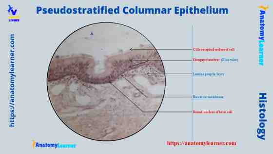
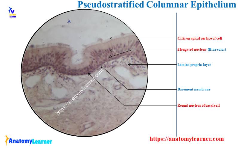
Identification of pseudostratified columnar cells under a microscope
Have you ever seen the pseudostratified columnar cells under a light microscope? No problem if you don’t know what the pseudostratified columnar cells look like under the light microscope. Here, I will help you identify the pseudostratified columnar under the microscope with its proper identifying characteristics.
Let’s see what these identifying features are –
For pseudostratified columnar with stereocilia (like epididymis) –
- The sample tissue shows numerous nuclei at various levels of the epithelium, but cells are not stratified (single layer).
- This is because the cells are of different shapes and heights, and many do not reach the surface of the epithelium. But, all of these cells attach to the epithelium’s basement membrane.
- You will find the stereocilia on these cells’ apical part that reaches the surface.
So, this is a slide of pseudostratified nonciliated columnar epithelial (epididymis). You will not find any mucous secretory goblet cells in this epithelium section.
Identifying points for the ciliated pseudostratified columnar cells –
- The sample tissue section also shows numerous nuclei at different levels as cells possess various shapes and heights.
- Some of the columnar cells of this epithelium reach the surface, and they possess true cilia.
- There are mucous secreting goblet cells (flask-shaped cells) present in the epithelium of the provided sample tissue.
- It also shows a thick basement membrane, a lamina propria, and numerous blood vessels.
So, this is a ciliated pseudostratified columnar epithelial slide. The main difference between the ciliated and nonciliated pseudostratified columnar epithelial is present on the cell’s apical surface. You will find the goblet cells and the cilia on the apical surface of the ciliated pseudostratified columnar cells.
Types of pseudostratified epithelium
There are two types of pseudostratified epithelium – pseudostratified columnar and transitional epithelium. All cells contact the underlying basement membrane in both pseudostratified epithelia, but not all reach the free surface.
Again, the pseudostratified columnar divides into two –
- Ciliated pseudostratified columnar and
- Noncilitaed pseudostratified columnar cells.
I think you already know the basic difference between ciliated and nonciliated pseudostratified columnar cells. The various shapes and heights of cells with their nuclei will be found in both pseudostratified epithelia.
As the cells are vary shaped and height, you will find the nuclei at different levels under the light microscope. Thus it provides the false stratification of the nucleus of cells.
You will see the cilia in the ciliated pseudostratified columnar epithelium under the light microscope. Many parts of the respiratory tract like the nasal cavity, nasopharynx, larynx, trachea, auditory tube, and large bronchi contain the ciliated pseudostratified columnar cells.
The mucosa secretory goblet cell occurs in pseudostratified epithelial and intermixes with the ciliated columnar cells. Thus it forms the respiratory epithelium in the respiratory tract of an animal. The cilia of the pseudostratified cell act as a mucociliary escalator that entraps and rids airways of foreign particles by sweeping and coordinating ciliary motion.
Again, the nonciliated pseudostratified columnar cells possess the same features as ciliated cells without the cilia. Instead of cilia, some of the cells possess stereocilia. These pseudostratified columnar cells lack goblet cells and are found in the animal’s reproductive tract. You may find the stereocilia in the lining epithelium of epididymis, the uterine horn of an animal.
Transitional epithelium
The transitional epithelium is a pseudostratified type of epithelium with many appearances. It lines the hollow organs capable of considerable distension, such as the renal pelvis, ureter, urinary bladder, and urethra.
You may also find the transitional epithelium in an animal’s larynx, palpebral conjunctiva, and nasopharynx. The cells increase in size from the basal layers to the superficial layers of the transitional epithelium.
Again, the shape of the transitional epithelium depends on the degree of organ enlargement at fixation. You will find the pillow-shaped large cell on the surface when the epithelium is under a little tension.
But, in the stretched condition, you will find flattened and elongated cells, and thus the height of the epithelium decreases. You may know more about transitional epithelium from another article by an anatomy learner with a labeled diagram.
Pseudostratified columnar epithelium location or examples
So, the location of the pseudostratified columnar epithelium is in the respiratory tract and reproductive tract. Most of the respiratory tract of an animal is lined with the pseudostratified ciliated columnar. The reproductive tract of an animal line with the nonciliated pseudostratified columnar cells. But, some pseudostratified columnar cells of the reproductive tract may contain stereocilia.
Fine, let’s know the location or example of pseudostratified columnar cells (ciliated) –
- The lining of the nasal cavity (nose) and paranasal sinuses
- Nasopharynx and larynx
- Lining of trachea
- Some parts of the large bronchi of the respiratory tract
Again, the pseudostratified columnar cells with stereocilia (long microvilli) are seen –
- In the lining of the epididymis of a male animal
You will also find the nonciliated pseudostratified columnar cells in –
- Some parts of the auditory tube,
- The lining of the ductus or vas deference, and
- Some part of the male urethra lining
So, I think you got a good idea of the location of pseudostratified columnar cells in the animal body. Now, you may know the details of these pseudostratified columnar cells from an animal’s different parts or structures, or organs.
Pseudostratified in the nasal cavity
An animal’s right and left nasal cavities to consist of a two-component – dilated vestibule and internal nasal cavity. You know the skin of the nose enters into the nares that possess sweat, sebaceous glands, and coarse, moist hairs. These hairs of the nasal cavity filter out the particular material from the inspired air.
You will find a two-chamber in the nasal cavity of an animal within its skull that is separated by a nasal septum. There are three bony shelflike projections at the nasal cavity’s lateral wall, known as the nasal conchae or turbinates. You may know the details of anatomical features of a nasal cavity from the below-mentioned article by an anatomy learner.
- Anatomical features of the nasal cavity (example with dog nose)
You will find the pseudostratified ciliated columnar cells in the lining of a nasal cavity. These are the respiratory epithelium and consist of tall, densely packed columnar cells that possess the apical cilia.
These nasal cavity cells are responsible for moving mucous along the epithelial surface. along with the pseudostratified columnar cell of a nasal cavity, you will also find the following cells –
- Goblet cells – mucous secreting cells that contain the basal nucleus.
- Basal cells – rounded cells
- Brush cells –these are the columnar cells and possess the apical microvilli instead of cilia.
- Small granule cells or dense core granule cells – contain small secretory granules and vesicles.
- Clara cell – sometimes may present in the respiratory epithelium (describe details with the epithelial cells of the trachea).
Olfactory epithelium of nasal cavity
You will also find the pseudostratified columnar lining on the olfactory region of a nasal cavity. This olfactory epithelium contains the modified bipolar neuron, olfactory neuron cell, supporting cells, and basal cells.
The olfactory neuron cells of the olfactory epithelium respond to odoriferous substances. Again, the supporting cells are the columnar cells with a broad cylindrical apex. The basal cells are small, spherical, and cone-shaped cells of the olfactory region of a nasal cavity.
Pseudostratified columnar epithelium trachea
You will also find the pseudostratified columnar epithelium in the trachea lining under a light microscope. So, there is a presence of respiratory epithelium that contains the pseudostratified columnar cells along with the goblet cell, basal cell, brush cell, small granule cell, and Clara cells.
You already know the features of all these respiratory epithelium cells except Clara cells. The Clara cells are present in the lower part of the trachea. You will also find these Clara cells in the bronchi (numerous).
These cells possess a smooth apical surface, and the dome protrudes towards the lumen of the trachea. The electron microscope view reveals numerous smooth endoplasmic reticulum, rough endoplasmic reticulum, and secretory vesicles.
The Clara cell secrets a phospholipid-rich lipoprotein surfactant that reduces the surface tension in air passageways. Again, the basement membrane of the trachea is thick under the light microscope.
In the trachea histology slide, you will also find the submucosa that shows the loose to moderately dense fibroelastic connective tissue with numerous tubule acinar glands. Again, in muscularis mucosa, you will see the C-shaped hyaline cartilage that bridges by smooth muscle at the posterior free end.
The adventitia of a trachea histology slide contains the loose connective tissue with numerous blood vessels. Learn more about trachea histology with a labeled diagram from another article by an anatomy learner.
Histological features of the trachea (lining epithelium and other different layers) with a labeled diagram.
Pseudostratified columnar of paranasal sinuses
Paranasal sinuses are the bilateral closed cavities in the frontal, maxillary, ethmoid, and sphenoid bones of the animal’s skull. The paranasal sinuses are also lined by the thinner respiratory epithelium (pseudostratified columnar cells) and fewer goblet cells.
Again, the lamina propria of the paranasal sinuses contain few glands and continue with the underlying periosteum. These paranasal sinuses communicate with the nasal cavity through the small opening.
With the activities of the ciliated epithelial cells of the paranasal cells, mucous drains into the nasal passages.
The caudal part of the vomeronasal organ will also contain the ciliated pseudostratified columnar cells. But, the middle part of the vomeronasal organ contains the nonciliated pseudostratified columnar cells.
If you don’t know what vomeronasal organs are, then it is for you – this is a tubular, blind-ending bilateral organ that locates in the mucosa of the ventral part of the nasal septum. This vomeronasal organ consists of the internal epithelial duct, middle propria submucosa, and external cartilaginous support, and it opens rostrally into the incisive ducts.
Pseudostratified in nasopharynx
The pharynx can be viewed as consisting of a nasopharynx in an animal’s respiratory system. In the nasopharynx mucosa, you will find two different types of epithelium lining.
Most area of a nasopharynx is lined with pseudostratified ciliated columnar cells. But, the lining epithelium of the caudodorsal part contact each other while swallowing food particles. So, if you view the caudodorsal part of the nasopharynx under a light microscope, you will find the stratified squamous epithelium lining.
Again, in the nasopharynx, you will find the loose to moderate dense fibroelastic connective tissue with serous, mucous, and mixed glands in its lamina propira. There are also prominent lymphatic nodules in the dorsal part of the nasopharynx. These lymphatic nodules are the pharyngeal tonsil of the nasopharynx.
You will not find any tunica muscularis layer in the nasopharynx. But, the nasopharynx surrounds by the skeletal muscle of the head and neck.
Pseudostratified columnar in the larynx
Under the light microscope, the lining epithelium of the larynx may vary from pseudostratified columnar epithelium to nonkeratinized stratified squamous. Most of the area of larynx lines with pseudostratified columnar cells. But, the mucosa of a vocal cord area, epiglottis, and upper part of the larynx lines with the nonkeratinized stratified squamous epithelium.
Except in the vocal cord, you will find the loose to moderate fibroelastic connective tissue and serous, mucous, and mixed glands in the lamina propria. Again, in the vocal cord’s lamina propria, dense fibroelastic connective tissue underlies a thin region of loose connective tissue.
The submucosa of the larynx consists of dense connective tissue, blood vessels, lymph vessels, and submucosal glands. Again, there is hyaline cartilage in the inner part of the tunica submucosa (in the thyroid, cricoid, and arytenoid).
But, you will find the elastic cartilage with dense fibroelastic connective tissue in the tunica muscularis layer of the epiglottis, cuneiform, corniculate, and tip of the arytenoid cartilage.
The lining epithelium of bronchioles
The bronchioles have three parts – primary or larger, terminal, and respiratory. The mucosa of a larger bronchiole consists of pseudostratified ciliated columnar cells with few scattered goblet cells. You will find numerous ciliated Clara cells in the mucosa of a larger bronchiole.
But, in the mucosa of the terminal and respiratory bronchioles, you will not find the pseudostratified columnar cells. The lining epithelium of the terminal part of the bronchiole is ciliated simple cuboidal. At the same time, the lining epithelium of respiratory bronchiole is ciliated and nonciliated simple low columnar or cuboidal with patches of simple squamous epithelium.
Pseudostratified columnar cells with stereocilia in the epididymis
When you view the duct of epididymis under a light microscope, you will see the lining of pseudostratified columnar cells. But, there are cilia on the apical surface of this pseudostratified epithelium. Instead of the cilia, you will find some long microvilli (known as stereocilia) involving secretion and absorption.
In these pseudostratified cells, you will see some tall columnar cells that reach the free surface. Again, some basal cells are in the microscopic view of an epididymis slide. These basal cells of the mucosa of an epididymis don’t reach the free surface.
But, the epithelial lining of the epididymis is not always found to be the same. You may only find the tall columnar cells (ciliated) in some parts of the epididymis, especially in the head.
Beneath the lining epithelium (pseudostratified columnar cells), there is a layer of circularly arranged smooth muscle fibers. So, you may tell the smooth muscle fiber to surround each duct of the epididymis.
Identifying features of epididymis under a light microscope (epithelial lining of mucosa and other features from different layers)
The epididymis labeled diagram shows the outer connective tissue capsule (identified). It also shows the pseudostratified columnar cells variety with stereocilia. The provided epididymis histology slide image identifies the circularly arranged smooth muscle fibers.
The sectioned tissue also shows the clump of spermatozoa in the lumen of the epididymal ducts.
Nonciliated pseudostratified columnar epithelium in ductus deferens
The ductus deferens (vas deferens) is a muscular tube extending from the epididymis’s lower end to the prostatic urethra. You will find the tunica mucosa, tunica muscularis, and tunica adventitia on the wall of a ductus deferens of an animal.
The mucous membrane of a ductus deferens shows numerous longitudinal folds so that the lumen appears to be stellate in section. Again, the lining epithelium may vary from the simple columnar to the pseudostratified columnar epithelium in the distal part of the tube under a light microscope.
In addition, you may also find the ciliated pseudostratified columnar lining cells in the extra-abdominal part of the ductus deferens. A thin lamina propria supports these epithelia with many elastic fibers.
You may know more about the histological features of a ductus deferens from another article by an anatomy learner.
- Histology and identifying features of ductus deferens with labeled diagram (including the lining epithelium from the mucous membrane)
There are inner longitudinal, middle circular, and outer longitudinal smooth muscle layers in the tunica muscularis of a ductus deferens. So, the tunica muscle layer of a ductus deferens is thicker than other organs.
The outermost layer is tunica adventitia, which comprises collagen fibers, blood vessels, and nerves. In the ductus deferens labeled diagram, I tried to show you the pseudostratified columnar cells from the small irregular lumen.
Again, this diagram shows a thick layer of smooth muscle arranged in inner longitudinal, middle circular and outer longitudinal patterns. In addition, the diagram also shows the thin adventitia that contains the fibroelastic connective tissue.
Pseudostratified columnar cell description under the microscope
I know you already got a clear idea of the description of a pseudostratified columnar cell under a light microscope. The pseudostratified columnar comprises a single layer of cells, but their nuclei are located at various levels because the cells are irregular in size and shape.
Thus this pseudostratified columnar appears to have several layers. The cells (columnar cells) that extend from the basement membrane to the surface of pseudostratified columnar are ciliated or nonciliated. They also possess mucous secretory goblet cells.
The basal cells of the pseudostratified epithelium attach to the basement membrane but do not reach the free surface. By division and differentiation, basal cells replace the other epithelial cells.
But, under the light microscope, you will not see the boundary of the pseudostratified cells. You will only see the nuclei of these cells at the different levels. Again, under the electron microscope, the exact figure of the pseudostratified epithelium will be seen.
Pseudostratified columnar epithelium with goblet cells
Goblet cells are the primary example of the unicellular exocrine gland. These goblet cells may find among the columnar or pseudostratified columnar cells. They line the digestive tract and part of the respiratory tract. The goblet cells produce the mucous.
The microscopic figure of the goblet cell reveals that there is a thin basal region that sites on the basement membrane. Again, they possess the expanded apical portion (the theca) that faces the digestive or respiratory tract lumen.
You will find the membrane-bound secretory droplets at the apical part of the goblet cells. The microscopic figure of the goblet cells also shows the cytoplasm to the cell’s periphery and the nucleus towards its base.
The mucous release from the goblet cells is regulated and stimulated by chemical irritation and parasympathetic innervation. This results in the exocytosis of the entire secretory contents of the goblet cells.
Do you know the main functions of goblet cells? It lubricates and protects the epithelial sheet of the digestive and respiratory tracts.
Pseudostratified columnar epithelium with cilia
You may have a question what are the cilia of pseudostratified columnar epithelium? Well, the cilia are long hair-like projections of the plasma membrane. Whereas the stereocilia are very long, thick microvilli, nonmotile, and may show branches. Stereocilia increase the surface area for absorption.
Again, the microvilli are a minute figure-like projection of the plasma membrane, increasing the surface area for absorption. They also transport the absorbed material and participate in the digestion of carbohydrates.
Under the light microscope, you will easily see the cilia on the apical surface of the pseudostratified columnar cell. In the living state, cilia can be seen to be motile.
Again, under the light microscope, it is very hard to observe the structure of cilia. You may only understand the structural feature of cilia under an electron microscope.
You will find a shaft at the free end of the cilium. Again, the base is the attached portion of the shaft with the cell surface. The shaft of a cilium becomes tapper in its tip.
A cilium comprises an outer covering formed by the extension of the cell membrane and an inner core exonumia. The exonumia is formed by the microtubules arranged in a definite manner.
The electron microscope view shows a central pair of tubules in the cilium structure that is surrounded by nine pairs of tubules. The outer tubules are connected to the inner pair by radial structures. Other projections pass outwards from the outer tubules.
At the cilium base, an additional tubule is added to each outer pair. The microtubules of the cilia bind with the protein dynein and nexin.
Significance of cilia of pseudostratified cells
You will find a variety of significance in the cilia of the pseudostratified epithelium. Some of the significant functions are listed below –
- Cilia of the respiratory epithelium help to move the secretion in the trachea and bronchi towards the pharynx.
- The ciliary action helps the movement of the ova through the uterine tube and spermatozoa through the male reproductive tract.
- The olfactory cilia of the respiratory organ perform sensory functions.
Pseudostratified columnar cells labeled diagram and drawing
You already got different labeled diagrams on the pseudostratified cells. Again, I will show you a hand drawing pseudostratified columnar cell.
Here, in the pseudostratified columnar hand drawing image, all structures like cells, nucleus, goblet cells, basal cells, and cilia are presented clearly. You may follow this image and may draw the pseudostratified columnar cell quickly.
Again, you may get more hand-drawing pseudostratified columnar cells on social media for anatomy learners.
Frequently asked questions on pseudostratified columnar cells
Now, let’s get the short and quick answers to the frequently asked questions on pseudostratified columnar cells (ciliated and nonciliated).
Where is pseudostratified columnar epithelium found?
The ciliated pseudostratified columnar epithelial is found in the different parts of the respiratory tract (nasal cavity, paranasal sinuses, auditory tube, trachea, nasopharynx, and larger bronchioles). Again, a pseudostratified columnar cell with stereocilia is found in the mucosa lining of the epididymis.
But, the nonciliated pseudostratified columnar epithelium is found in some parts of the auditory tube, ductus deferens, and some parts of the male urethra. I have already described these structures in detail in this article; please read this article from start to end.
What are the functions and location of pseudostratified columnar epithelium?
The pseudostratified epithelium is secretory, whereas the short basal cells are the stem cells that constantly replace the tall columnar cells. Again, the cilia of the pseudostratified cells clearance the mucous. In addition, the stereocilia of the pseudostratified columnar help in absorption.
What are the characteristics of a pseudostratified columnar epithelium?
The main characteristics of pseudostratified columnar cells are – the presence of a single layer of tall columnar cells and short basal cells. So, the size and shape of the pseudostratified epithelium may vary.
They contain single layers of nuclei but locates at different levels, and thus they provide the state of stratification. Within the tall columnar and basal cells, you will also find the mucous secretory goblet cells.
What does pseudostratified columnar epithelium do?
The main function of the pseudostratified columnar cells is secretion, protection, and absorption.
Conclusion
The pseudostratified columnar epithelium is a single layer of irregular cells (tall columnar, short basal cells, and goblet cells) that rest on the basement membrane. But, the nuclei of these pseudostratified epithelia are located at various levels that appear to have several layers.
Some of the pseudostratified columnar cells possess the cilia; some possess the stereocilia. The tall columnar cells of the pseudostratified epithelial rest on the basement membrane, reach the free surface and possess the cilia. Again, the short basal cells are attached to the basement membrane but do not reach the free surface. The mucous secretory goblet cells are among the tall columnar and basal cells.