The cat digestive system includes a mouth cavity, pharynx, alimentary canal, and different accessory organs. There are two major divisions in the mouth cavity of a cat – vestibule and mouth cavity proper. The alimentary canal of a cat starts with the esophagus and ends at the large intestine.
Again, the accessory organs include teeth, tongue, liver, gallbladder, pancreas, and three major types of salivary glands. In this single article, I will try to summarize the anatomy of all the organs from the cat digestive system with a labeled diagram.
In addition, I will provide some of the pictures of the dissected cat at the end of this article. You will learn the unique features of the cat’s digestive system organ and how they differ from other animals like goats, horses, or humans.
Cat digestive system
First, I would like to provide a basic idea of the different parts of the cat digestive system. The digestive system of any animal consists of these structures and organs that function in prehension, transport and break down of feed particles.
The organs or parts of the cat’s digestive system are not so complicated as those of birds or small mammals. You might have a good anatomical idea of the major component like mouth, pharynx, alimentary canal, liver, and pancreas from the digestive system. But, let’s try to identify all of the parts, organs, and structures from the cat’s digestive apparatus.
Cat digestive system parts and functions
The labeled diagram of the cat digestive system shows the following parts, structures, and organs –
- A mouth cavity of the cat,
- The pharynx of the cat,
- A long alimentary canal, and
- Different accessory organs of the cat digestive apparatus
Again, in the mouth cavity of a cat’s digestive apparatus, you will find the following structures, organs, and parts –
- Vestibule – includes the lip, cheek,
- Mouth cavity proper – includes teeth, gum, tongue, and salivary glands.
There are different types of papillar structures (like filiform, fungiform, vallate, conical, marginal) present on a cat’s tongue. Again, there are the parotid, mandibular, sublingual, and zygomatic salivary glands in the mouth cavity proper of a cat (they will enlist under the accessory organs).
The pharynx of a cat includes three major parts –
- The nasal pharynx of the cat,
- A oral pharynx of the cat, and
- The laryngeal pharynx of the cat.
Again, the alimentary canal of the cat’s digestive apparatus includes the following parts, organs, and structures –
- The esophagus of a cat,
- The stomach (simple) of a cat – includes two major parts – glandular and non-glandular,
- Small intestine – includes duodenum, jejunum, and ileum,
- The large intestine of the cat’s digestive apparatus – includes the cecum, colon, and end parts.
- The colon of a cat possesses three major divisions – ascending, transverse, and descending. Again, some other parts of the cat’s colon will be described in this article’s specific part.
- The main accessory organs of the cat’s digestive apparatus are –
- Liver of a cat,
- The pancreas of a cat,
- The spleen of a cat, and
- Salivary glands (listed earlier).
You will see a close relationship between the respiratory organs and a cat’s digestive organs. Now, let’s explore and identify the organs mentioned earlier from the dissected samples of a cat.
Functions of the digestive organs of a cat
This is not my main goal to provide all the functions of the specific organs from the cat’s digestive apparatus. Here, I will summarize all the organs’ functions in short.
The digestive organs like a lip and tongue help to prehension of feed, again the teeth help them to masticate or grind,
All the feed is then transported through the esophagus to the stomach of the cat,
The breakdown of the grinding feed particles occurs,
Again, the temporary storage and absorption occur in the intestine of a cat,
Finally, the waste product discharges from the end part of the cat’s alimentary canal.
Again, the liver, pancreas, spleen, and salivary glands have specific functions in digesting and absorption of the feed particles (directly or indirectly).
Special features of the cat’s digestive organs
Though you will learn the anatomical features of the specific organs from the cat’s digestive apparatus, here, I will only include the unique characteristics.
The orifice of the cat’s mouth is extensive, and the upper lip covers the lower lip. You will find the papillar salivates just opposite the third premolar tooth. Again, the papilla incisive locates behind the central incisor teeth.
There are 30 permanent teeth in the dental formula of a cat. They have fewer premolar and molar teeth compared to dogs or other common domestic mammals.
The cat tongue occupies most of the space within the oral cavity. There is a longitudinal groove (median sulcus) in the dorsal surface of a cat’s tongue. The body and root of the cat tongue are less mobile than the apical part.
The cat has several paired salivary glands, along with the diffuse salivary tissue in the oral cavity. You will find encapsulated V-shaped parotid salivary gland in the base of a cat’s ear. Again, the mandibular gland is also the oval and encapsulated organ in a cat.
The pharynx is an intersection between the digestive and respiratory systems, a Y-shaped area in a cat (when viewed with a sagittal section).
The cat esophagus has three distinct parts – cervical, thoracic, and abdominal. Again, a cat’s stomach is a J-shaped organ that lies mainly on the left side of the body.
The cat intestine is not highly modified and constitutes the alimentary canal’s relatively simple tubular constitution. You will find a short, comma-shaped cecum in a cat that divides from the proximal part of the large intestine.
Special features of accessory organs
The liver of cat digestive system divides into right, left, quadrate, and caudate lobes. Again, the right and left lobe of the cat liver subdivides into the lateral and medial parts by a fissure. But, sometimes, you may find the quadrate and right medial lobe in the cat’s liver.
A cat’s pancreas is a pale, lobulated organ that consists of a body and right and left lobes. Again, the body of the pancreas is nestled in the bend of the cranial part of the duodenum. From this body, the right and left lobes of the pancreas are origins.
The spleen of a cat is a large lymphoid organ discussed with the digestive system because it occurs in the peritoneal cavity and the viscera. You will find a large, tongue-like spleen that lies on the left side of the body and attaches to the ventral layer of the greater omentum.
Cat digestive system anatomy
This part will discuss the anatomical facts of different organs, parts, and structures in detail from the cat digestive apparatus. But, you may also get the full guide on the different digestive organs’ anatomy in the cat and dog anatomy learning section.
Now, let’s start with a cat’s oral cavity, which has two parts – the vestibule and oral cavity proper. The cat’s oral cavity is bounded rostrally and laterally by the lips and cheek. It extends caudally to join the oropharynx at the caudal border of the hard palate.
You know the vestibule of the oral cavity is the space peripheral to the teeth and bounds by the lips and cheeks. There are some diffuse salivary glands, sebaceous glands, and apocrine glands present on the lip of a cat.
In the upper lip of a cat, there is a median groove called the philtrum. You will also find the additional salivary tissue in the cat’s cheek. The buccinator’s muscle primarily supports the check of a cat.
The oral cavity proper of the cat is bound dorsally by the hard palate. This is formed by incisive, maxillary, and palatine bone processes. You will find the hard palate in a cat with seven transverse ridges.
There is a papilla incisive just caudal to the central incisor teeth of the cat. You will find the sublingual caruncles located on the oral cavity floor to either side of the rostral end of the frenulum lingual.
Now, let’s discuss the different parts, organs, and structures from the mouth cavity of a cat with the labeled diagrams.
The lip and cheek of a cat
The lips are the thick skin fold that bound the entrance to the mouth cavity. You will find the hair on the outer surface and mucous membrane on the inner surface of a cat’s lip. As you know, there is a median deep external groove on the upper lip of a cat.
A thick fold (frenulum) binds the jaw along the inner surface of the median external groove. You will find numerous papillae on the inner surface of the lip.
The cat’s lower lip is also joined to the jar by the frenulum at the median groove. You will find different muscles in the cat’s lip like – orbicularis oris, levator labii superioris, angular or levator labii superioris proprius, and buccinators.
You will also see the myrtoformis, moustachier, and quadratus labii inferioris muscles in a cat’s lip structure.
The orbicularis oris is a very thin layer of muscle that make the upper lip thicker than the lower lip of the cat. Again, the labii superioris muscles erect the whiskers and help raise the cat’s upper lip.
The caninus or levator anguli oris muscle raises the cat’s upper lip. There is a thin buccinator muscle against the mucous membrane of the cat’s upper lip. This muscle joins with the orbicularis oris and lies beneath the caninus muscle of the lip.
Myrtoformis muscle of the cat’s lip elevates the upper lip. The quadratus labii inferioris is a thin flat band extending along the cat’s lower lip length. This quadratus muscle depresses the cat’s lower lip.
The cheek of the cat
The cheek of a cat is relatively thin and small compared to the dog. Cat’s cheek extends from the lips caudal to the ramus of the mandible. You will also find the hair on the outer surface of the check of a cat. Again, the inner surface of the cat’s cheek is smooth and folded.
On the inner surface of the cat’s cheek, you will find the opening ducts of the different salivary glands like – the duct of parotid or steno’s duct, duct of the molar gland, and duct of the infraorbital salivary gland.
Cat teeth anatomy
The cat teeth are highly modified for grasping, puncturing, and tearing. You will not find the first premolar and lower first or second molar tooth in the dental formula of a cat. Again, there is only a single molar tooth on each side of a cat’s upper and lower jaw.
You will find 6 incisors, 2 canine, 5 premolar, and 2 molar teeth on each side of the jaw. So, there are, in total, 30 teeth in a cat.
The number of teeth is fewer than that of other domestic mammals. But why does this occurs in a cat? This is due to the absence of premolar and molar teeth in a cat.
The teeth of the cat are highly adapted for a carnivorous diet. Six incisor teeth are embedded in the alveolar border of the premaxilla and mandible. These are the very small teeth in the cat that possess very sharp edges.
Again, the small, wedge-shaped incisors are adapted for the nipping. In addition, the elongated conical canine teeth are for stabbing and holding the prey. You will also see the blade-like molariform teeth in a cat’s mouth.
There is a large space in between the lower canine and premolar teeth. This space is known as the diastema, absent in human and ruminant animals.
The teeth are implanted in the alveolar border of the three different bones – premaxilla, maxilla, and mandible. There are three distinct parts in every tooth of a cat – root, neck, and crown. In the dog teeth anatomy article, you will get a details description of the root, neck, and crow of a tooth.
Deciduous teeth of a cat
You will not find any teeth on the bird of a kitten. Later there appear 26 (twenty-six) teeth in the dental formula of a kitten. You will find twelve incisors, 4 (four) canines, and 10 (ten) molar teeth in a kitten.
Again, all these teeth are replaced by permanent teeth, and then you will see 30 teeth.
Cat tongue anatomy
The tongue is a very mobile and muscular organ in the cat digestive system that plays an important role in the cat’s life. You will see the frenulum lingual that attaches to the tongue on the anterior floor of the oral cavity.
The cat tongue also possesses three main parts – root, body, and apex, where the body and root are fewer mobiles than the apex. Again, the cat tongue’s dorsal surface is structurally different from that of the dog tongue.
You will see a dorsal longitudinal groove (median sulcus) on the dorsal surface of the cat tongue. The dorsal surface of the cat tongue possesses numerous highly cornified caudally directed filiform papillae. Again, there are other papillae like fungiform, vallate, and foliate on the cat tongue.
The fungiform are the less numerous mushroom-shaped, circular papillae located at the rostral two-thirds of the dorsum of the tongue. They remain scattered and distributed within the filiform papillae.
You will find the large, round vallate papillae arranged in a V-shaped configuration near the root of the tongue. Again, foliates are the leaf-shaped papillae that locate on the posterolateral aspect of the cat’s tongue.
A cat’s tongue’s fungiform, vallate, and foliate papillae show the taste bud in microscopic examination.
In addition, the bulk of the cat’s tongue consists of intrinsic skeletal muscle, connective tissue, and adipose tissue. The intrinsic muscle of the cat tongue includes genioglossus, styloglossus, and hyoglossus.
You will find the motor innervation in the cat tongue from the hypoglossal nerves. Again, the cat’s tongue is a highly vascular organ that supplies by the lingual arteries.
Muscles of the cat tongue
The genioglossus muscle lies beneath the geniohyoid bone and origins from the medial surface of the cat’s mandible. Muscle fibers pass dorsally in a fan-like manner and form a flat verticle plate. Do you know the function of the genioglossus muscle in a cat?
This muscle of the cat’s tongue draws its root forward, and it also draws the tip backward.
The hyoglossus muscle of a cat’s tongue origins from the ventral surface of the body of the hyoid bone. Again, the fibers from the genioglossus muscle interchange with the styloglossus and the lateral part of the tongue. This muscle retracts the cat’s tongue and depresses it.
The styloglossus muscles of a cat’s tongue originate from the mastoid process of the temporal bone. Its fiber passes towards the tip of the cat’s tongue. This muscle also retracts the cat’s tongue and raises it.
Salivary glands of a cat
A cat’s salivary glands are paired and located along the lateral surface of the head beneath connective tissue and skin. You will find several salivary glands in a cat’s digestive apparatus (accessory organs) –
- Buccal or molar glands,
- V-shaped parotid salivary glands,
- Ovoid mandibular salivary glands,
- The monostomatic and polystomatic sublingual salivary glands, and
- Zygomatic salivary glands of the cat.
The parotid is the largest salivary gland in a V-shaped cat and locates at the base of the ear. They are the large, diffuse, lobulated structure associated with the overlying connective tissue.
The parotid gland duct emerges from the midpoint of the anterior surface of the gland and opens opposite the third upper premolar tooth. Another parotid salivary gland duct name is Stenon’s or Steno’s duct.
You will find the ovoid and encapsulated mandibular or submaxillary salivary gland in the cat that locates just ventral to the parotid and posterior to the angular process of the mandible. These are the lobular glands, but the lobes are less diffuse and gove a smooth appearance.
The duct of the mandibular salivary gland emerges from the anterior edge and opens at the base of a small papilla just anterior to the lingual frenulum. Another name for the mandibular salivary glands duct is Wharton’s duct.
In addition, the sublingual glands are the smallest, which are conical in shape and possess a smooth surface. The sublingual glands of a cat divide into monostomatic and polystomatic parts. You will see the sublingual ducts run parallel to the mandibular duct and open at the oral cavity floor in the vicinity of the mandibular duct.
Two minor salivary glands of a cat
If you explore the cat dissected sample, you will find two other salivary glands – molar and zygomatic or infraorbital glands. You will find the brownish-gray and granular molar salivary glands of a cat.
This molar gland of the cat locates at the angle of the jaw and immediately beneath the skin. Again, the duct of the molar salivary glands opens on the inner surface of the cheek.
The zygomatic or infraorbital salivary glands are the small glands present in the salivary system of a cat. These zygomatic salivary glands lie on the floor of the orbit of a cat’s eye. The duct of the zygomatic salivary glands opens into the posterolateral part of the roof of the cat’s mouth cavity.
Hard and soft palates of a cat
The hard and soft palates are also included in the cat digestive system because it is part of the oral cavity. The cat’s oral cavity roof is formed by the two palates – hard and soft. The hard palate forms the cranial part of the roof, whereas the soft palate forms the caudal part of the roof.
In the hard palate structure, you will find a bony shelf. This bony shelf comes from the palatine bone and palatine processes of both maxilla and premaxilla bones. Again, the hard palate covers with a series of folds (sever or eight – known as transverse ridges).
You will see rows of papillae in between the transverse ridges of the cat’s hard palate. There is a papilla at the midline of the most cranial ridge. The Stenson’s duct of a cat is open on each side of the papilla located on the cranial ridge.
Soft palate of the cat
The soft palate of the cat consists of connective tissue and muscle that extend from the caudal end of the hard palate to its free edge. So, it forms the caudal part of the cat’s palate and attaches to the caudal part of the palatal plates.
The soft palate also attaches to the ventral border of the perpendicular palatine, pterygoid process, and hamuli of the sphenoid bone. Again, the caudal end of the soft palate lies at the level of the epiglottis.
You will see two mucosal folds passes to the floor of the pharynx and from the caudal and cranial pillars of the fauces. Again, within these folds, there are two reddish lobulated tonsils.
There are two major muscles in the cat’s soft palate structure – tensor veli palatine and levator veli palatine muscles. The tensor veli palatine muscle of the cat’s soft palate originates from the ventral surface of the sphenoid bone. This muscle stretches the soft palate of the cat.
The levator veli palatine of the cat’s soft palate is a flat triangular muscle lying within the tensor veli palatine muscle.
The pharynx of a cat
The pharynx is a space shared by the digestive and respiratory systems of a cat. It extends from the oral cavity to the larynx of a cat and subdivides into nasopharynx, oropharynx, and laryngopharynx. When viewed in the sagittal section, you will see a Y-shaped area in the cat pharynx.
There are some intervening muscles – longus capitis, longus coli, and levator scapulae ventralis that separate the dorsal wall of the pharynx from the base of the skull. Again, the hyoid bones and laryngeal cartilages support the lateral and ventral walls of the cat’s pharynx.
The nasopharynx lies dorsal to the soft palate and is also part of a cat’s respiratory system. Again, the oropharynx lies ventral to the soft palate and continues with the oral cavity. In addition, the laryngopharynx of the cat is that portion of the pharynx that continues from the tip of the epiglottis to the glottis.
The dorsal part of the cat’s nasopharynx extends from the internal nasal choanae or nares to the free end of the soft palate. This nasopharynx is strictly related to the respiratory system; no feed particle does not pass through this nasopharynx.
You will see two openings of the auditory tube in the lateral wall of the nasopharynx. These auditory tubes connect the air-filled cat’s middle ear cavity with the nasopharynx. Functionally, you will find the important role of the nasopharynx in the equalization of the air pressure.
Oropharynx of the cat
The cat’s oropharynx is a space bounded laterally by the palatoglossal arches. This oropharynx of a cat extends from the base of the tongue to the free end of the soft palate.
There are fauces that transition between the oral cavity and pharynx. Feed particles or air passes through the oropharynx to the trachea or esophagus of the cat. You will see small palatine tonsils lying in the tonsilar fossae. These tonsilar fossae with the tonsils are located at the oropharynx’s dorsolateral aspect.
The caudal part of the nasopharynx continues as the small pharynx proper that is cranially bounded by the epiglottis and margin of the soft palate. You will find different types of muscle in the pharynx of a cat.
Let’s know a little about the anatomy of the pharyngeal muscles from cat anatomy.
Muscles of the cat’s pharynx
So, what are the common muscles in the cats’ pharynx? Well, you will find the following muscles in the cat’s pharynx –
- Glossopharyngeal muscle of the pharynx,
- Constrictor pharynges inferior muscle,
- Constrictor pharynges medius muscle,
- Stylopharyngeus muscle of the pharynx,
- Pterygopharyngeus muscle of the pharynx.
The glossopharyngeal muscle originates from the lateral part of the genioglossus and midventral part of the styloglossus. It inserts into the median dorsal raphae of the cat’s pharynx and constricts the pharynx.
The constrictor pharynges inferior muscle is a thin sheet that covers the caudal end of the cat’s pharynx. It originates from the lateral surface of the thyroid and cricoid cartilages and inserts on the median longitudinal raphe on the dorsal surface of the pharynx.
This constrictor pharynges inferior muscle and also constrict the pharynx.
The constrictor pharynges medius muscle is also a thin sheet of muscle covering the pharynx’s middle part. This muscle originates from the hyoid bone and is inserted into the median dorsal raphe of the pharynx.
The stylopharyngeus muscle of the cat’s pharynx originates from the tip of the mastoid process of the temporal bone. You will find a flat, triangular pterygopharyngeus muscle in the cat’s pharynx that originates from the tip of the hamular process of the pterygoid bone.
The pterygopharyngeus muscle of the cat’s pharynx also inserts into the median dorsal raphe of the pharynx. This muscle also constricts the pharynx of the cat.
The esophagus of the cat
The esophagus is the first part of the alimentary canal of the cat digestive system. It is a muscular tube capable of being great expansion. Again, the cat’s esophagus passes through the mediastinum dorsal to the trachea.
A cat’s esophagus divides into three major divisions – cervical, thoracic, and abdominal parts. At the beginning of the cervical part, it runs over the dorsal surface of the trachea. Again, the caudal part of the cervical esophagus slides off to the trachea’s left side and enters into the thoracic inlet.
In addition, you will find a close association between the recurrent laryngeal nerve and the left carotid sheath with the trachea (on the left side). The cervical part of a cat’s esophagus is normally flattened.
The thoracic part of the cat’s esophagus includes dorsally in the cranial mediastinum. Again, it returns to the dorsal surface of the trachea. It also lies posterior mediastinum and ventral to the aorta while passing through the thoracic cavity.
The esophagus then passes the right side of the aortic arch and crosses the dorsal aspect of the tracheal bifurcation. Again, it continues through the caudal mediastinum to the esophageal hiatus in the diaphragm.
You will find a close relationship with the vagus nerve at the heart level.
There are muscular, submucosa, and mucosal layers in the cat’s esophagus structure. You will also find the longitudinal folds in the inner surface of the cat’s esophagus.
But, there are no glands in the mucosa of the cat’s esophagus. You will find the mucosal gland in the submucosa of the pharyngoesophageal part of the cat’s esophagus.
Exception in cat esophagus
Generally, in the structure of the muscular layer of the cat’s esophagus, you will find smooth muscle. But, the part of the esophagus cranial to the base of the heart possesses the striated muscle in its muscular layer.
So, there you may find a change in the mucosal pattern in the esophagus at the heart level. You will find the longitudinal folds and the transverse mucosal folds in this part of the esophagus.
The cat’s esophagus passes through the right crus of the diaphragm at the esophageal hiatus. Again, it continues as a short abdominal part and joins to the stomach at the cardia. You will find a loose attachment of the esophagus to the border of the hiatus by its adventitia.
Again, there is a serosal covering on the short abdominal part of the cat’s esophagus. You may learn more about the structure of the cat’s esophagus from the microscopic study. Here, I have already published an article related to the microscopic features of the animal esophagus in detail with the properly labeled diagrams.
Peritoneum of cat
In a cat’s abdominal and pelvic cavities, you will find the peritoneum. This is a serous membrane that covers the internal organs of the cat’s abdominal and pelvic cavities. The covering of the serous membrane or peritoneum starts from the wall of the cavities onto the surface of the digestive and urogenital organs.
TThe cat’s peritoneum has two different parts– parietal and visceral parts. The part of the peritoneum that lines the abdominal and pelvic cavities is the parietal peritoneum. Again, the part of the peritoneum that covers the surface of the specific organs of the abdominal and pelvic cavities is the visceral peritoneum.
As there are two layers in the peritoneum and ultimately continue with each other, you will find a peritoneal cavity between these two layers. Again, you will find the peritoneal fluid in the peritoneal cavity of the cat.
There are different modifications of the cat’s peritoneum like mesentery, omentum, and ligament that are described in the ruminant digestive system anatomy. You will see different blood vessels that passage through the two layers of the peritoneum and supply the specific organs of the digestive and pelvic cavities.
Again, some organ remains against the body wall and covers only the peritoneum on one surface. These are the retroperitoneal organs; the best example of the retroperitoneal organ of a cat is a kidney.
Cat stomach from the digestive system
The stomach of the cat digestive system is another important and unique organ compared to the ruminant. You will see a J-shaped simple stomach lying mainly on the left side and possessing two major curvatures – greater and lesser. Again, you will find three parts in the cat’s stomach: cardiac, pyloric, and funds.
You know a cat’s stomach is the expanded or widest part of the alimentary canal. Grossly, you will see two main parts – the dorsal expanded part and the narrow ventral part. The dorsal expanded part of the cat’s stomach lies left and dorsal and has a communication with the esophagus.
Again, the narrow ventral part of the cat’s stomach lies right and ventrally, connecting with the first part of the duodenum. The convex left margin of the cat’s stomach is the greater curvature, whereas the concave right margin is the lesser curvature.
You know the peritoneum attaches to the stomach, which is called the omentum. So, the peritoneum that attaches to the lesser curvature is the lesser omentum. The lesser omentum of the cat’s stomach occurs between the liver and the lesser curvature of the stomach.
Again, the peritoneum covering the greater curvature of the cat’s stomach is the greater omentum. You will see a triangular gastrocolic ligament that extends from the greater omentum to the mesocolon.
The greater curvature moves caudoventrally during the cat’s stomach filling, but the lesser curvature remains fixed in position.
Now, let’s identify the three major divisions or ends or parts of the cat’s stomach.
Parts of the cat’s stomach
The part of a cat’s stomach below the cardiac sphincter that communicates with the esophagus is cardiac. Again, the pyloric part is the narrow part (opposite the cardiac) that connects to the intestine (duodenum).
You will see a pyloric sphincter, a muscular valve located in the distal end of the pyloric region. This pyloric sphincter regulates the movement of the stomach contents into the intestine.
Again, you will find a large inflated part of the stomach between these two parts (cardiac and pyloric). So, this inflated part of the cat’s stomach is the fundus and body. But, which specific part is the fundus and body?
The upper inflated part of the stomach is the fundus, and the lower inflated part is the body of the cat’s stomach.
Grossly, you will see the longitudinal mucosal folds on the inner surface of the fundus and pyloric part of a cat’s stomach. But, the prominence of these longitudinal folds depends on the degree of distension.
The greater omentum and different types of ligaments hold the cat’s stomach in its position. There is a gastroduodenal ligament that connects the stomach with the duodenum. Again, the gastrolienal ligament connects the stomach with the spleen.
Microscope features of a cat stomach
The wall of a cat’s stomach consists of mucosa, well-developed submucosa, a laminated tunica muscular layer, and a serosal covering. You know the entire stomach of a cat is glandular, and gastric folds or rugae on its mucosal surface.
These irregular folds of the cat’s stomach are oriented predominantly in a longitudinal direction. You may see the prominent longitudinal folds in the empty stomach of a cat.
The mucosal lining epithelium may change from the stratified squamous to the simple columnar epithelium in the cat stomach. Again, the mucosa of the cat’s stomach contains different glands in specific regions.
You will see the seromucous glands in the cardiac region of the cat’s stomach that form a narrow annular zone immediately distal to the cardiac opening. The lining is thicker at the fundus and body of the cat’s stomach, which contains the tubular gland. They contain the acid-producing parietal and enzyme-releasing chief cells.
There is lighter and thinner mucosa in the pyloric region of the pylorus part, and they contain branched mucous-producing glands.
Again, the tunica muscular layer of a cat’s stomach may vary, but the normal pattern is – outer longitudinal and inner circular smooth muscle layers. But, in the greater curvature of the cat’s stomach, you will see the intervening third layer of the internal oblique muscle layer.
In addition, the serosa of the cat stomach is formed by the visceral peritoneum and closely invested in the tunica muscular layer.
Three major branches of the celiac artery supply the cat’s stomach.
Small intestine from cat digestive system
The intestine from a cat digestive system is not highly modified and contains two major parts – the small intestine and the large intestine. The cat’s intestines attach to the dorsal body wall by the mesentery. In a cat’s intestine structure, you will see mucosa, submucosa, muscularis, and serosa layers.
The mucosal surface of the different parts of the cat’s intestine possesses longitudinal folds of variable height. You will also find the mucosal gland throughout the small and large intestine mucosa. Again, cats possess few gross patches of aggregated lymph nodules in the wall of a small intestine.
In addition to the longitudinal folds of the cat’s intestine, you will see a carpet-like covering of the intestinal villi. The cat’s small intestine intestinal villi greatly increase the absorptive surface.
You will see the major nerves and vascular network at the submucosa of the cat’s intestine. Again, the muscular layer of the cat’s intestine consists of a thinner outer longitudinal and an outer thicker circular layer of smooth muscle.
The cat’s small intestine consists of the duodenum, jejunum, and ileum, extending from the pylorus to the cecolic junction. Again, the length of the small intestine of a cat is very interesting, and it has a length about three times that of its body.
Now, let’s discuss the different parts of a cat’s small intestine.
Duodenum of a cat
The short proximal part of the cat’s small intestine is the duodenum. It extends from the pyrolus to the position of the duodenocolic fold. A cat’s duodenum is fixed in position by its short mesentery (mesoduodenum).
You will find three major parts in the cat’s duodenum – ascending, flexure (cranial and caudal), and descending parts. A short cranial part of the duodenum courses cranially from the pylorus.
Then it turns sharply to the right and caudally to form the cranial flexure. Finally, it continues as the descending part of the duodenum at the right side of the abdomen.
At the level of five lumbar vertebrae, the descending duodenum turns to the left, then cranially at the caudal duodenal flexure. Again, it continues as the ascending duodenum and passes cranially to the duodenojejunal flexure. The duodenojejunal flexure marks the junction of the duodenum and jejunum of the small intestine.
You will find duodenal glands at the proximal part of the cat’s duodenum. The bile and pancreatic ducts obliquely penetrate the mesenteric border of the descending duodenum just distal to the cranial duodenal flexure.
The area where these two ducts open on the duodenum is known as the ampulla of Vater. This is an oval space in the dorsal wall of the cat’s duodenum.
Microscopically, you will find all four layers of a typical tubular organ in a cat’s duodenum wall. You may read the details of the microscopic features of the animal’s duodenum from another article by an anatomy learner.
Anatomy of a cat jejunum
The jejunum is the longest part of the cat’s small intestine, which remain colis and form loops. You will see a relatively long mesentery so that the jejunum of a cat can freely move into the occupied spaces available within the abdomen.
The jejunal loops of the cat’s jejunum typically fill the middle part of the abdominal cavity. It fills between the stomach and liver and caudally around the urinary bladder.
But, the jejunum may be displaced craniodorsally by the gravid uterus (in late gestation) and compressed into whatever space is available in the abdomen.
Ileum from cat digestive system
The ileum is the short and terminal part of the cat’s small intestine. It lies caudal to the abdominal cavity and is separate from the ventral wall of the abdomen by the greater omentum. You will find a uniform diameter in the cat’s ileum, but the caudal part may possess a thin wall compared to the cranial part.
The microscopic features of the jejunum and ileum possess almost similar structures. So, it is very hard to differentiate the jejunal part from the ileum part based on the microscopic view. But, grossly, you may differentiate the ileum from the cat’s jejunum.
The area where the proximal edge of the ileocecal fold joins the cat’s small intestine can be considered the junction of the ileum and jejunum. You will see a small portion of the small intestine that is straight; then, it continues as the cecum (a portion of the large intestine).
Again, the ileum passes into the caudal end of the colon (second portion of the large intestine), where the ileocolic valve guards the opening. This ileocolic valve is the marked projection of the mucosa and transverse layer of the smooth muscle of the ileum into the colon.
The large intestine of a cat
The large intestine of the cat digestive system divides mainly into the colon and end part. But, here, you will find a blind pouch formed by the cranial end of the colon – cecum. So, you may say there are three different parts in the large intestine of a cat.
You know the water absorption, fermentation, rotting of undigested material, and vitamin synthesis occurs in the cat’s large intestine. The vitamin is absorbed across the tunica mucosa of the cat’s large intestine.
Now, let’s discuss the different parts of the cat’s large intestine with the labeled diagrams.
Cat cecum anatomy
So, anatomically, the proximal part of the cat’s large intestine forms a blind diverticulum or pouch. This small blind diverticulum is the cecum of a cat. You will not find any appendix in the cat’s large intestine. But, in ruminant, the cecum ends in a slight conical projection that may be considered the vermiform appendix.
The size of the cat’s cecum is very short, but you will find a large accumulation of lymphoid nodes on the inner surface. Again, the cecum continues with the colon at the cecocolic junction. This cecocolic junction of a cat’s large intestine occurs at the level of the ileal papilla.
The colon of a cat
From the cat cecum, the colon continues cranially on the right aspect of the body as ascending part. You will see three major parts in the cat’s colon – ascending, transverse, and descending.
The ascending colon of a cat makes a left-hand turn and crosses the left abdominal cavity that forms the transverse part of the colon. Again, the transverse colon curves caudally and continues as the descending colon.
You will see the mesocolon that suspends the large intestine from the cat’s parietal peritoneum of the dorsal wall.
At the second lumbar vertebra level, you will find the initial part of the ascending colon of a cat. Then the ascending colon turns to the left at the right colic flexure. Again, immediately cranial to the root of the mesentery, the transverse colon continues from right to left. Then again, it turns caudally at the left colic flexure.
In addition, the descending colon continues caudally along the left abdominal wall and curves medially to continue as the end part of the cat’s large intestine at the pelvic inlet. Please find the anatomical and microscopical features of the end part of a cat’s large intestine.
Accessory organs of cat digestive system
You already know the different accessory organs from the cat’s digestive apparatus. The liver, pancreas, gallbladder, and spleen are considered animals’ digestive system accessory organs.
First, let’s try to identify all the accessory organs from the cat’s digestive apparatus with the help of the labeled diagrams. Then learn the anatomical facts of the accessory organs with the diagram.
These accessory organs play an important role in different ways. The major function of the cat’s liver is the formation and elimination of bile. Again, the liver plays an important role in hematopoiesis during fetal life.
The pancreas excretes sodium bicarbonate, an important enzyme necessary for carbohydrate, fat, and protein digestion.
Now, let’s know some of the important anatomical facts about the accessory organs from the cat’s digestive apparatus.
Cat liver anatomy
The liver is the largest internal and most important accessory organ in a cats digestive system. The cat liver directly rests below the diaphragm. You will see four main lobes in a cat liver – right, left, quadrate, and caudate, where the right and left lobes again divide into a lateral and medial part.
So, you will find six lobes in total in a cat’s liver. But, occasionally, the quadrate and right medial lobes of a cat’s liver are fused. If you want to know the exact location of a cat liver, then this information is for you –
The cat liver locates almost entirely within the intrathoracic part of the abdominal cavity. You will see mainly two surfaces – convex diaphragmatic surface and concave visceral surface.
The convex diaphragmatic surface of a cat’s liver intimately relates to the diaphragm. Again, the concave visceral surface relates to the parietal surface of the stomach and other organs of the cat.
You will see two distinct borders in a cat liver – a round and a sharp border. The rounded border of the cat liver is directed dorsally, whereas the sharp border is directed caudolaterally and caudoventrally.
Again, you will find an esophageal notch on the dorsal border of the cat’s liver. A short abdominal part of the cat esophagus crosses through this esophageal notch of the liver.
Lobes and ligaments of cat’s liver
First, let’s identify the falciform ligament of the cat liver. The falciform ligament of a cat liver remains in the deep fissure that extends from the liver to the ventral body wall. This falciform ligament divides the right and left lobes into left medial and left lateral parts.
You will see the quadrate lobe adjacent to the right of the falciform ligament of the cat’s liver. The right medial lobe of the cat liver partially joins with the quadrate lobe.
A cystic fossa (a semicircular depression) is located between the quadrate and right medial lobe. Within this fossa, you will find the cat’s gallbladder.
The right lateral lobe of the liver locates just posterior to the right medial lobe. Again, the caudate lobe locates just posterior to the right lateral lobe of the liver.
The falciform ligament of the cat liver continues as the coronary ligament and attaches to each side of the diaphragm. From the free surface of the falciform ligament, you will find the round ligament. It represents the remnant of the umbilical vein during the cat’s fetal life.
A hepatorenal ligament extends between the caudate lobe of the cat’s liver to the kidney. You will also see the lesser omentum that connects the liver to the lesser curvature of the stomach and duodenum.
Cat gallbladder
As I told you before, the gallbladder of a cat locates at the deep fossa within the liver (in between the quadrate and right medial lobes). You will easily see the cat gallbladder from both the parietal and visceral surfaces of the liver.
Several ducts are present between the liver and gallbladder of a cat. From the left and right lateral lobe, you will see many hepatic ducts that converge to form two prominent hepatic ducts.
The smaller end of the gallbladder continues as the cystic duct. Again, the hepatic and cystic ducts form the common bile ducts that extend parallel to the hepatic portal vein.
The common hepatic ducts and relatively small artery empties into the duodenum (ampulla of Vater). Again, you will also find the pancreatic duct that opens in this part of the duodenum.
The gallbladder and surrounding liver tissue may be stained an olive green which is the typical color of the bile. The duct release bile under the influence of digestive tract hormones. And the fatty foodstuff in the intestinal contents also influences on releasing bile.
You will also find the lymph vessels and nerves that form a very rigid tendinous structure at the right lateral border of the hepatoduodenal ligament.
Cat pancreas anatomy
The pancreas is a flattened, lobulated, and glandular structure in the cat digestive system. You will find a body and two lobes (right and left) in the cat’s pancreas. The body of the pancreas is nested in the cranial part of the duodenum.
From the body of a cat’s pancreas, two lobes diverge. You will see the right lobe and the body remain between the layer of the mesoduodenum and are closely associated with the duodenum. It extends caudally along the mesenteric border of the duodenum.
The left lobe is thicker and extends between the layer of the deep wall of the greater omentum. You will find two ducts in the cat’s pancreas – the main pancreatic duct and the accessory pancreatic duct.
The cat pancreas’ larger or main pancreatic duct collects pancreatic fluid from the two halves. This short and broad duct joins the common bile duct in the hepatopancreatic ampulla on the duodenum.
You may sometimes find an accessory duct in the cat’s pancreas that opens into the duodenum at the caudoventral of the ampulla of Vater. That means it drains into the gastrosplenic portion of the cat intestine.
But, it isn’t easy to find the accessory pancreatic duct in a cat’s pancreas. Sometimes this duct may be absent in some the cat breeds.
The spleen of the cat
The spleen of a cat is a lymphoid organ, but it is often discussed with the digestive apparatus. This is because the cat spleen occurs in the peritoneal cavity and the viscera.
The Cat spleen is a deep red, flattened, and elongated organ. The left side of the spleen lies on the ventral layer of the greater omentum of the stomach.
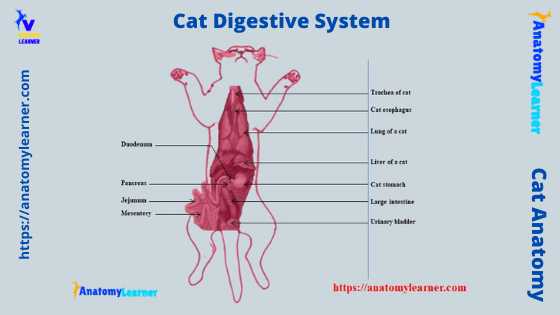
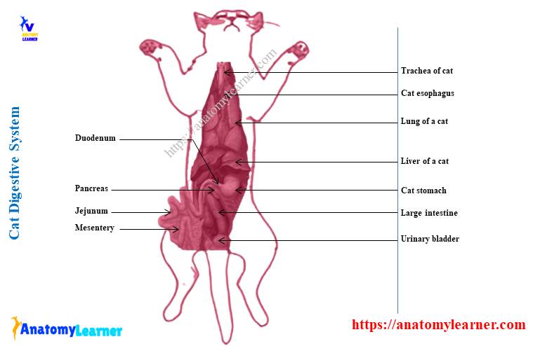
Cat digestive system labeled diagram
I know you already got the different labeled diagrams on the cat digestive system organs. Now, it is the final turn for you to identify all the organs from the cat’s digestive apparatus with the help of labeled diagrams.
Here, in the cat digestive organs labeled diagrams, I tried to show you the different parts of the vestibule (lip and cheek) and different parts of the oral cavity proper. You will see the tongue, different types of teeth, gum, and three different major salivary glands in the cat digestive organs labeled diagrams.
Again, the labeled diagram also shows different parts of the alimentary canal that extends from the esophagus to the last part of the large intestine. Here, the diagram also shows a close relationship of the esophagus with the trachea of a cat.
All the parts from the cat’s small intestine (duodenum, jejunum, and ileum) are identified here in the cat digestive apparatus labeled diagram. Again, the different parts of the large intestine are also shown in the labeled diagram.
You will also see the major accessory glands like the liver, gallbladder, pancreas, and spleen in the labeled diagram sample.
Again, you may find more labeled diagrams on the cat digestive system here on the social media of anatomy learners.
Cat digestive system dissection
I have dissected a cat the previous year, where I explore the organs from different systems. I follow the standard procedure of animal dissection to explore all the cat’s organs.
The details guide on cat dissection and organs from its digestive system (picture) is provided here. All the organs are identified in the cat dissected sample pictures.
Frequently asked questions on cat digestive apparatus
In this part, I will try to enlist the common questions on the cat digestive apparatus asked by the cat anatomy learners. I hope you will get interested in knowing the information I provided below.
How long does it take for food to pass through a cat’s digestive system?
The time for passing intake feed through a cat’s digestive tract may vary. It usually takes ten to twenty hours for feed to pass through the digestive tract.
What is unique about a cat’s digestive system?
There are many unique features present in the cat digestive system. From the tongue to the last part of the large intestine, you will find some unique features that help you make a difference between the cat and ruminant.
Throughout this article, I have already provided the unique features of the cat’s digestive organs. The tongue is a mobile and muscular organ that possesses numerous papillae on its dorsal surface.
A cat has only thirty teeth, whereas the ruminant or dog possesses more than these numbers. The cat esophagus possesses three distinct cervical, thoracic, and abdominal divisions.
The cat’s stomach is a J-shaped expanded part of the alimentary canal. There is less modification on the different parts of a cat’s small intestine. The lobes of the cat liver (six in number) are somewhat different than these of the ruminant.
How do I know if my cat is having a digestive problem?
Your cat may show vomiting and loss of appetite with other clinical symptoms if it has any digestive problems. Other clinical symptoms include lethargy, abdominal distension or swelling, fever, and dehydration.
If you want to be a good veterinary practitioner, you might learn the different organs system from a cat. You may learn about other different organ systems from the cat, like –the respiratory system, endocrine system, and urinary system of a cat.
Conclusion
I hope you got the basic idea of the different organs from the cat digestive system. The main three parts of the cat digestive apparatus are – the oral cavity, alimentary canal, and different accessory organs. Again, the different organs, parts, or structures from these three major divisions are identified on the labeled diagrams.
The alimentary canal of a cat includes the esophagus and different parts of the small and large intestine. From the accessory organs of the cat digestive system, you might have a good piece of knowledge on the liver, gallbladder, pancreas, and spleen.