The cecum of a horse is the first part of its large intestine. You will find a great variation in the horse’s cecum anatomy compared to the cow’s. Let’s see – what is the different about the cecum of the horse?
Quick answer: a horse cecum is a massive comma-shaped sac-like structure. It extends curvedly from the sublumbar region to the xiphoid cartilage. You will find 4 longitudinal bands and 4 rows of sacculations in the structure of a horse cecum.
This article will show you the horse cecum anatomy with its unique features. You will confidently identify the horse cecum from live samples and differentiate it from others.
So, if you want to learn the different or unique features of a horse cecum, let’s continue this article.
What is the cecum in a horse?
The large intestine is the terminal segment of the horse’s gastrointestinal tract. Again, the large intestine divides into 3 divisions, where the cecum is the first part.
The horse cecum is a larger cul-de-sac between the small intestine and the colon. It is very remarkable in size, shape, and position in a horse compared to dogs and cows.
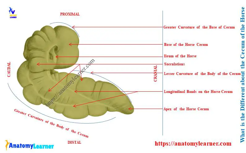
Here, I tried to show the shape and position of the horse cecum in the labeled diagram. You will find the different shapes and positions if you compare them with the dogs and cows.
In ruminants (cows), you will find the cecum as a blind sac-like structure that cranially continues with a colon. Again, the caudal blind end of the cow cecum is the apex that extends up to the right side of the pelvic inlet.
That means the blind end of the cow cecum directs caudally. Sometimes you may find little ventral curviness in a few ruminants.
You will find the diagram and location of the cow cecum in the below-mentioned article –
The dog shows a long closed tubular and curved cecum in the first segment of the large intestine. Its cranial end opens into the starting points of the dog’s colon.
What is the different about the cecum of the horse?
Let’s see the different in the cecum of a horse –
- The cecum of a horse is a big conical or comma shape sac-like organ,
- You will find a base, a longer body, and a blind apex in the horse cecum structure,
- This cecum extends in a curved manner from the sublumbar region to the xiphoid cartilage along the abdominal floor,
- The blind end apex is directed forward, and the body rests on the ventral wall of the abdomen,
- There are four longitudinal bands and four sacculations in the structure of a horse cecum,
- Both the ileocecal and cecocolic orifices locates in the lesser curvature at the base,
Again, Table 1 will help you to find out the differences in horse cecum while comparing it with dogs and cows –
| Cecum | Horse | Cow | Dog |
| Shape | Conical curved Or Comma shape | Long blind sac | Long close tubular And curved sac |
| Size | 1.25 meters (125 cm) | 70 – 80 centimeters | 12 – 15 centimeters |
| Capacity | 25 – 30 liters | 6 – 8 liters | 1.5 – 3 liters |
| Direction | Cranially | Caudally | Caudally |
| Longitudinal bands | 4 bands | Absent | Absent |
| Sacculation | More | Less | Indistinct |
So, the shape and the extension of a horse’s cecum show the unique features of dogs and cows.
Let’s now see the anatomical features of the horse cecum from the next section of the article.
What is the size of the cecum of a horse?
The average length of the horse’s cecum is 1.25 meters (125 centimeters) to 1.75 meters. Again, the capacity of the horse cecum varies from 25 to 30 liters.
The cecum is conical in form and is curved in the horse. Thus, it looks like a comma shape.
Both ends of the horse cecum are blind. You will find two orifices at the upper end or concave curvature (ileocecal and cecocolic orifices).
Horse cecum anatomy
For description purposes, the horse cecum anatomy divides into a base, a longer body, and an apex. Let’s see the following features from the horse cecum anatomy labeled diagram –
- The base of the horse cecum
- Body of the equine cecum,
- The apex of the horse cecum,
- Longitudinal bands on the cecum,
- Sacculations on the cecum body, and
- Ileocecal and cecocolic orifices,
All these features mentioned above are identified in the horse cecum labeled diagram. I also provide more labeled diagrams on social media on the horse cecum structure here.
Let’s see the summary of the base, body, and apex of the equine cecum –
- Base – is the bulbous beginning of the cecum in the right paralumbar fossa,
- Body – is the continuation of the base cranially along the right abdominal wall and floor of the abdominal cavity,
- Apex – is the tapered end of the cecum that lies on the floor of the abdominal cavity just caudal to xiphoid cartilage,
- Ileocecal orifice – is the ileal opening into the base of the cecum, and
- Cecocolic orifice – is the opening at the base of the cecum to the ascending colon of the horses.
Base of the equine cecum anatomy
The base of the equine cecum is dorsal and strongly curved. Here, the greater curvature faces the dorsal, and the lesser curvature faces the ventral to the base.
Cranially, the base of the cecum extends on the right side of fourteen ribs. Again, you will find the caudal extension of the base of the cecum up to the tuber coxae.
The base of the cecum attaches to the terminal part of the ileum and the beginning part of the colon. Here, I found the rounded blind end at the base that faces ventrally.
You will also find various attachments of the cecum’s base with surrounding organs. It attaches to the pancreas and right kidney with the connective tissue and peritoneum.
A small part of the abdomen also attaches to the base of the equine cecum. No other unique features in the horse cecum make it different from others.
Body of the horse cecum
The body is the longer part of the cecum and lies between the base and apex. It extends ventrally and cranially from the base of the cecum.
You will find most of the part of the cecum’s body on the ventral wall of the horse’s abdomen. The lesser curvature of the body is parallel to the costal arch.
If you don’t know what the costal arch is; below-mentioned article might help you to understand –
You will find the attachment of the cecum’s body with the first part of the colon. It attaches to the colon dorsolaterally by caeco-colic folds.
I will clearly show you the longitudinal bands from the body of the equine cecum. Again, I will discuss these bands of the cecum in the next section of the article.
The apex of the equine cecum anatomy
The apex of the equine cecum is free and possesses a blind end. It consequently may vary in position.
The apex of the cecum primarily lies on the abdominal floor, usually to the right of the median plane. Again, the cranial extension of the apex may go up to the xiphoid cartilage of the sternum.
Relationship of the horse cecum
The right (parietal) aspect of the horse cecum related to the below-mentioned organs or parts –
- Right abdominal wall,
- With few areas of the diaphragm,
- Little area of the duodenum and liver,
The left (visceral) aspect of the equine cecum lies against the below-mentioned organs or parts –
- The left and terminal parts of the colon,
- Root of the great mesentery and small intestine,
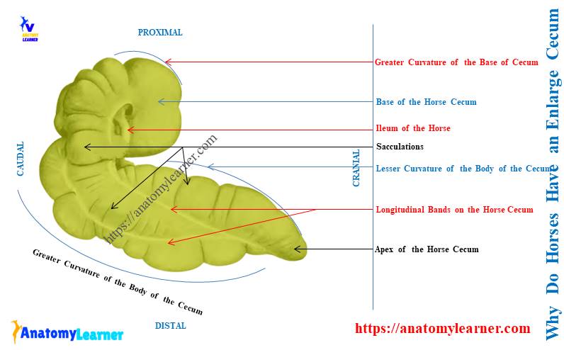
- Bands and sacculations on the horse cecum structure
The horse cecum possesses 4 longitudinal bands on dorsal, ventral, right, and left surfaces. These 4 longitudinal bands cause four rows of sacculation in the cecum.
The longitudinal bands of the equine are known as the taeniae caeci. In contrast, another name for the sacculations is haustra.
Here, the ventral band of the cecum is almost exposed or free. This band begins from the higher part of the base. It runs along the medial aspect of the greater curvature and joins with the medial band at the apex.
The cecum’s dorsal longitudinal band extends from the terminal part of the ileum to the apex. It passes along the lesser curvature of the cecum.
The medial band of the equine cecum passes along the medial part of the lesser curvature of the base. It inclines ventrally and goes forward, and finally ends by joining with the ventral band.
The lateral band arises from the attachment of the right ventral part of the colon with the cecum. This band of the cecum covers the vessels, lymph glands, and fat.
The lateral longitudinal band of the equine cecum includes ventrally and passes cranially. This band ends at the apex, or sometime you may find them as a faded structure that did not reach the apex.
What is the ileo-caecal orifice in a horse?
The ileo-caceal orifice opens the ileum into the cecum in a horse. It locates in the lesser curvature of the base of the horse cecum.
You will find this orifice about two or three inches to the right of the median plane of the horse’s body.
The fold of the mucous membrane surrounds the ileo-caceal orifice. You will also find the circular muscle layer within this fold of the ileo-caceal orifice. This circular muscular layer is known as the sphincter ilei.
What is a caeco-colic orifice in a horse?
The caeco-colic orifice is the colon’s opening into the horse’s caecum. You will see a comparatively small caeco-colic orifice between the horse’s caecum and colon.
It is only one to two inches long and possesses a narrow oval outline. You will also find a thick muscular fold in the caeco-colic orifice of the horse.
This thick fold locates at the ventral margin of the orifice. It consists of muscular rings and is known as the sphincter caeca.
Conclusion
So the answer to the question – ‘what is the different of the cecum of the horse’ was revealed. The comma-shaped external appearance of the horse cecum anatomy is the first point to differentiate it from others.
Again, 4 longitudinal bands, 4 rows of numerous sacculation, and direction are other important features to identify the equine cecum. Now you might observe these identifying features from the horse cecum anatomy from the real sample.