This guide might help you identify all the gross anatomical features of the different organs of the animal urinary system. The external and internal features of the urinary system organs are different in various animals.
Quick overview: the organs of the animal urinary system consist of two kidneys, two ureters, a urinary bladder, and a urethra.
I will focus on identifying the gross features of the animal’s kidney, ureter, bladder, and urethra. But you will find the list of detailed guides on these organs below.
What is the urinary system of the animals?
The urinary system of animals is the system of urinary organs consisting of kidneys, ureters, bladder, and urethra. They are similar in origin and structure and perform urine production and excretion.
The learning objectives of the animal’s urinary system are –
- To know the significant terminology related to the urinary organs,
- Identify the organs or parts of the urinary system and describe their surface and topographic location from the animals,
- Compare the external and internal gross anatomy of the urinary organs of different animals (especially cows, sheep, and dogs) and
- Know the innervation and arterial supply of the animal’s urinary organs
Animal urinary system parts
First, let’s identify the parts of the animal urinary system. It includes the following organs –
- Right and left kidneys – they are variable in location and gross features in different animals,
- Right and left ureters – the right ureter is comparatively longer than the left ureter,
- Urinary bladder – it is a thick wall muscular sac-like structure that works as the reservoir for urine and
- Urethra – the length of the urethra varies in male and female animals,
Here, the diagram shows the different organs or parts of the animal’s urinary system. Let’s find the individual diagram of these urinary organs below.
Guides on the animal urinary organs
Table 1 shows the list of articles that help you to learn the anatomy of the animal urinary organs –
| Animal urinary organs | Complete Guides on the Animal Urinary Parts |
| Animal Kidneys | The cow kidney anatomy – external and internal features of the bovine kidneys |
| Dog kidney anatomy – the right and left canine kidneys with their location and diagrams | |
| Ureters of animals | Right and left ureter of the animals |
| Urinary bladder | Dog urinary bladder anatomy with the diagram |
| Urethra of animals | Male and female urethra of the animals |
But you may find other guides on urinary organs here on Anatomy Learner.
Animal urinary system anatomy and diagram
Each animal has paired kidneys with variable external and internal anatomical features. They remove the waste products and perform certain functions of the animal’s body.
The system of tubules in each kidney forms the single musculo-membranous ureter. Each of the ureters passes caudally and emptys into the urinary bladder.
The urinary bladder of the animal is a distensible reserve for storing urine. In empty condition, it is located on the floor of the animal’s pelvic cavity. Again, it may be found in the abdominal cavity partly when the urine fills the bladder.
Finally, the urinary bladder discharges the urine through another muscular tube called the urethra.
Now, let’s identify the gross anatomy of the kidneys, ureters, bladder, and urethra of animals.
Gross features of the ox kidney – identification
Let’s find the below-mentioned gross features of the ox kidneys –
- Superficial polygonal lobes of the kidneys (have variable sized fissure and fills with fats),
- Brownish red color kidneys,
- Right (smaller) and left (larger) kidneys of the ox,
Both kidneys of the ox are embedded in a large amount of the peritoneal fat. This is also known as the adipose capsule of the animal’s kidney.
How do we differentiate the ox’s right kidney from the left kidney?
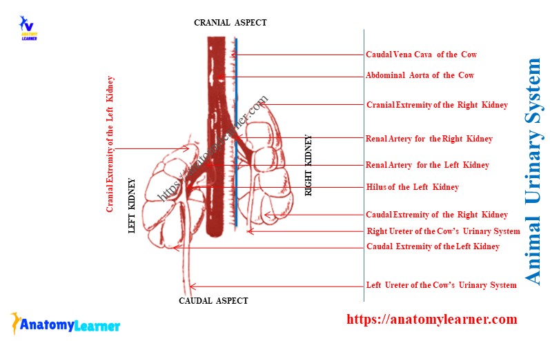
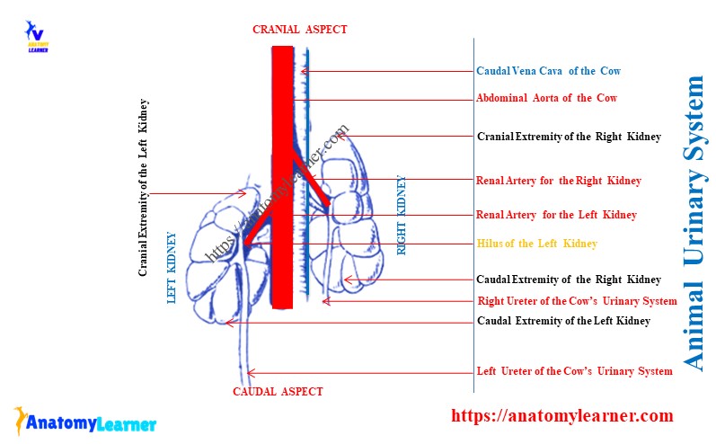
The right kidney of the ox is light and less heavy than the left kidney. Again, the right kidney has 2 surfaces and 2 borders. Meanwhile, the left kidney of the ox has 3 surfaces and 3 borders.
Let’s know the differences between the ox’s right and left kidneys in detail –
Right kidney of the ox:
It is an elongated and elliptical kidney of the ox. This kidney is a little dorsoventrally flattened.
- Location: the right kidney of the ox lies ventral to the thirteen ribs and the first two or three lumbar transverse processes. Thus, this kidney of the ox is palpable in the paralumbar fossa.
- Surfaces: possess 2 surfaces – dorsal and ventral. Here, the dorsal surface of the ox’s right kidney is rounded and in contact with the sublumbar muscles. The ventral surface of the right kidney is less convex and has contact with the liver, duodenum, and colon.
- Hilus of the right kidney: the hilus of the ox’s right kidney is located on the anterior part of the ventral surface (near the medial border).
- Borders of the ox’s right kidney: has 2 borders – lateral and medial. The lateral border of the ox’s right kidney is convex. Meanwhile, the medial border of the right kidney is straight and parallel to the caudal vena cava.
- Extremities of the right kidney: has 2 extremities – cranial and caudal. The cranial extremity of the right kidney is pointed and occupies the renal impression of the cow liver.
Related articles: cow stomach anatomy – (description of the rumen, reticulum, omasum, and abomasum part of the ruminant stomach).
Left kidney of the ox:
The position of the ox’s left kidney is variable. When the rumen is empty, the left kidney of the ox lies on the floor of the pelvis and partly left to the median plane.
But, when the rumen full, it pushes the kidney caudally. Thus, it crosses the median plane and locates the right, caudally, and ventral to the third to fifth lumbar vertebrae.
Related articles: – cow lumbar vertebrae with the labeled diagram.
The unique features of the ox’s left kidney –
- Extremities of the ox’s left kidney: it has cranial and caudal extremities. The cranial extremity is small, and the caudal extremity is rounded and larger.
- Surfaces of the left kidney: it possesses 3 surfaces – dorsal, ventral, and ruminal. Here, the dorsal surface is convex and possesses the hilus. Again, the ventral surface is related to the intestine, and the ruminal surface has contact with the rumen.
- Hilus of the left kidney: the hilus of the ox’s left kidney is located in the craniolateral part of the dorsal surface.
- Borders of the ox’s left kidney: have 3 borders – lateral, medial, and ventral.
Here, the lateral border is formed between the ruminal and dorsal surfaces. The medial border of the left kidney is formed between the dorsal and ventral surfaces. Finally, the ruminal and ventral surfaces form the ventral border of the left kidney.
Internal structures of the ox kidney
Let’s identify the following internal structures from the ox’s kidneys –
- External fibrous capsules,
- The darker inner cortex and outer pale medulla of the ox kidneys,
- Renal pyramid with the renal papillae (renal pyramid are the triangular masses consisting of uriniferous tubules and connective tissue),
- Funneled-shaped minor calyx (where the renal papillae open),
- Thin-walled and wide major calyx and
- Renal sinus of the ox kidney (formed by the hilus of each kidneys),
You will not find the renal pelvis in the structure of the ox kidneys.
Kidneys in the other animals
In sheep and goats: the kidneys are bean-shaped and smooth externally. These are the major differentiating points of the small ruminant kidneys from the large ruminants.
Internally, you will also find the following –
- The renal pelvis of the sheep or goat kidneys (dilated origin of the excretory duct),
- The renal crest of the goat kidneys (the medial central part of the medulla forms a concave ridge that projects into the pelvis) and
- Area cribrosa – present numerous small openings at which the renal tubules open into the renal pelvis,
Horse kidney identification
Location of the kidneys: the right kidney of a horse lies ventral to the dorsal part of the last two or three ribs and the first lumbar vertebral transverse process. Again, the left kidney of the horse lies ventral to the last rib and the first two or three lumbar transverse processes.
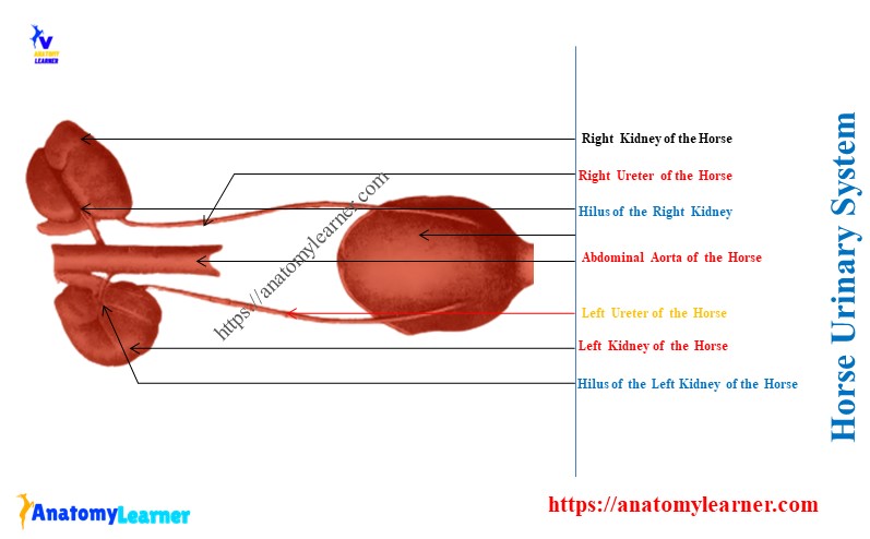
Let’s find the below-mentioned gross features of the horse kidneys –
- The right kidney of the horse – is the heart or equilateral triangular shape,
- The left kidney of the horse – is bean-shaped,
- Lobulation on the external surface: has no lobulation like the cow’s kidneys,
- Location of the renal hilus: at the middle of the medial border of each kidney,
- Features of the renal pyramid: the renal pyramid is not distinct in the horse kidneys and
- The internal structure of a horse kidney: it possesses the renal sinus and renal crest,
Gross structure of the dog kidney – identification
Let’s identify the gross features of the dog kidneys –
- Right and left kidneys of the dog: bean shape and possess smooth surface externally,
- The right kidney of the dog lies under the bodies of the first three lumbar vertebrae,
- The left kidney of the dog lies under the bodies of the third to fifth (3 – 5) lumbar vertebrae,
- Location of the hilus: at the middle of the medial border of each kidney,
What are the unique gross features of the ureter, bladder, and urethra of animals?
Ureter of the ox: there are right and left ureters in the ox’s urinary system. Each of the right and left ureters of the animal’s urinary system is formed by the union of the major calyx at the renal pelvis.
The length of the right and left ureters of the ox is a little variable. They pass caudally, and both open the caudodorsal surface of the urinary bladder.
The horse’s right and left ureters have no peculiar anatomical features. However, the renal pelvis of horses is more dilated than other domestic animals.
What is the urinary bladder of an animal?
- It is a thin wall Musculo-membranous sac located in the ventral wall of the pelvic cavity,
- The ox’s urinary bladder has three parts – apex or vertex, body, and neck,
The cranial rounded blind end of the ox’s urinary bladder is the apex or vertex. Again, the body of the ox’s urinary bladder is rounded and dorsoventrally little compressed. Finally, the caudal, narrow tubular part of the ox’s urinary bladder is the neck, which continues with the urethra.
Conclusion
This guide identifies all the gross anatomical features of the animal urinary system. The animal’s right and left kidneys, ureters, urinary bladder, and urethra are the organs of its urinary system.
Good knowledge of the urinary organs confirms an understanding of their specific functions. Now, you should practically identify all the gross anatomy from the animal’s urinary system organs.