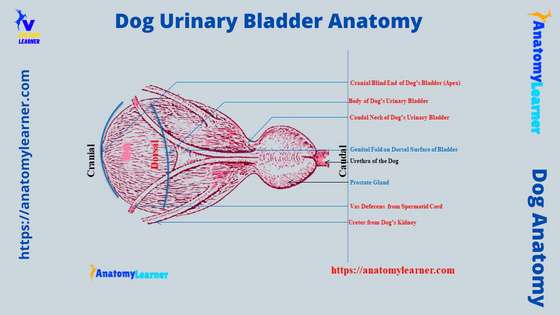
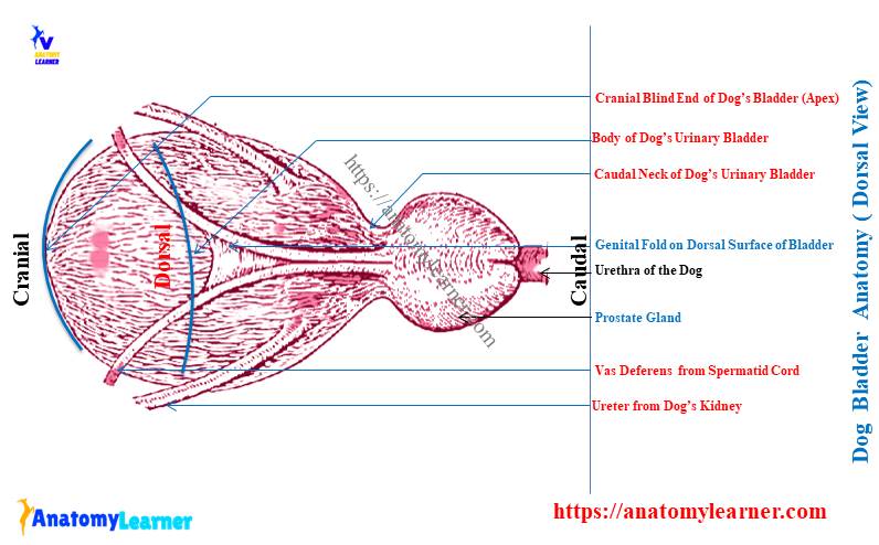
The dog urinary bladder anatomy consists of its shape, size, location, and structure. It is a thick-walled hollow musculomembranous sac that serves as a reservoir for urine.
The shape, size, and position depend upon the amount of urine it contains. Here, I will describe the anatomical facts of the dog’s urinary bladder with the labeled diagram so that you can quickly identify all the features from the actual sample.
Different ligaments hold the dog’s urinary bladder within the pelvic cavity. Again, 2 important structures (ureters and vas deferens) pass over the dorsal surface of the urinary bladder and open on its neck.
You will also find the relationship of different organs in both the male and female dog urinary bladder. So, I will show you the relative position of the dog’s bladder with the diagram both from male and female dogs.
I hope you will enjoy these anatomical facts about the dog’s urinary bladder.
Dog urinary bladder anatomy
The shape of the dog’s urinary bladder may vary from oval to elongated appearance. Again, the size of the urinary bladder may vary in different dogs.
Averagely, the diameter of the dog’s urinary bladder varies from 2 – 15 centimeters. In comparison, the length of the bladder may vary from 3 – 17 centimeters (average).
First, let’s know what you should learn from the dog’s urinary bladder. You might learn the followings from the dog bladder anatomy –
- Position of the dog’s urinary bladder,
- Three different parts of the dog’s urinary bladder,
- Layers of the dog’s bladder,
- Ligaments of the dog’s urinary bladder,
- Blood vessels and nerves innervate to the dog’s urinary bladder,
Now, I will describe all these topics from the dog’s urinary bladder. But, it will be better if you try to identify the below-mentioned anatomical features from the male dog urinary bladder –
- Cranial blind end or apex of the bladder,
- Rounded dorsoventrally compressed body of the dog’s bladder,
- The caudal narrow tubular neck of the bladder,
- Detrusor muscles of the dog’s bladder,
- Trigon of the dog’s urinary bladder,
- Median and lateral ligaments of the urinary bladder,
- Round ligament of the dog’s bladder,
- Genital fold on the dorsal surface of the dog’s bladder,
- Opening of both right and left ureters on its corresponding caudodorsal aspects,
- Opening site of 2 vas deferens at the neck of the bladder,
- A prostate gland at the neck of dog’s bladder with seminal colliculus, and
- Starting part of the dog’s urethra at the neck of the bladder,
The labeled diagram identifies all these structures from the dog’s urinary bladder. I hope these features help you understand the dog’s urinary bladder structure.
Unique features of the dog urinary bladder
Now, I will enlist the most important and unique anatomical features of the dog urinary bladder. This might also help you to get a summary of the canine urinary bladder anatomy.
Okay, let’s see the particular anatomical facts of the dog’s bladder within a few minutes –
- The dog’s urinary bladder is a hollow musculomembranous ovoid to elongated structure that locates in the pelvic cavity (but, the position may vary),
- It may divide into cranially located apex, rounded body, and caudal neck,
- There are three different muscles layers in the structure of the dog’s bladder (detrusor muscle; please see the bladder histology layers section),
- You will find a triangular area near the neck of the dog’s urinary bladder known as the trigone of the bladder,
- A median, lateral, and round ligaments are the main structure that contributes to holding the dog’s bladder in its position (on pubis),
- The internal pudendal, obturator, and umbilicus are the main arteries that supply the dog’s urinary bladder,
Now, it’s time to know the details of the dog’s bladder features with the labeled diagram. First, let’s start with the dog’s urinary bladder location.
Dog bladder anatomy location
The empty dog’s urinary bladder remains in a contracted condition on the floor of the pelvic cavity (on the pubis bone). Again, the distended urinary bladder (filled with urine) of a dog gets projected to the abdominal cavity from the floor of the pelvis.
In empty conditions, the dog’s bladder becomes an elongated shape. Whereas in the distended state (filled with urine), it becomes somewhat oval.
So, here you should have a clear idea of the formation of the pelvic floor and the boundary of the cavity. You may know the details on the pelvic cavity and formation of the dog’s pelvic floor from the below-mentioned article –
- Dog pelvis anatomy with the labeled diagram,
Again, let’s discuss a little about the dog’s pelvis with a few lines.
The dog’s pelvis is formed by the fusion of bilateral ischium and pubis bones. They join together in the midventral part of the body and form the pelvic symphysis.
The pubic bone from both the right and left side joins to form the pubic symphysis of the pelvis. In contrast, the ischium bones from both the right and left sides join and form the ischial symphysis.
The cavity within the hip bones (ilium, ischium, and pubis) and sacrum form the pelvic cavity. Here, the boundary of the dog’s pelvic cavity contains the followings –
- Dorsally – a body of the dog’s sacrum bone,
- Ventrally – pubis bones or iliopubic eminence of the pubis bone,
These form the pelvic inlet in the pelvic cavity of a dog. The lateral wall of the dog’s pelvic cavity forms with the ilium and ischium bones along with the different pelvic muscles. Finally, the boundary of the pelvic outlet consists of –
- Dorsally – last sacral vertebrae (sometimes first 1 -2 caudal vertebrae), and
- Ventraly – ischial arch of the ischium bones,
Parts of dog urinary bladder
For description purposes, the dog urinary bladder divides into three main parts –
- Blind cranial end or apex or vertex of the bladder,
- Middle rounded body of the urinary bladder, and
- A narrow tubular caudal neck,
Here, the blind cranial apex of the dog’s urinary bladder possesses cicatricial tissue in its middle. This is the remnant of the urachus that communicated between the fetal bladder and allantois.
Sometimes in some dogs, this urachus may persist after birth. A urinary fistula may develop in the dog’s urinary bladder in this condition. As a result, urine may pass through the umbilicus.
Now, let’s see the features of the body of the dog’s urinary bladder. It is rounded and dorsoventrally compressed in the dog.
Here, the dorsal surface of the body is convex, whereas the ventral surface is almost flat. You will find the relationship between the different organs and structures of the pelvic cavity with the body (of the urinary bladder) in the next part of this article.
Now, the neck is the caudal part of the dog’s urinary bladder. It is a narrow tubular structure with a thick muscular layer on its wall. Finally, this narrow tube of the urinary bladder continues with the urethra.
Organs related to dog urinary bladder
While studying the dog urinary bladder anatomy, you will see other different organs or structures surrounding it. The urinary bladder of a dog covers the visceral surface of the peritoneum.
Cranial to the pubis bone, the visceral layer of the peritoneum of the ventral surface of the dog’s urinary bladder is separate from the parietal peritoneum of the abdominal wall. Between these two peritoneal layers, you will find the greater omentum of the stomach.
You may know more about the greater and lesser omentum or the structure of the dog’s stomach from the below-mentioned suggested article –
- Dog stomach anatomy with the labeled diagram,
The caudal ventral surface of the dog’s urinary bladder lacks the peritoneum covering.
You will see a distinct peritoneal fold (median ligament of the dog’s urinary bladder) that attaches the ventral surface of the bladder to the linea alba and pubis symphysis. Other different ligaments will discuss in the dog bladder ligaments section.
I hope you have a good piece of knowledge on the linea alba of the dog. It is the white fibrous connective tissue at the midventral surface of the dog’s body. This white fibrous connective tissue or linea alba is formed by the fusion of fibers from the 2 obliquus abdominis and transverse abdominal muscles.
Now, let’s see the different organs or structures that have a direct or indirect relationship with the urinary bladder of the dog. First, let’s see the labeled diagrams from the male and female dogs, where I tried to show the location of the urinary bladder and other organs.
Urinary bladder in male and female dogs
In both male and female dogs, you will find the urinary bladder on the pelvic floor in case of an empty condition. Again, in distended condition, you will see the little projected portion of the urinary bladder into the abdominal cavity.
But, there is little variation in the relative position of the different organs that have a relationship with the urinary bladder in both males and females.
In the case of a dog male, the dorsal surface of the urinary bladder is related to the genital fold, prostate glands, and vas or ductus deferens. Whereas in the female, the cervix and the body of the uterus are in contact with the dorsal surface of the urinary bladder.
Again, in both male and female dogs, you will find contact with the small intestine (jejunum and ilium) and the descending segment of the colon to the dorsal surface of the bladder. Again, the space on each lateral side of the urinary bladder occupies the different parts of the small intestine.
Layers of dog urinary bladder wall – detrusor muscle
The wall of the dog’s urinary bladder consists of three distinct muscles layer. You will see the following arrangement of the muscles bundle in the dog’s urinary bladder wall –
- Inner longitudinal muscle layer in the bladder’s wall,
- A relatively thick middle circular smooth muscle layers in the bladder’s wall, and
- The outer longitudinal muscle layer in the dog’s urinary bladder’s wall,
This pattern of the muscles arrangement is poorly understood if you see it grossly. So, if you study the microscopic figure of the dog’s urinary bladder under a light microscope, you will easily understand these arrangement patterns of the muscles.
For details on histology of the urinary bladder wall, you may go through the following article that I have already published here in anatomy learner –
- Urinary bladder histology slide (male and female) with the labeled diagram,
In this article, I tried to show you the different layers of the urinary bladder (from tunica mucosa to tunica serosa or adventitia). The three layers of the bladder’s muscle are clearly visible in the labeled diagram provided in that article.
Microscopic anatomy of dog’s bladder wall
But, to save you time, I will also show you the different histological features of the dog’s urinary bladder (wall) here. Let’s see the urinary bladder wall labeled diagram where I identified different layers and structures.
Here, this diagram shows four distinct layers – tunica mucosa, submucosa, muscularis, and serosa. Here, in the tunica mucosa layer, you will find the numerous folds that line with transitional epithelium.
You will also find the transitional epithelium in the ureter and renal pelvis of the dogs.
There is the thickest tunica muscularis layer identified in the bladder’s wall labeled diagram. Here, the outer and inner longitudinal muscle layers and the middle circular layer are clearly visible in the diagram.
Finally, you will see the tunica serosa layer in the labeled diagram, which lines with the mesothelium (simple squamous epithelium).
Detrusor muscle and trigone in dog bladder
At the urethra and bladder (neck part) junction, all the muscle fibers form an oblique appearance. This urinary bladder muscle of the dog is also known as the detrusor muscle.
The wall of the dog’s urinary bladder is irregularly folded when it becomes empty. But, in the distended urinary bladder, the irregular fold will disappear.
Near the neck of the dog’s urinary bladder, there is a triangular area internally. This triangular area near the neck of the bladder is trigone and composed of the mucous membrane.
At the apex of the trigone, you will see the opening of the urethra. Again, there is the opening of the ureter at the base of the urinary bladder trigone.
In this area (trigone of the urinary bladder), you will not see any mucosal folds. But, there are a few poorly developed ridges present in the trigone.
These ridges of the bladder trigone cover the urethral crest.
Ligaments of the bladder dog anatomy
The peritoneum that attaches to the dog’s urinary bladder is modified to form the ligament. Here, the peritoneum from the lateral and ventral surface of the dog’s bladder attaches to the lateral pelvic wall and the ventral abdominal wall.
In the dog bladder anatomy, you will find 3 important ligaments that fix the position of the urinary bladder –
- A large median ligament of the dog’s urinary bladder,
- The lateral ligament of the dog’s urinary bladder, and
- A round ligament of the dog’s urinary bladder,
These ligaments of the dog’s urinary bladder are made of double layers of peritoneum. They (double layers) separates by the intercalated blood vessels, nerves, lymphatics, and adipose tissues.
You will also find some other structures like the ureters, vas deferens, and vestiges of embryonic structure that separate these double layers of the peritoneum.
Again, you will find some other structures that help the dog’s urinary bladder fix its position on the pelvic floor. Now, let’s discuss these important ligaments from the dog urinary bladder structure.
Median ligament of dog urinary bladder
Another name of the median ligament of the dog’s urinary bladder is the ventral ligament. It is the largest peritoneal fold that reflects from the ventral surface of the urinary bladder.
This median ligament attaches to the pelvic symphysis and the linea alba of the abdominal wall. It extends up to the umbilicus of the middle linea alba.
The shape of the median ligament is triangular in dogs, and it remains at the midventral portion of the bladder. Again, the size of the median or ventral ligament may vary in different dogs.
You will see the greatest height at the caudal part of the median ligament. In addition, the cranial part of the dog’s urinary bladder median ligament is narrow. It forms an acute angle with the abdominal wall at the umbilicus.
The caudal extension of the median ventral ligament of the bladder may vary in male and female dogs. In the male dog, this ventral median ligament of the bladder ends at the level of the transverse plane through the middle of the prostate gland.
You know the prostate gland of the male dog locates at the neck (dorsolaterally) of the urinary bladder.
In addition, the ventral median ligament of the female urinary bladder ends approximately at the level of the caudal vestibular junction.
Finally, you may tell the ventral median ligament of the urinary bladder to connect the ventral surface of this organ with the floor of the pelvis in both male and female dogs.
You may see the urachus and the umbilical arteries in the structure of the medial fetal ligament. You know the urachus is the remnant of the embryonic allantois.
This structure of the median ligament typically disappears shortly after birth. A vestigial fibrous urachus may find in this ligament.
Lateral ligament of dog bladder anatomy
You will also find 2 lateral ligaments in the dog urinary bladder, which are also triangular in shape. These 2 lateral ligaments connect the lateral surfaces of the dog’s urinary bladder to the respective lateral pelvic walls.
These lateral ligaments are in the form of a membrane and comprise a rounded cord-like structure in its margin. This cord-like structure of the lateral ligament is the round ligament of the bladder and ureter.
If you understand the structure of the round ligament of the dog’s bladder, you might go back to the formation of the lateral fetal ligament.
Here, in the dog fetal lateral ligament of the bladder, you will see the association of the large umbilical artery. It extends cranially along the rudimentary urinary bladder and course with the median ligament.
So, the large umbilical artery, lateral ligament, and median ligament of the dog fetus run with the urachus to the umbilicus. You have already got the idea about the urachus, which is the stalk of the embryonic allantois.
This structure connects the apex of the rudimentary urinary bladder to the allantoic part of the fetal membrane. I hope you know the fetal circulation in dogs or other animals.
From the fetal circulation, you may know the bilateral umbilical arteries carry blood from the fetus to the placenta. And you also know that these are the component of the umbilical cord of the fetus.
After birth, the umbilical arteries separate and become fibrous cords. It runs between the bladder and the umbilicus.
You will find the narrow lumened umbilical arteries in between the internal iliac and bladder in a young dog. Again, there are some of the smaller cranial arteries that supply the apex and body of the dog’s urinary bladder.
Round ligament of bladder dog
The round ligament of the dog’s urinary bladder is the remnant of the umbilical arteries. It attaches at the free margin of the lateral ligament of the dog’s urinary bladder as a rounded cord-like structure.
The ureter and the round ligaments of the female dog’s urinary bladder crosses at the right angle to each other. You will find this crossing at the junction of the broad and lateral ligament.
Now, the lateral ligament of the female urinary bladder joins with the broad ligament of the uterus and the lateral wall of the pelvis.
In the male dog, the ureter and vas deferens cross each other near the entrance of the ureters into the urinary bladder. Here, the vas deferens join the meso-vas deferns (a fold of the peritoneum).
Now, the vas deferens and mesovas deferens runs dorsocaudally, just dorsal to the ureters in the lateral ligaments of the urinary bladder. Finally, they (2 structures) end at the prostatic urethra of the dogs.
What is the genital fold on the dog’s urinary bladder?
A short peritoneal fold locates the dorsal surface of the dog’s urinary bladder. This short peritoneal fold connects between the two vas or ductus deferens. And this is the genital fold on the dog’s urinary bladder.
But, in the female dog, this genital fold connects between the two uterine tubes. You will also find two pouches in between the genital fold and lateral ligaments of the urinary bladder.
Vessels in dog’s urinary bladder
The dog’s urinary bladder receives the main arterial supply from the caudal vesical arteries. These are the branches of the prostatic artery of dogs.
Again, the prostatic artery of the dogs comes from the internal pudendal artery. In addition, you know this internal pudendal artery of the dog is a branch of the internal iliac artery.
Finally, the internal iliac artery is the branch of the last part of the dog’s abdominal aorta. You may learn the details branching pattern of the abdominal aorta (descending aorta) from another article by an anatomy learner here.
But, please start to learn from the ascending aorta of the dog that arises from the left ventricle of the heart. In the previous article, I have already shown the branching pattern of the different vessels of the dog’s body.
A small cranial vesical artery arises from the umbilical and supplies to the apex or vertex of the dog’s urinary bladder.
You will see the venous plexus on the dog’s urinary bladder. The dog’s bladder’s venous plexus primarily drains into the pudendal vein. Finally, they join with the caudal vena cava of the dog’s venous system.
In addition, the caudal vena cava of the dog opens into the right atrium of the heart. You may also learn the details of the venous supply in the dog’s body from another article published here on anatomy learners previously.
Nerves in the dog bladder anatomy
The nerve innervation of the dog bladder anatomy is very complex. It receives autonomic innervation from both the sympathetic and parasympathetic general visceral efferent neurons.
Here, the primary function of sympathetic innervation on the dog’s urinary bladder is urine storage. In addition, evacuation of stored urine from the urinary bladder is the primary function of parasympathetic innervation.
You will find the sympathetic preganglionic cell bodies in the lateral horn of the first 4 lumbar spinal cord segments. Now, the preganglionic axon enters the abdomen through the lumbar sympathetic trunk and splanchnic nerves.
They synapse on the cell bodies of the postganglionic axon of the caudal mesenteric ganglion or in the wall of a dog’s urinary bladder. Now, the postganglionic axon leaves the caudal mesenteric ganglion in the hypogastric nerves and runs with the pelvic plexus.
The postganglionic axon, along with the hypogastric nerve, innervates to the detrusor muscles of the dog’s bladder. Again, the preganglionic axons run into the pelvic nerve and supply to the dorsal part of the dog’s urinary bladder.
Finally, you will also find the innervation of the pudendal nerve in the dog’s urinary bladder.
Dog urinary bladder labeled diagram
Now, I will show you the different features of the dog’s urinary bladder with the labeled diagram. In the first diagram of the dog’s urinary bladder, I tried to show you the exact location of the pelvis.
Again, it shows the other organs or structures related to the dog’s bladder. Here, I also identify the base, body, and apex of the dog’s bladder in the diagram.
I try to show the courses of the ureter and vas deferens of the dog in the labeled diagram. Again, the opening of these tubes (ureter and vas deferens) are identified from the dog bladder labeled diagram.
Here, near the neck of the urinary bladder, it shows the location of the prostate gland and the opening of the prostatic duct. Again, the different ligaments and genital folds of the dog’s urinary bladder are identified in the labeled diagram.
You will also find more labeled diagrams on the dog’s urinary bladder here on the social media of the anatomy learner.
Now, let’s compare the urinary bladder of the other different species, like horses, goats, and pigs, to the dogs.
The bladder of horse, pig, and rabbit
You will not find any more distinct differences in the bladder structure of horses, pigs, and rabbits compared to the dogs. The basic structure and shape are almost more or less similar to the dogs.
The bladder of the horse is comparatively smaller than these of the dogs. One exceptional thing happens to the horse’s urinary bladder. You know, in the distended condition of the dog’s urinary bladder, it extends up to the abdominal cavity.
But, in the case of horse urinary bladder, it does not extends up to the abdominal cavity in the distended condition.
Now, let’s see the features of the pig’s urinary bladder. Here, the pig’s urinary bladder is comparatively larger than the dogs. The peritoneum does not cover the whole part of the pig’s urinary bladder.
The urinary bladder of a rabbit shows some exceptional features, but the basic structure of its bladder is similar to that of dogs.
You will find a thinner muscular wall in the rabbit’s urinary bladder compared to the dog. Again, the rabbit’s urinary bladder is a little more elongated than the dogs.
Suggested reading for you –
If you want to know the different internal organs of the horse, pig, and rabbit, you may read the below-mentioned articles –
- Internal organs of the horse with the labeled diagram (focus on the thorax, abdomen, and pelvic cavity),
- The anatomical features of the pig’s internal organs with the labeled diagram, and
- Internal organs (topographic location) of the rabbit with the labeled diagram,
You will not find any urinary bladder in any bird. Here, the ureter directly opens into the urodeum of the bird. I hope you will love to learn these internal organs anatomy from these common popular animals.
Common inquiries on the dog bladder anatomy
Now, I will enlist some common inquiries on the dog bladder structure. But, ensure you read the information above on the male and female dog’s urinary bladder.
These questions about a dog’s bladder are essential for first-year veterinary students new to learning dog anatomy. So, if you are a first-year veterinary student or even a veterinary practitioner, you should go through the questions below.
These are the common questions on the dog’s urinary bladder that veterinary students in their examination frequently face –
How do you know if your dog has a bladder problem?
Before answering this question, you might have a piece of good knowledge about the abnormalities of the dog’s urinary bladder. Commonly you may find the urinary stone in different locations of the dog’s bladder.
The most common site for the formation of the urinary stone is the neck of the dog’s bladder. But, you know, the stone may also form and store in a different segment of the urethra.
Again, the dog’s urinary bladder may show different abnormalities, like the diverticula and urachal crest in the bladder. You may also find the persistent urachus and strictures of the neck of the dog’s urinary bladder.
Finally, an enlarged prostate gland may also happen and causes the dilation of the dog’s urinary bladder. So, you will find a wide range of symptoms in the urinary bladder of a dog with a different specific problem.
If urolithiasis occurs (formation of stone) in the neck or any other area of the bladder, it will show some specific features. More abdominal distension will occur than normal in this urolithiasis condition.
The abdomen becomes tighter with time passing and caseation of the urination. Finally, this condition leads to animal discomfort ness and restlessness.
Where is a female dog’s bladder located?
The female dog’s urinary bladder locates on the floor of the pelvic floor. But, the relative position of the other organs is different compared to the male dogs.
Again, you know the ischium and pubis bones of the dog’s hip form the pelvic floor at the midventral position of the body. Like the male dog, the ischium and pubis bones from both the right and left lateral aspect form the pelvic symphysis (ischial and pubic symphysis).
You will also see the medial ventral ligament on the ventral surface of the female dog’s urinary bladder that attaches to the pelvic floor. Dorsally, you will see the contact with the small intestine, part of the descending colon, uterine horn, and uterus body with the female dog’s urinary bladder.
The right and left lateral aspects of the female urinary bladder’s apex have contact with the different segments of the small intestine.
So, you can understand that there is no basic difference in the location of the female dog’s urinary bladder in the pelvic cavity. The difference is only found in the surrounding organs in the female dog’s pelvis.
Can you feel a dog’s bladder?
Yes, you can feel a dog’s urinary bladder easily. But, you need to be an expert for it to find the urinary bladder of the dogs.
First, you might know the exact location (topographic location) of the dog’s urinary bladder. Again, you might have a clear concept of the formation of the dog’s pelvic floor.
If the dog’s urinary bladder is empty, then it is very hard to feel or hold from the pelvic cavity. But, in distended conditions, you may easily feel the urinary bladder in between the last part of the abdominal cavity and the floor of the pelvis.
What is the trigone of the bladder in dogs?
The trigone of the urinary bladder of the dogs is a triangular area. It locates near the neck of the dog’s urinary bladder.
You may easily understand this triangular area from the internal surface of the bladder’s neck. This triangular area of the neck (trigone) possesses the apex caudally and base cranially.
In the base of the dog bladder’s trigone, you will find the orifice for the ureter. That means the 2 ureters open on the base of the trigone of the urinary bladder.
Again, the apex of the trigone is identified with the orifice for the urethra. So, the urethral opening may find in the apex of the bladder trigone.
You know all the internal surfaces of the urinary bladder (tunica mucosa) possess mucosal folds. But, here in the trigone of the dog’s urinary bladder, you will not find any mucosal folds.
Instate mucosal folds; you may find some ridges that may cover the urethral crest. These ridges indicate the boundaries of the trigone of the dog’s urinary bladder.
What is the size of a dog’s bladder?
The urinary bladder of a dog is an oval or elongated musculomembranous sac-like structure. It varies in size, form, and position in the same breed of dog.
A urinary bladder of a normal-sized dog may contain an average of 100 – 200 ml of urine. As it is an oval or elongated-shaped tubular organ, you will find both the length and diameter.
Here, the average length of the dog’s urinary bladder may vary from 3 – 15 centimeters. The diameter of both male and female dogs’ urinary bladders may vary from 1 – 15 centimeters.
So, the actual size of the dog’s urinary bladder depends on these individuals.
What are the specific ligaments of the dog’s urinary bladder?
In the dog urinary bladder, you will mainly find 2 important ligaments – median (ventral) and lateral ligaments. But, the lateral ligament of the dog’s urinary bladder is also from the round ligament.
So, you may consider 3 main ligaments in the dog’s urinary bladder. Here, the median or ventral ligaments of the dog’s urinary bladder connect the flattened ventral surface of this organ with the pelvic floor.
In addition, 2 lateral ligaments attach to the lateral aspect of the dog’s bladder to the lateral pelvis muscle (wall). In the lateral ligament of the bladder, you will see the free margin, and it contains the remnant of umbilical arteries (in the form of a rounded cord).
This is the round ligament of the dog’s urinary bladder that is also considered an important structure.
Conclusion
So, from this short guide, you got the basic idea of the dog urinary bladder anatomy. You should know the exact location of the dog’s bladder from both male and female dogs. The bladder is located on the pelvic floor for male and female dogs and contains 3 essential parts.
Here, the 3 essential parts of the dog urinary bladder anatomy are the cranial neck, middle, rounded body, and caudal neck. The topographic location is necessary for all the structures that pass over and 2 lateral aspects of the dog’s urinary bladder.
Now, you should practice and identify all the anatomical facts from the male and female dogs’ urinary systems from the actual samples. I hope the provided labeled diagrams on the dog’s urinary bladder might help you identify all the features from the samples.