The dog cephalic vein is the only large superficial structure of the thoracic limb used for venipuncture. Different dorsal and palmar proper digital veins join from the cephalic vein at the forearm of a dog.
This vein is also commonly used for venipuncture in a dog, like the cephalic vein. And if you want to use a cephalic vein for venipuncture, you might know the exact location and other anatomical facts.
Quick answer: dog cephalic vein runs the cranial aspect of the forearm (radius ulna bones) and passes the craniomedial aspect of the elbow joint. So, the cranial aspect of the dog’s forearm is the best site to locate the cephalic vein perfectly to perform a successful venipuncture.
You will also find deep veins like radial, ulnar, medial, and axillary in the thoracic limb of a dog. These veins also connect with the cephalic vein and thus form a complicated venous channel in the thoracic limb of a dog.
But, I will try to make this venous channel simple for your better understanding of the dog’s thoracic limb. I will focus on the (superficial) course of the cephalic vein so that you may apply this knowledge practically to locate it from the live dog’s thoracic limb.
Again, I will also show you the association of the deep veins with the cephalic vein of a dog’s thoracic limb. So, if you want to learn the anatomical facts of a dog’s cephalic vein with the labeled diagram, let’s continue this article till the end.
Dog cephalic vein
You know the cephalic vein of any animal is a tributary (flow) of the external jugular vein. As this vein is practically required for venipuncture in animals, you might know the exact superficial location.
First, let’s see the short summary of the dog cephalic vein (the most superficial large vein) from the thoracic limb. You will easily identify the perfect superficial location of the cephalic vein after reading the below-mentioned summary.
Table 1 shows the summary of the cephalic vein course (superficial) from a dog’s thoracic limb –
| Regions | Cephalic Vein (Dorsal Aspect) | Cephalic Vein (Palmar Aspect) |
| Phalanges | Dorsal proper digital veins (Form digital venous arch at fetlock joint) Dorsal common digital veins (II, III, IV) | Palmar proper digital veins (Superficial palmar venous arch at fetlock) Palmar common digital veins (II, III, IV), |
| Metacarpal | Dorsal metacarpal veins (II, III, IV), | Palmar metacarpal veins (II, III, IV), |
| Carpus | Form dorsal venous rete of carpus with – Dorsal metacarpal, and Dorsal metacarpal veins, | Form deep palmar venous arch by – Palmar metacarpal veins, Radial, and ulnar veins, Intertarsal vein, CONTINUE AS – CEPHALIC VEIN |
| Forearm | Accessory cephalic vein | Cephalic vein join with accessory cephalic |
| Cephalic vein | Continue cranial (dorsal) aspect of Forearm | N/A |
| Elbow joint | Craniomedial aspect and joins with – Median cubital, Brachial vein | N/A |
Here, in table 1, I show the course of the cephalic vein from the digits to the elbow joint. But, in the description section, I will show you the course of the cephalic vein from the dog’s thoracic limb (how this vein joins with the cranial vena cava).
A short course of the cephalic vein
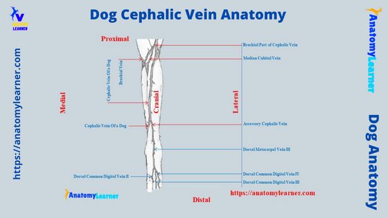
Let’s see the diagram on the short course of the cephalic vein from the dog’s thoracic limb. This might help you to identify the superficial location of the cephalic vein, which is perfect for a venipuncture.
You will find the dorsal proper digital veins on the dorsal aspect of the digits. These dorsal proper digital veins course proximally and form the dorsal common digital veins II, III, and IV.
But, at the fetlock joint of the dog’s thoracic limb, you will see the digital venous arch that actually continues as dorsal common digital veins. Now, you will find the dorsal metacarpal veins II, III, and IV at the metacarpal.
These dorsal metacarpal veins and branches of radial vein join at the carpus and form the dorsal venous rete of the carpus. These anastomoses of the veins continue proximally as the accessory cephalic vein at the distal part of the forearm.
On the other hand, palmar proper digital veins continue proximally as the palmar common digital veins II, III, and IV. The course proximally and form the palmar metacarpal veins II, III, and IV.
Now, you will see the deep palmar venous arch, where you will find the ulnar, interosseous, and radial veins at the carpal. The radial veins continue as the cephalic vein, which joins with the accessory cephalic vein at the distal extremity of the dog’s forearm.
Now, the cephalic vein runs proximally on the forearm (Cranio medial aspect). Then it passes from the craniolateral angle of the elbow joint and continues as the brachial part of the cephalic vein.
All these summaries of the cephalic vein from a dog’s thoracic limb are presented here in the diagram –
Dog cephalic vein anatomy
Table 1 and the diagrams provided in this article might help you locate the superficial segment of the cephalic vein from the dog’s thoracic limb. I will show you the detailed dog cephalic vein anatomy with the labeled diagram.
Here, I will show the course of the cephalic vein from the digits to the cranial vena cava. As other deep veins are associated with this cephalic vein, I will also show you the course or branches of these specific structures.
For description purposes, you may divide the full course of thoracic limb venous channel into the followings –
- Veins in the forepaw of a dog,
- Deep veins in the dog’s thoracic limb, and
- Superficial veins in the dog’s thoracic limb,
These might complete the full course of the cephalic vein from the dog’s thoracic limb. And you will also know the other different small or larger superficial and deep veins from the dog’s thoracic limb.
But keep in mind you always need the superficial course or location of the cephalic vein to perform a successful venipuncture (drug administration or blood collection). From the above information, you may identify the superficial location of the cephalic vein from the dog’s thoracic or fore limb.
Let’s see the exact location of the cephalic vein (suitable for venipuncture) from the dog’s thoracic limb.
Canine cephalic vein location
The canine cephalic vein extends from the mediopalmar surface of the carpus to the Cranio lateral aspect of the humerus. But, this canine cephalic vein locates more superficially at the craniomedial part of the radius and ulna bones (forearm of the dogs).
The canine cephalic vein is the actual continuation of the radial vein. But, if you see the full course or diagram, this radial vein of a dog arises from the superficial palmar arch.
Here, the superficial palmar venous arch locates at the fetlock joint of the dog’s thoracic limb. From this palmar venous arch palmar proper digital vein continue as the palmar common digital vein.
You will learn the details of the radial vein of the dog’s thoracic limb in the next section of this article.
At the level of the (olecranon) elbow joint, the canine cephalic vein runs cranaiolaterally and continues proximally. But, here, the cephalic vein goes deep to the cleiodobrachialis muscle.
Finally, the cephalic vein joins with the omobrachial vein at the level of a proximal end of the humerus bone (arm region). You know, the omobrachial vein now joins with the external jugular vein, which also drains into the cranial vena cava of the heart.
On the other hand, the cephalic vein continues as the axillobrachial vein (after the omobrachial vein). Now, this axillobrachial vein anastomoses with axillary and subscapular veins at the level of the shoulder joint (caudally).
The axillibrachial vein of a dog joins with the axillary vein. But, some main deep veins (like the brachial vein) also join with the axillary vein. You will learn the details of the axillary vein of a dog in the deep vein description section of the article.
How to draw (collect) blood from a dog cephalic vein
It is more convenient to collect (draw) blood from a dog’s cephalic or saphenous veins than the jugular vein. Following is the short procedure to draw blood from a dog cephalic vein –
- Hold the thoracic limb (forearm) in the right position,
- Use a tourniquet or create pressure (hold tightly with grip) on the vein,
- Socking and performing the venipuncture (drawing blood),
As the cephalic vein passes obliquely superficially from the tarsus to the elbow joint (Cranio medially), you may choose this area (forearm). For this, you should raise the vein first to draw blood from it.
So, you should hold the forearm in the right position and hold the vein with a tight grip. You may create a slight rotation at the medial angle of the dog’s elbow joint if you want.
These will help to raise the cephalic vein perfectly. Now, you may sock over the vein to better understand the appearance.
After that, you will quickly understand the course of a vein (as it rises) and perform venipuncture. So, this is the easiest way to draw or collect blood from the cephalic vein.
Now, you will know the different veins from the forearm deep and superficial veins from the dog’s thoracic leg. First, let’s understand the other veins from the forepaw of the dogs (both dorsal and palmar).
Veins in the forepaw of a dog
The veins of the forepaw of a dog divide into dorsal and palmar sets. First, let’s see the dorsal and palmar veins from the dog’s forepaw and try to identify them from the labeled diagram.
First, let’s see the dorsal and palmar veins from the forepaw of a dog. On the dorsal aspect of the forepaw of a dog possess the following veins –
- Dorsal proper digital veins II, III, IV, and V (axial and abaxial),
- Dorsal common digital veins I, II, III, and IV, and
- Dorsal metacarpal veins I, II, III, and IV,
But, you will find different anastomoses in different regions of the dorsal surface of the dog’s forepaw. Again, a minute dorsal venous rete may find on the dorsal surface of the carpus.
Now, let’s see the palmar veins from the dog’s forepaw. You will find the below-mentioned veins on the palmar surface of the dog’s forepaw –
- Palmar proper digital veins II, III, IV, and V (axial and abaxial),
- Palmar common digital veins II, III, and IV, and
- Palmar metacarpal veins II, III, and IV,
You will find 2 major venous arches on the palmar aspect of the dog’s forepaw – superficial and deep palmar venous arches. Now, let’s see the formation and course of the dorsal and palmar veins from the forepaw of the dogs.
Dorsal veins of dog’s forepaw
The distribution pattern of every single vein from the dog’s forepaw is complicated. But, here, I will show you the simple distribution pattern of these dorsal veins.
All these dorsal proper digital veins (II, III, IV, and IV) run proximally on the dorsal aspect of the digits. They receive the communicating branches from the palmar proper digital veins.
You will see the digital arches (anastomosing of veins) near the fetlock joint. They occur on the axial surface of digits II and III and the abaxial surface of digits IV and V.
You will find some small veins (venules) connected with the arch and collect blood from the corium and claws of the dog’s forepaw. Again, at the level of the first digit, you may find a small anastomosis between the dorsal proper digital veins and dorsal common digital veins.
Here, the dorsal common digital veins II, III, and IV continue proximally on the extensor tendon. These veins also receive the different communicating branches from the palmar proper digital veins.
The dorsal common digital vein IV runs proximally and joins with the dorsal common digital vein III. Now, these veins again join with the dorsal common digital vein II and form the accessory cephalic vein in a dog.
You will see the delicate dorsal metacarpal veins I, II, III, and IV on the dorsal aspect of the dog’s metacarpals. They lie on the grooves of the dorsal aspect of the metacarpal bones.
Here, you will see two major anastomoses – the middle and distal third of the metacarpal (distally) and on the dorsal carpus (proximally). The dorsal one is the dorsal venous rete of the dog’s carpus.
Palmar veins of dog’s forepaw
Again the distribution of the palmar veins in the dog’s forepaw is very complicated. Here, the palmar proper digital veins form different digital arches.
They run proximally to the palmar surface of the interphalangeal joints and phalanx. They collect blood from the distal extremity of the dog’s digits.
Palmar proper digital veins III and IV divide into axial and abaxial branches on the first phalanx. Again, the palmar proper digital veins communicate with the branches of dorsal proper digital veins and form the arches.
They run proximally and form the palmar common digital veins II, III, and IV. Again, these common palmar digital veins immediately form the superficial palmar venous arch.
Here, in the superficial palmar venous arch, you will find the contribution of radial and fourth metacarpal veins. Again, the palmar metacarpal veins II, III, and IV run over the superficial palmar venous arch.
There is a deep palmar venous arch that lies deep to the origin of the interosseous muscles. You will see the connection of the deep palmar venous arch with the radial vein medially.
Again, it anastomoses with the ulnar and interosseous branches of the common interosseous vein.
Dog superficial cephalic vein with branches
Along the superficial course of the dog cephalic vein, you will find other different veins that directly connect with it. If you see the thoracic limb’s vein labeled diagram, you will find the below-mentioned veins that have a direct relationship with the cephalic vein –
- Radial vein of the dog,
- Median cubital vein of the dog’s thoracic leg,
- Brachial part of the cephalic vein,
- Axillobrachial vein of the dog, and
- Omobrachial vein of the dog,
Let’s describe the anatomical facts of the veins mentioned above from the dog’s thoracic limb.
A radial vein in dog
Dog radial vein passes the superficial palmar arch and runs proximally on the palmar aspect of the interosseous muscles. From the palmar surface, the cephalic vein arises from the radial vein as the main venous channel.
But, the cephalic vein also receives other smaller channel and continue proximally. At the beginning of the distal fourth of the antebrachium, the cephalic vein also receives the accessory cephalic vein.
And you have already seen that the accessory cephalic vein receives dorsal common digital veins II, III, and IV on the dorsum of the metacarpal. Then it passes proximally over the carpal joint and cranial aspect of the antebrachium and becomes the cephalic vein.
Median cubital vein of a dog
You will see the median cubital vein at the level of the elbow joint of the dog (cranio lateral aspect). This vein extends between the superficial brachial vein at the flexor angle of the elbow and the cephalic vein of the arm region.
The length and diameter of the dog’s median cubital vein are less compared to the cephalic vein. It (median cubital vein) obliquely crosses the distal end of the biceps brachii muscle.
Then the median cubital vein runs proximolaterally to anastomoses with the cephalic vein. This anastomoses between the median cubital and cephalic veins occur near the lateral border of the biceps brachii muscle.
Brachial part of the dog cephalic vein
The diagram shows that the cephalic vein continues proximally and passes the antebrachium. It crosses the flexor angle of the elbow joint and becomes the brachial part.
Again, this vein runs proximally and crosses the cleidobrachialis muscle. It joins with the axillobrachial vein on the lateral surface of the triceps brachii muscle.
A branch of the cephalic vein proximomedially deep to the cleidobrachialis muscle at the level of the middle antebrachium. It enters into the external jugular vein between the omobrachial and axillary veins. With the jugular vein, the cephalic and omobrachial veins form the communicating branches.
Axillobrachial and omobrachial veins of the dog
The dog’s thoracic limb vein labeled diagram shows the axillobrachial is the continuation of the cephalic vein. This axillobrachial vein passes over the lateral head of the triceps brachii muscle.
It anastomoses with axillary and subscapular veins just caudal to the shoulder joint. This vein receives many small venules from the triceps brachii muscle. Finally, the axillobrachial vein terminates in the axillary vein.
On the other hand, the omobrachila vein leaves the axillobrachial vein just proximal to the cephalic vein (distal extremity of the arm). First, it runs superficially on the deltoideus muscle, then arches cranially and medially.
The omobrachial vein crosses the brachiocephalic muscle and enters into the lateral surface of the jugular vein. Here, the omobrachial vein doesn’t receive any small venules from the skin or muscle of the neck.
Deep veins of the dog’s thoracic limb
First, let’s see what veins are considered as deep from the dog’s thoracic limb. If you explore the venous channel from the dog’s thoracic limb, you will find the below-mentioned deep veins –
- Dog radial vein,
- A deep ulnar vein in the dog’s thoracic limb,
- The small median vein in the dog leg,
- Dog’s brachial vein, and
- An axillary vein in dog’s thoracic limb,
I have already described the anatomical facts of dog’s radial vein. So, let’s discuss the other deep veins from the dog’s thoracic limb.
The deep ulna and small median veins of the dog
The deep ulnar vein of a dog arises from the palmar branch of the interosseous vein at the carpal joint. This vein passes proximally in the deep digital flexor muscle of the dog leg.
The deep ulnar vein of a dog receives some small branches from the caudal antebrachial muscles. Again, it joins with the common interosseous vein in the proximal antebrachium.
The small median vein in the dog’s thoracic limb arises from the superficial palmar venous arch. This small median vein passes proximally through the carpal canal.
It joins with the radial and antebrachial veins at the middle to the proximal antebrachium region. Finally, this small median vein of the dog continues with the brachial vein.
Brachial and axillary veins of the dog
The dog brachial vein receives the common interosseous, ulnar, cranial, and caudal interosseous veins. You will see the large median cubital vein that connects the brachial vein with the dog cephalic vein.
Again, the dog brachial vein receives the following veins at the brachium region –
- Transverse cubital vein,
- Superficial brachial vein,
- Collateral ulnar vein, and
- Bicipital and deep brachial veins,
Now, the brachial vein continues as the axillary vein proximally. Here, the dog axillary veins also receive other small and larges branches or veins.
It receives the small cranial circumflex humeral vein cranially and thoracodorsal caudally. Caudal to the sog shoulder joint, you will also find the subscapular and caudal circumflex humeral veins that join with the axillary vein.
Small thoracic and external thoracic veins also join with the axillary vein of the dog. Finally, the axillary vein becomes subclavian at the level of the first rib.
Then it joins with the external jugular vein and forms the brachiocephalic vein in a dog.
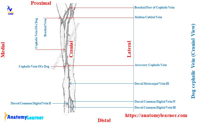
Dog cephalic vein labeled diagram
Now, I will show you the dog cephalic vein labeled diagram (which is essential for venipuncture). In the diagram, I tried to show (view) you the position of the cephalic vein from the dorsal cranial medial aspect of the radius and ulnar bones.
Again, I show you the veins (dorsal common digital veins) from the dorsal surface of the forepaw of a dog. The diagram also shows the caudal part of the cephalic and accessory cephalic veins at the distal part of the antebrachium region.
At the level of the elbow joint (olecranon Process), the diagram shows the different small branches or veins, like the median cubital that joins with the cephalic vein. The diagram (more here) also shows a little portion of the brachial vein (deep vein of the dog’s front leg).
More inquiries on a cephalic vein
Now, let’s see the commonly asked questions on the canine cephalic vein by anatomy learners. Here, you will find the questions on the canine cephalic vein along with the concise answer.
Let’s see the questions or inquiries on the canine cephalic vein from the learner below. But, it is recommended to learn the details and anatomical facts of the cephalic vein and other superficial and deep veins from the dog’s thoracic leg.
Again, you may also learn the details anatomical facts of dog saphenous veins from another article of anatomy learners –
- Dog saphenous vein – lateral and medial saphenous with the labeled diagram.
Okay, let’s get into the answer and question section of cephalic vein anatomy.
Where is the cephalic vein in a dog?
In short, the cephalic vein in a dog locates on the cranio medial aspect of the forearm or antebrachium (radius and ulna bones). This cephalic vein is the best option for a successful venipuncture in dogs.
The cephalic vein begins on the mediopalmar surface of the carpus, where it is a continuation of the radial vein. Proximally, the cephalic vein runs below the cleidocephalicus muscle and joins with the axillobrachial vein.
What does the cephalic vein drain into a dog?
The cephalic vein drains the deoxygenated blood from the periphery of the thoracic limb into the heart. You may see the distal formation of the cephalic vein from the diagram or information provided in the article earlier.
These veins collect blood from the periphery and different muscles and run proximally. The cephalic vein proximally joins with the omobrachial and axillobrachial and finally drains into the external jugular vein.
How do you hold a dog for cephalic venipuncture?
The normal sitting position or the lateral recumbency is better for cephalic venipuncture. I have already provided the procedure for collecting blood from the cephalic vein of the dogs (blood collection section of this article).
If you hold the dog in a normal sitting or lateral recumbency position, you may quickly raise its vein from the thoracic limb.
Where is the best place to draw blood from a dog?
To draw blood from a dog, you may choose three options: cephalic, saphenous, and jugular. In my sense, cephalic and saphenous veins are more suitable for drawing blood from dogs compared to the jugular vein.
But, if you want, you may also collect the blood from the dog’s jugular vein. You might have basic knowledge of their anatomy to collect blood from any of these veins from the dog.
Conclusion
The dog cephalic vein is the main superficial large vein of the thoracic limb. This vein is also suitable for a successful venipuncture like the saphenous vein.
The dog cephalic vein becomes more superficial from the carpus to the elbow joint of the thoracic limb. More specifically, this cephalic vein lies cranio medial to the antebrachium of the dog. All the provided information and diagram might help you learn the course details and the anatomy of the cephalic vein perfectly.