The dog jugular vein is another essential neck structure that may use for venipuncture. There are external and internal jugular veins in the right and left lateral aspects of the dog’s neck.
If you want to perform venipuncture on a dog jugular vein, you might know the exact location on the neck. Here the external jugular vein is more superficial than the internal, and thus you might choose it for a venipuncture.
Quick answer: the jugular vein runs the ventrolateral aspect of the dog’s neck. It lies just deep in the dog’s neck’s skin and is suitable for a successful venipuncture.
Here, I will show you the anatomical facts of the jugular vein (both external and internal) in the dog’s neck. Again, you will find the full guide where I will provide the formation of this vein in a dog with their courses.
Finally, I will show how you may easily collect blood from this external jugular vein, both from the canine and feline. So, if you want to know the anatomical facts of the canine jugular vein and collect blood easily from it, let’s continue this article till the end.
Dog jugular vein
First, let’s see the diagram where I tried to show a dog jugular vein from both lateral aspects. This might provide a clear concept of the external and internal jugular veins from the dog’s neck region.
If you notice in the diagram, the maxillary and linguoficial veins join caudally and form the external jugular vein. Then the external jugular vein passes along the lateral aspect of the dog’s neck.
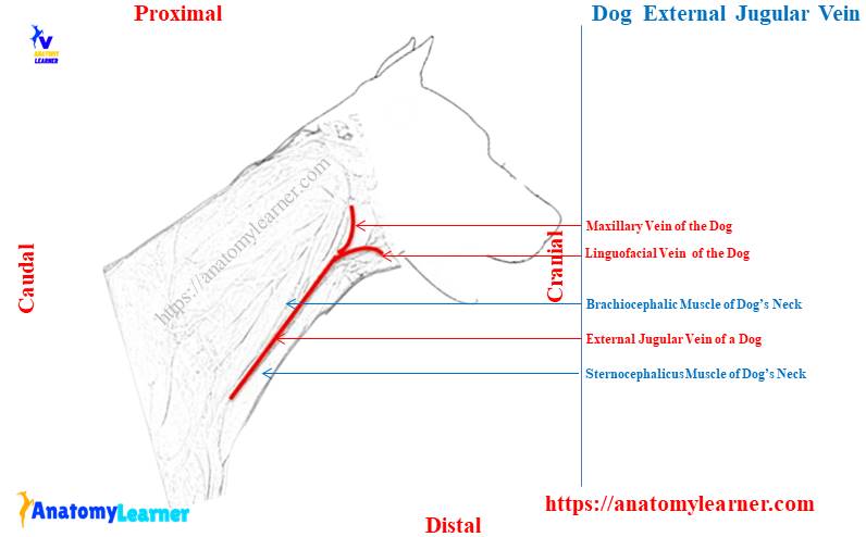
On this course, this vein receives blood from other veins like – omobrachial, superficial cervical, cephalic, and subclavian. Then the jugular vein joins with the brachiocephalic vein.
Finally, the canine brachiocephalic vein directly connects with the cranial vena cava, which ultimately opens on the right atrium of the heart. Similar features are also found in the other lateral aspect (for the left external jugular vein) of the dog’s neck.
Again, if you notice the internal jugular vein on the dog’s neck, they run parallel to the external jugular veins. The right and left internal jugular veins of the dog to receive blood from different regions at the neck.
These internal jugular veins receive mainly blood from the cranial thyroid, middle thyroid, and retropharyngeal areas. Finally, they (both right and left internal jugular veins) drain blood directly into the brachiocephalic vein.
And you know, blood from the dog brachiocephalic vein drains into the cranial vena cava and follows by the right atrium of the heart.
You might choose the external jugular vein (right or left) for a successful vein puncture in the dog’s neck. But you may also choose cephalic or saphenous veins for a successful venipuncture in the dog.
Dissection and exploring jugular vein
If you want to dissect and explore the jugular vein from the canine neck, you might have a good piece of knowledge on the anatomical facts of this neck. The below-mentioned article will provide the basic concept of the different structures and organs of the dog neck –
- Dog neck anatomy – bones, muscles, glands, and other different organs with the labeled diagram,
Here, the internal jugular vein is closely related to the trachea, esophagus, thyroid glands, and internal muscles. Again, the external jugular vein has an indirect relationship with the esophagus, trachea, and thyroid gland.
If you want to explore the jugular vein, you should know the superficial structures of the dog’s neck. You might have a good concept of the superficial muscles of the dog’s neck.
Followings are the important muscles from the dog’s neck that are related to the jugular vein (when dissection and exploration perform) –
- Brachiocephalicus muscle of the dog’s neck,
- Sternocephalicus muscle of the dog’s neck,
- Omohyoideus muscle of the dog’s neck, and
- Other structures that are related to the jugular vein – skin, fascia, and some organs in the dog’s neck,
Above, these structures from the dog’s neck help to form the jugular furrow. And you know, within this jugular furrow, the jugular vein passes.
So, before dissection of the neck region to exploit the jugular vein, you should know its boundary. Let’s see the boundary of the dog jugular furrow from the labeled diagram with its contents.
Jugular furrow dog
The jugular furrow of a dog’s neck is a passage from where the jugular vein runs. You will find the following structures that surround the dog jugular furrow –
- Dorsally – brachicephalicus muscle of the neck,
- Ventrally – sternocephalicus muscle of the neck,
- Medially – omohyoideus muscle (deep), and
- Laterally – skin and fascia of the dog’s neck,
Within this dog’s jugular furrow, you will find the external jugular vein, carotid artery (deep to vein), and vagosympathetic trunk (part of the vagus nerve and sympathetic fibers). The labeled diagram identifies all these contents from the dog’s jugular furrow.
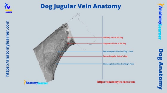
Dog jugular vein anatomy
Here, I will show the external dog jugular vein anatomy with the labeled diagram. This external jugular vein is considered the main channel for returning venous blood from the head.
If you see the diagram, this jugular vein begins with the linguofacial and maxillary veins union. They join at the caudal to the mandibular salivary gland or at the transverse plane through the cricoid cartilage of the trachea.
The diagram and length of the canine jugular vein vary in different species. The canine jugular vein’s diameter is about 1 centimeter, whereas the length is about 10 centimeters.
Generally, the jugular vein of animals doesn’t possess any valves, but in older dogs’ jugular veins, you may find non-functional valves. Now, the jugular vein passes through the jugular furrow of the dog’s neck.
Below the superficial fascia, the jugular vein runs caudally and crosses the lateral surface of the cleidocephalicus muscle obliquely. The jugular vein directly lies deep into the skin from the upper part to the middle of the neck.
So, this area (ventral part of the middle neck) is considered the best site for a successful jugular venipuncture in the dog. The canine jugular vein runs towards the shoulder joint from the middle of the neck.
Now, at the cranial border of the dog’s shoulder, this jugular vein receives the omobrachial and cephalic veins. You know these 2 veins (omobrachial and cephalic) come from the brachium of the dogs.
Again, the jugular vein receives superficial cervical and main subclavian veins at different levels. You will see the connection of the internal jugular vein on the medial aspect of the internal jugular vein (IJV) at the level of the shoulder joint.
Canine internal jugular vein
The canine internal jugular vein has no clinical importance compared to the external vein. Again, the diameter of the canine internal vein is less (approximately 1 millimeter) than the external jugular vein.
This internal jugular vein formed in the tympanooccipital fissure. Now, the canine internal vein runs along with the internal carotid artery.
Then, it lies in the covering of the common carotid artery in the dog’s neck (ventrolateral aspect). You will see the complex anatomical course of the canine internal jugular vein from its origin to the ends.
On its course, this internal jugular vein receives an anastomosing branch from the laryngeal tributary of a lingual vein. It also receives the cranial thyroid vein at the level of the caudal part of the larynx.
This cranial thyroid vein comes from the cranial pole of the dog’s thyroid lobe. You may also find another anastomotic connection of the internal jugular vein with the external one.
A medial retropharyngeal branch of the vein also drains blood into the internal jugular vein. The anatomy of the canine internal jugular vein also shows that the small middle thyroid vein drains blood into this vein.
And you know, this small middle thyroid vein comes from the caudal poles of both thyroid glands. Again, the caudal thyroid vein terminates directly into the brachiocephalic vein of the dogs.
Finally, the dog internal jugular vein terminates in the caudal part of the (1) external jugular vein. Sometimes you may find the termination of the internal jugular vein in the brachiocephalic vein of the dogs (rare).
So, the anatomical facts (especially location and course) of both the dog’s external and internal jugular veins are shown. Now, you may know how maxillary and linguoifacial veins are formed in the dog.
Lingofacial and maxillary veins of the dog
You may also know little anatomical facts about other different veins that are related to both the internal and external jugular veins of dogs. Just caudal to the mandibular salivary gland, you will find 2 major veins (linguofacial and maxillary) that join together to form the external jugular vein.
First, let’s know how the linguofacial vein is formed or collects blood and drains into the jugular vein. Followings are the major veins that help to form the linguofacial vein in dogs –
- Lingual vein of the dog, and
- Facial vein of the dog,
Again, the lingual and facial veins receive blood from other different veins. Table 1 shows the different veins that form the lingual and facial veins in the dog.
| Major veins | Receive these veins |
| Lingual vein | Ascending pharyngeal vein Cranial laryngeal vein Submental vein Sublingual vein |
| Facial vein | Deep facial vein Dorsal nasal vein Lateral nasal vein Infraorbital vein Superior labial vein Inferior labial vein Angular vein |
Except for these mentioned veins, you will also find other veins that drain blood into lingual and facial veins. Again, the lingual and facial veins join to form the linguofacial vein that also receives some veins (like cranial thyroid and medial retropharyngeal veins).
Again, the maxillary vein receives the major veins that are also shown in Table 2 –
| Major vein | Maxillary vein receives |
| Maxillary vein | Superficial temporal vein Transverse facial Rostral auricular vein Deep temporal vein Dorsal external ophthalmic vein Medial auricular vein Auricular veins – caudal, lateral, and intermediate |
You will find some venous plexus that have a direct connection with the linguofacial and maxillary veins of the dogs.
Facial veins that contribute to forming jugular vein
As I told you before, the venous system of the head and face region are very complicated in a dog. So, in this article, it is very hard to describe every single vein that forms the linguofacial part of the jugular vein properly.
Rather, I would like to provide a short anatomical description of the major veins from the face and lingual surface of the dog. Let’s see the following veins that contribute to the main facial vein in dogs –
Inferior labial vein – this vein runs along the ventral border of the buccinator muscle of the dog. The inferior labial vein receives an anastomosis from a submental vein.
Now, this vein also runs along the margin of the digastricus muscle and over the lateral surface of the mandible.
Superficial labial vein – this vein runs along the dorsal margin of the buccinator’s muscle. At the rostral end of the zygomatic arch, this superficial labial vein joins with the facial vein laterally.
You know, the superficial labial vein drains blood from the superior lip and dorsal part of the cheek.
Deep facial vein – this is a major deep vein in the dog’s face that receive numerous anastomoses. The deep facial vein arises from the ventral part of the orbit and adjacent pterygopalatine fossa.
Then it lies along the cranial part of the masseter muscle, just ventral to the zygomatic arch. You will find anastomosis with the superficial temporal vein that obliquely crosses (right to the left)the lateral surface of the zygomatic arch.
Angular vein of dog – this is a small vein in the dog that comes from the commissure of the lip. This vein joins with the facial vein just caudal to the lip commissure.
Dog facial veins (upper part)
Dorsal and lateral nasal veins – these two veins are small and enter into the main facial vein. They drain blood from the dorsal and lateral walls of the dog’s nasal cavity.
Infraorbital vein – this infraorbital vein is moderately long in dogs and joins with the ventral aspect of the facial vein. The infraorbital vein lies dorsal to the infraorbital foramen and runs along with the infraorbital nerve and muscles.
This infraorbital vein will unite with the main facial vein at the rostral part of the pterygopalatine fossa.
Inferior palpebral vein – this vein drains blood from the inferior eyelid (inner) at the lateral commissure of an eye. This vein joins with the dorsal surface (margin) of the facial vein on the side (2) of the dog’s maxilla.
External ethmoid vein – this is a small vein that passes through the ethmoidal foramen. The external ethmoid vein joins with the dorsal external ophthalmic vein.
Dorsal and ventral ophthalmic veins in dog – the ventral branch of the ophthalmic vein of the dog joins with the dorsal segment of the main ophthalmic vein. Here, you will find different anastomosis that joins with both the dorsal and ventral segments of the ophthalmic vein.
Again, the ventral ophthalmic vein turns ventrally and receives a branch from the third eyelid. Finally, they join with the main facial vein and drain blood into dog jugular vein through the lingofacial vein.
Here, the dorsal segment of the ophthalmic vein extends to the orbital fissure. Thus, it lies in the caudal two third of the orbit of a dog.
Lingual vein formation in dog
Followings are the major veins that contribute to forming lingual veins in the dog –
Cranial laryngeal vein – this is a small vein that collects blood from the cranial part of the larynx. It leaves the larynx just ventral to the cranial aspect of the thyroid cartilage.
You will find a great communication of the cranial laryngeal vein with the ascending pharyngeal vein. They also join with the lingual vein separately. You will also find a communicating branch to the internal jugular vein.
Ascending pharyngeal vein – this ascending pharyngeal vein passes between the vagosympathetic trunk and the internal carotid artery. This ascending pharyngeal vein of the dog terminates in the hyoid venous arch.
You will also find a communicating branch from the ascending pharyngeal vein to the internal jugular vein.
Hyoid venous arch – this is an unpaired and large vein that lies ventral to the basihyoid bone of the dog. This vein joins with both the right and left lingual veins just caudal to the termination of the sublingual vein.
The hyoid venous arch also receives the branch from a submental vein on each side. You will see anastomosis of the hyoid venous arch with the cranial laryngeal vein.
A submental vein in dogs – this vein receives blood from the mylohyoid and geniohyoid muscles. This vein runs caudally along the ventral part of the body of the dog’s mandible.
The lingual vein in dog – now the lingual vein joins with the facial vein and forms the lingofacial vein in the dog. It starts at the level of the tongue’s apex and runs caudally.
On the course of the lingofacial vein of the dog, it receives small branches from the cranial thyroid and medial retropharyngeal veins.
How maxillary vein is formed in a dog?
In the formation of the maxillary vein in a dog, you will find the below-mentioned major veins’ contributions –
Auricular veins – you will see different auricular veins that have a great contribution to draining blood into the maxillary vein. The labeled diagram identifies all the auricular veins (like caudal, deep, lateral, and intermediate).
Here, the lateral and intermediate auricular veins join to form the caudal auricular vein in the dog. Again, the deep auricular vein drains blood into the caudal auricular vein.
Superficial temporal vein – this vein passes within the mandibular foramen and receives branches from the medial aspect of the mandible. You will also see a small emissary vein that passes through the retroarticular foramen.
Different venous plexus also contribute to forming the maxillary vein in the dog. Now, the maxillary vein passes within the alar canal and joins with the lingofacial vein to form the jugular vein.
How to draw blood from a dog by yourself
This is very important for the clinician or practitioner to collect blood from the dog for different diagnostic purposes. There are 3 major veins that you may use to collect blood from the dog by yourself.
If you want to draw blood from the dog, you might know the exact location of the major superficial veins. First, let’s know the major sites from which the blood of dogs can collect.
Dog blood draw sites
As I told you before, there are 3 major sites from where you may easily collect blood from the dog. Again, you may also use these sites to the administration of drugs.
The followings are the 3 major sites that are used to collect blood from the dogs –
- Jugular vein of the dogs (superficial vein in the neck),
- Cephalic vein of the dogs (vein in the fore leg), and
- A saphenous vein in the dogs (hind leg vein),
You may know the exact location of the cephalic and saphenous vein from the below-mentioned articles –
Dog saphenous vein anatomy – location and blood collection procedure with the labeled diagram, and
Dog cephalic vein anatomy – location and procedure of blood collection with diagram,
As mentioned earlier, these articles might help you find the exact blood drawing sites from the dog’s fore and hind limbs.
Drawing blood from a dog jugular vein
You may follow the below-mentioned procedure in drawing blood from the dog jugular vein –
- Let’s locate the exact location of the superficial jugular vein from the dog’s neck,
- You may locate this superficial jugular vein at the level of temporomandibular articulation (or caudal to the mandibular salivary gland),
- Press this area with the thumb and passes caudally,
- Hold the vein with a tight grip (just ventral to the midline of the lateral aspect of dog’s neck),
- You may use a sock to visualize this vein perfectly,
- The vein will raise, and you should insert the needle vertically,
Thus, you may easily collect blood from the jugular vein of the canine.
Feline jugular vein anatomy – cat jugular vein
The feline jugular vein anatomy is almost similar to the dog’s. But, you may find a little variation in the branches of different veins that form the feline or cat external jugular vein.
Here, the external jugular vein of a cat runs along the ventrolateral aspect of the neck and passes caudally. The temporomandibular articulation may be considered a landmark for superficially locating the cat jugular vein.
Again, the internal jugular vein of a cat joins directly with the medial aspect of the external jugular vein. Finally, they join with the brachiocephalic vein and drain blood into the cranial vena cava of the cat.
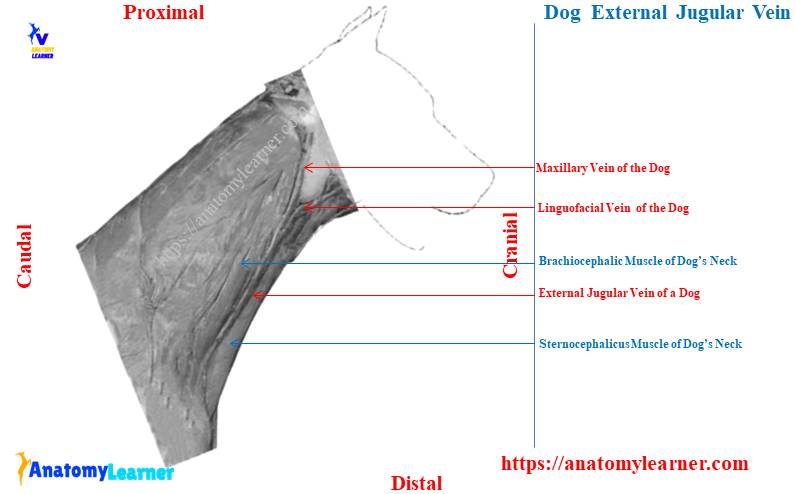
Dog jugular vein diagram
Now, I will provide different labeled diagrams on the dog jugular vein. Though you have already found different diagrams on canine jugular vein throughout the article, the following diagrams provide a clear concept.
First, let’s see some of the important muscles (brachiocephalicus, sternocephalicus, and omohyoideus) muscles from the dog’s neck. All these muscles are identified in the dog neck labeled diagram, which actually formed the jugular furrow.
The external jugular vein that passes within this jugular furrow is identified in the labeled diagram. Again, the different small branches of veins that drain blood into the external jugular vein are also identified in the diagram.
A small portion of the lingofacial and maxillary veins are shown in the diagram. Let’s find more diagrams on the canine jugular vein on social media of anatomy learners.
Frequently asked questions on dog jugular vein
Anatomy learners ask numerous questions about the dog jugular vein. Here, I will enlist these questions on the canine jugular vein that these learners frequently ask.
I will try to provide concise answers to the frequently asked questions on the canine jugular vein. Okay, let’s see the questions and their concise answers on the jugular vein –
How to hit the jugular vein on a dog?
First, you should restrain the dog by holding its neck and mouth. You might also need an assistant to hold the dog’s legs (front and back legs).
Now, you should locate the jugular vein from the dog’s neck perfectly. Just press the vein with your thumb and runs caudally (on the course of a jugular vein).
Hold the jugular vein with a tight grip, and then the vein will raise. Now, you may easily hit the jugular vein and collect blood from it.
Where is the jugular vein in a dog?
The jugular vein is situated at the ventral part of the lateral aspect of the dog’s neck. You will find the same jugular vein on both sides of the lateral aspect of the dog’s neck.
This vein runs just caudal to the mandibular salivary gland of the dogs. Or, you may consider the temporomandibular articulation of the dog’s neck as a landmark to locate this vein.
Do dogs have two jugular veins?
Yes, dogs have two jugular veins on their neck. They extend from the temporomandibular articulation to the point of the shoulder.
All the anatomical facts of the canine jugular vein have been described in this article. You may get the basic idea of their courses from both the right and left lateral aspects of a dog’s neck.
How big is a jugular vein in a dog?
The diameter of the dog jugular vein is about 1 centimeter, and the length is about 10 – 12 centimeters. You may easily find this vein in the superficial area of the dog’s neck.
Conclusion
So, you need to know the dog jugular vein anatomy to locate it perfectly from the lateral aspect of the neck. For a successful venipuncture in the jugular vein, you have a clear concept of the structures of a jugular furrow.
The right and left external jugular veins of the dog course in a similar pattern (but have a few exceptional ones). As the external jugular vein of a dog runs superficially from the temporomandibular articulation to the point of the shoulder, this might be a great site for drawing blood from the dogs.